. 2004 Mar;113(5):764-73.
doi: 10.1172/JCI19528.

Increased CD36 protein as a response to defective insulin signaling in macrophages

Affiliations

Increased CD36 protein as a response to defective insulin signaling in macrophages

Chien-Ping Liang et al. J Clin Invest. 2004 Mar.

Abstract

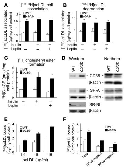
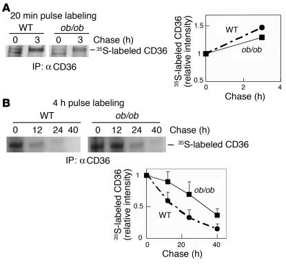
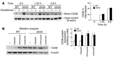
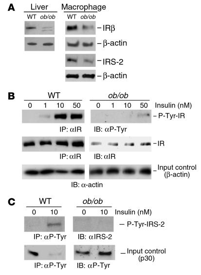
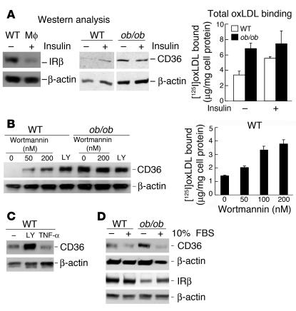
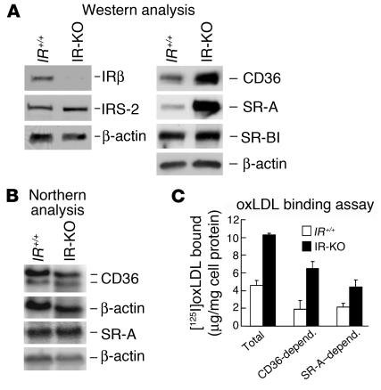
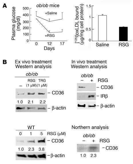
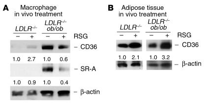
Accelerated atherosclerosis is a major cause of morbidity and death in insulin-resistant states such as obesity and the metabolic syndrome, but the underlying mechanisms are poorly understood. We show that macrophages from obese (ob/ob) mice have increased binding and uptake of oxidized LDL, in part due to a post-transcriptional increase in CD36 protein. Macrophages from ob/ob mice are also insulin resistant, as shown by reduced expression and signaling of insulin receptors. Three lines of evidence indicate that the increase in CD36 is caused by defective insulin signaling: (a) Treatment of wild-type macrophages with LY294002, an inhibitor of insulin signaling via PI3K, results in an increase in CD36; (b) insulin receptor knockout macrophages show a post-transcriptional increase in CD36 protein; and (c) administration of thiazolidinediones to intact ob/ob mice and ob/ob, LDL receptor-deficient mice results in a reversal of macrophage insulin receptor defects and decreases CD36 protein. The last finding contrasts with the increase in CD36 that results from treatment of macrophages with these drugs ex vivo. The results suggest that defective macrophage insulin signaling predisposes to foam cell formation and atherosclerosis in insulin-resistant states and that this is reversed in vivo by treatment with PPAR-gamma activators.


Figures

Figure 1
Figure 1
Enhanced uptake of modified LDL in ob/ob versus WT mouse peritoneal macrophages is mediated by increases in cell surface expression of CD36 and SR-A. (A–C) [125I,3H]acLDL cell association (A), degradation (B), and cholesteryl ester formation (C) are higher in ob/ob than in WT macrophages following acLDL loading. One representative experiment of three independent experiments each using pooled macrophages from five WT and seven ob/ob mice is shown. Short-term treatment (5 hours) of macrophages with either insulin or leptin does not change these parameters. C → CE (vertical axis, C), cholesterol to cholesteryl ester. (D) Protein expression of scavenger receptors CD36 and SR-A is increased while SR-BI expression is decreased in ob/ob versus WT macrophages, as determined by Western analysis (left). Protein extracts were prepared from pooled macrophages of five WT and five ob/ob mice. One experiment representative of four independent experiments is shown. CD36 and SR-A mRNA was not upregulated in ob/ob versus WT macrophages, as shown by Northern analysis (right). Northern analysis was performed with random-primed CD36, SR-A, and actin cDNA probes using total RNA isolated from pooled macrophages of ten mice of each strain. One experiment representative of three independent experiments is shown. (E) Specific binding of oxLDL to ob/ob macrophages is elevated. (F) Effects of fucoidan and anti-CD36 antibody on oxLDL binding to ob/ob and WT macrophages. [125I]oxLDL binding assays were performed with pooled macrophages isolated from five mice of each strain, preincubated with the SR-A ligand fucoidan (20 μg/ml), mouse anti–CD36 IgA (20 μg/ml), or mouse control IgA (20 μg/ml, not shown). The decreases in total oxLDL binding in the presence of CD36 IgA or fucoidan were considered as binding mediated by CD36 or SR-A, respectively. CD36 contributes more to the increase in oxLDL binding to ob/ob versus WT macrophages than SR-A. One experiment representative of three independent experiments is shown. CD36-depend. and SR-A depend., oxLDL binding mediated by CD36 and SR-A, respectively.
Figure 2
Figure 2
CD36 protein turnover is decreased in ob/ob macrophages. Pooled WT or ob/ob macrophages were pulse-labeled with [35S]methionine/cysteine cell-labeling mix for 20 minutes (A) or 4 hours (B), then chased in medium with methionine/cysteine for the times indicated. 35S-labeled CD36 in total lysates was immunoprecipitated with anti-CD36 and subjected to SDS-PAGE followed by transfer to nitrocellulose membrane and autoradiography. 35S intensities were quantified by densitometric analysis, and each number was normalized for input (measured by Western analysis of β-actin in lysates), then to the relative intensity at chase time 0 hours. For 20-minute and 4-hour labeling experiments, one experiment representative of two and four independent experiments, respectively, is shown. Macrophages were isolated from five to seven mice of each strain.
Figure 3
Figure 3
CD36 is internalized similarly in WT and ob/ob macrophages and is increased in lysosome- and proteasome-inhibited WT but not in ob/ob macrophages. (A) CD36 in ob/ob macrophages is endocytosed at a rate similar to that of WT cells. Cell surface proteins were biotinylated in pooled macrophages of five mice of each strain. One experiment representative of two independent experiments is shown. Biotinylated CD36 was allowed to internalize for different times, as indicated. Surface biotin was then removed with glutathione (+). Cells without glutathione (–) were used for the measurement of total biotinylated CD36 left in the cells at the time point. The total biotinylated CD36 at time 0 was used as a reference (100%) for the quantification of internalized CD36 (all corrected for input with β-actin). Percent internalization was calculated as described (27). (B) CD36 protein is increased by lysosomal and proteasomal inhibitors in WT but not in ob/ob macrophages. Pooled macrophages isolated from five mice of each strain were treated with calpeptin (30 μg/ml), chloroquine (50 μM), or lactacystin (1 μM) for 1 day. Total lysates were prepared, and Western analysis was performed. One experiment representative of three independent experiments is shown.
Figure 4
Figure 4
Short-term treatment with insulin or PI3 kinase inhibitors, or long-term treatment with glucose or fatty acids, does not increase CD36 protein expression in WT macrophages. (A–D) Pooled WT macrophages (A, B, and D) or WT and ob/ob monocytes (C) isolated from three to ten mice were incubated with insulin (1 μM) for 0.5 hours or with the PI3 kinase inhibitors wortmannin (200 nM) or LY294002 (20 μM) for 2 hours (A), with glucose or mannitol at the indicated concentrations for 2 days (B), with 400 mg/dl of glucose or mannitol for 1 day (C), or with oleic acid– or palmitic acid–BSA complexes for 1 day (D). Troglitazone (TRG; 1 μM) was used as a positive control. Treated cells were then used for oxLDL binding assays with the SR-A ligand fucoidan (50 μg/ml) added to the binding buffer or for total protein extraction followed by Western analysis. One experiment representative of two (A, B, and D) or three (B) independent experiments is shown. Mφ, macrophage; FA, fatty acid; Conc., concentration.
Figure 5
Figure 5
The expression and signaling of insulin receptor is downregulated in ob/ob versus WT macrophages. (A) Insulin receptor β-subunit (IRβ) expression is decreased in pooled ob/ob compared with WT macrophages isolated from six mice of each strain, as determined by Western analysis. One experiment representative of three independent experiments is shown. A similar expression pattern is also found in ob/ob liver. (B and C) Insulin-dependent tyrosine phosphorylation of IR (B) and IRS-2 (C) in ob/ob macrophages is defective, even at a higher insulin concentration (10 nM) than ob/ob plasma insulin levels (about 4 nM). Ex vivo tyrosine phosphorylation of IR or IRS-2 by IR tyrosine kinase in response to insulin in 32P-preincubated pooled ob/ob and WT macrophages isolated from five mice each was performed for 10 minutes. Total protein lysates were then subjected to immunoprecipitation with anti-IR or anti-phosphotyrosine (αP-Tyr) and separated by SDS-PAGE followed by membrane transfer and 32P autoradiography. The membranes were then probed with anti–P-Tyr or anti-IR (for IR) or with anti–IRS-2, respectively. In C (bottom), P-Tyr p30 whose tyrosine phosphorylation status was not affected by insulin is shown to reflect the amounts of initial extracts used for immunoprecipitation of phosphoproteins. One experiment representative of two independent experiments is shown. IP, immunoprecipitation; IB, immunoblotting.
Figure 6
Figure 6
Defects in insulin signaling in mouse macrophages increase oxLDL binding and CD36 protein expression. (A) Ex vivo effects of chronic high-dose insulin on insulin receptor β-subunit (far left) and CD36 protein expression (middle panels), and oxLDL binding to WT and ob/ob macrophages (far right). Macrophages were incubated with (+) or without (–) insulin (200 nM) for 1 day. Cells were used for oxLDL binding assays or Western analysis with the indicated antibodies. (B) The increase in CD36 protein expression in WT macrophages by the PI3 kinase inhibitor wortmannin is dose dependent. WT or ob/ob macrophages were treated with wortmannin at the indicated concentrations or another inhibitor, LY294002 (LY; 10 μM), for 1 day, followed by protein extraction and Western analysis (left) and by oxLDL binding assays with fucoidan (50 μg/ml) in the binding buffer (right). Inhibition of the insulin receptor effector PI3 kinase results in an increase in oxLDL binding to macrophages. (C) TNF-α has no effect on CD36 protein expression. WT macrophages were treated with LY294002 (LY; 10 μM) or TNF-α (10 ng/ml) for 1 day. Total lysates were prepared and Western analysis was performed. (D) CD36 protein expression is reduced by 10% FBS in ob/ob and WT macrophages. Insulin receptors are increased under this condition. All experiments were performed with pooled macrophages isolated from three to six mice of each strain indicated. One experiment representative of three independent experiments is shown.
Figure 7
Figure 7
Lack of insulin receptors in peritoneal macrophages leads to upregulation of CD36 protein expression and oxLDL binding. (A) Western analysis of macrophage IRβ subunit, IRS-2, CD36, SR-A, and SR-BI protein expression in IR knockout (IR-KO) mice rescued with the human IR transgene and in control littermate (IR+/+) mice. Both CD36 and SR-A protein are increased in IR-deficient macrophages. (B) Northern analysis of CD36 and SR-A mRNA levels in macrophages of IR-KO and IR+/+ mice. (C) Assay of oxLDL binding to macrophages from IR-KO and control IR+/+ was carried out with or without the addition of fucoidan (50 μg/ml), mouse anti-CD36 IgA (20 μg/ml), or control IgA (20 μg/ml; not shown) in the binding buffer. CD36-depend. and SR-A–depend., oxLDL binding mediated by CD36 and SR-A, respectively. All experiments were performed with pooled macrophages isolated from three to five mice of each strain. One experiment representative of three independent experiments is shown.
Figure 8
Figure 8
Treatment of ob/ob mice with rosiglitazone improves insulin resistance and normalizes oxLDL binding and CD36 protein expression in macrophages. (A) In vivo rosiglitazone (RSG) treatment reduces plasma glucose levels and oxLDL binding to macrophages in ob/ob mice. Plasma glucose levels (left) and macrophage oxLDL binding (right) were assessed in ob/ob mice treated with rosiglitazone or control saline. At the times indicated, plasma glucose levels from these mice were measured. After 3 weeks, macrophages were collected and an oxLDL binding assay was performed with the addition of fucoidan (50 μg/ml) in the binding buffer. (B) Differential patterns of CD36 protein expression in ob/ob macrophages in response to thiazolidinedione treatments in vivo and ex vivo. Left, Western analysis of CD36 protein expression in ob/ob and WT macrophages treated ex vivo with RSG or troglitazone (TRG) at the indicated doses for 1 day. Right, the expression of CD36 and IR β-subunit (IRβ) protein in macrophages from ob/ob mice described in A was determined by Western analysis. Macrophage CD36 protein was normalized in ob/ob mice, though its mRNA was increased by rosiglitazone in vivo, as measured by Northern analysis. Macrophage experiments were performed with pooled cells isolated from three to five mice of each strain indicated. One experiment representative of two independent experiments is shown.
Figure 9
Figure 9
In vivo treatment with rosiglitazone reduces macrophage CD36 protein expression in ob/ob LDLR–/– mice while increasing its expression in LDLR–/– mice. (A) Western analysis of macrophage CD36 and SR-A protein expression from mice with or without rosiglitazone (RSG) treatment for 3 weeks as described in Methods. (B) For comparison, the expression of CD36 protein in adipose tissue of mice described in A was determined by Western analysis. CD36 protein is upregulated by PPAR-γ activator in adipose tissue of both ob/ob LDLR–/– and LDLR–/– mice. All mice treated with RSG showed a similar increase in CD36 protein expression in adipose tissue, and results from one mouse of each strain were shown here. Macrophage experiments were performed with pooled cells isolated from three mice of each strain. One experiment representative of two independent experiments is shown.

References

    1. Haffner SM, Lehto S, Ronnemaa T, Pyorala K, Laakso M. Mortality from coronary heart disease in subjects with type 2 diabetes and in nondiabetic subjects with and without prior myocardial infarction. N. Engl. J. Med. 1998;339:229–234. - PubMed
    1. Ginsberg HN. Insulin resistance and cardiovascular disease. J. Clin. Invest. 2000;106:453–458. - PMC - PubMed
    1. Reaven GM. Multiple CHD risk factors in type 2 diabetes: beyond hyperglycaemia. Diabetes Obes. Metab. 2002;4:S13–S18. - PubMed
    1. Haffner SM. Lipoprotein disorders associated with type 2 diabetes mellitus and insulin resistance. Am. J. Cardiol. 2002;90:55i–61i. - PubMed
    1. Steinberg HO, Baron AD. Vascular function, insulin resistance and fatty acids. Diabetologia. 2002;45:623–634. - PubMed

Publication types

MeSH terms