. 2004 Mar 1;164(5):653-9.
doi: 10.1083/jcb.200307032.

Phosphoinositide binding and phosphorylation act sequentially in the activation mechanism of ezrin

Affiliations

Phosphoinositide binding and phosphorylation act sequentially in the activation mechanism of ezrin

Bruno T Fievet et al. J Cell Biol. .

Abstract

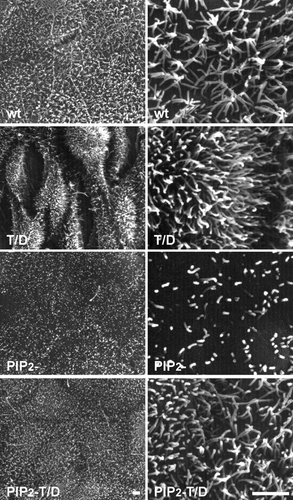
Ezrin, a membrane-actin cytoskeleton linker, which participates in epithelial cell morphogenesis, is held inactive in the cytoplasm through an intramolecular interaction. Phosphatidylinositol 4,5-bisphosphate (PIP2) binding and the phosphorylation of threonine 567 (T567) are involved in the activation process that unmasks both membrane and actin binding sites. Here, we demonstrate that ezrin binding to PIP2, through its NH2-terminal domain, is required for T567 phosphorylation and thus for the conformational activation of ezrin in vivo. Furthermore, we found that the T567D mutation mimicking T567 phosphorylation bypasses the need for PIP2 binding for unmasking both membrane and actin binding sites. However, PIP2 binding and T567 phosphorylation are both necessary for the correct apical localization of ezrin and for its role in epithelial cell morphogenesis. These results establish that PIP2 binding and T567 phosphorylation act sequentially to allow ezrin to exert its cellular functions.


Figures

Figure 1.
Figure 1.
PIP2 ezrin is restricted to the cytoplasm. (A) Localization of wt and PIP2 ezrin by double fluorescence with anti–VSV G antibody (ezrin VSVG) and phalloidin (actin) with or without preextraction with a 0.5% Triton X-100 buffer. Images of xz sections, and apical, median, basal focal xy sections are shown. wt ezrin is associated with actin of the apical microvilli. PIP2 ezrin is present in the cytoplasm and is totally extracted with detergent. Bars, 10 μm. (B) Association of wt, PIP2 , T/D, and PIP2 T/D ezrin with the cytoskeleton. Similar amount of total (Tot), soluble (Sol) or insoluble material (Ins) were analyzed by Western blotting with an anti–VSV G antibody. (C) Distribution of ezrin mutants in membrane and cytosol fractions. Total (Tot), cytosolic (Cyt), and membrane-associated material concentrated eightfold (Mb 8x) were immunoblotted with anti–VSV G antibody. The amount of ezrin was quantified by densitometric analysis of serially diluted fractions obtained from three independent experiments. Average values are given ± SEM. (D) wt ezrin is released from the membrane and extracted with Triton X-100 buffer (+ext) after ionomycin treatment (iono).
Figure 2.
Figure 2.
Membrane localization of Nter and NterPIP 2 ezrin. (A) Fluorescence was performed as in Fig. 1. Nter and NterPIP2 ezrin are localized at the membrane but in a nonpolarized manner. Bar, 10 μm. (B) Distribution of ezrin mutants in membrane and cytosol fractions. Total (Tot), cytosol (Cyt), and membrane-associated material (Mb) were immunoblotted with an anti–VSV G antibody.
Figure 3.
Figure 3.
Ezrin phosphorylation at T567 requires the interaction of the N-ERMAD with PIP2. (A) Immunoprecipitated wt and PIP2 ezrin were analyzed by Western blot with an anti–VSV G antibody (VSVG) or with an antibody recognizing ezrin phosphorylated at T567 (T567P). +PPase indicates than immunoprecipitated proteins were treated with λ-phosphatase. In contrast to wt ezrin, PIP2 ezrin is not phosphorylated at T567. (B) After transient transfection, wtPH and PIP2 PH ezrin were localized by immunofluorescence with an anti–VSV G antibody. Both proteins are localized at the plasma membrane and in microvilli. Bar, 10 μm. (C) Distribution of wtPH and PIP2 PH ezrin in membrane and cytosol fractions was obtained as in Fig. 2. (D) Immunoprecipitated wtPH ezrin is phosphorylated at T567, whereas PIP2 PH ezrin is not phosphorylated.
Figure 4.
Figure 4.
Unpolarized localization of T/D and PIP2 T/D ezrin at the plasma membrane. Fluorescence was performed as in Fig. 1. T/D ezrin was mainly located on the dorsal surface of the cells and in cell–cell contacts. PIP2 T/D ezrin was uniformly distributed at the plasma membrane. T/D and PIP2 T/D ezrin remained associated with the cortical actin cytoskeleton after extraction with the Triton X-100 buffer. Bars, 10 μm. (B) Membrane localization of T567D and PIP2 T/D ezrin and cytoskeleton association are preserved after ionomycin treatment followed by extraction with the Triton X-100 buffer before fixation.
Figure 5.
Figure 5.
Binding of ezrin to PIP2 is required for its function in epithelial cell morphogenesis. Scanning electron microscopy of confluent cells grown on filters for 4 d producing wt, T/D, PIP2 , and PIP2 T/D ezrin. Clones producing PIP2 ezrin formed a well-organized monolayer displaying shorter and more disperse microvilli on their cell surface as compared with cells producing wt ezrin. Cells producing PIP2 T/D ezrin did not show the aberrant morphology observed with cells producing T/D ezrin. Bars, 2 μm.

References

    1. Algrain, M., O. Turunen, A. Vaheri, D. Louvard, and M. Arpin. 1993. Ezrin contains cytoskeleton and membrane binding domains accounting for its proposed role as a membrane–cytoskeletal linker. J. Cell Biol. 120:129–139. - PMC - PubMed
    1. Barret, C., C. Roy, P. Montcourrier, P. Mangeat, and V. Niggli. 2000. Mutagenesis of the phosphatidylinositol 4,5-biphosphate (PIP2) binding site in the NH2-terminal domain of ezrin correlates with its altered cellular distribution. J. Cell Biol. 151:1067–1079. - PMC - PubMed
    1. Bretscher, A., K. Edwards, and R.G. Fehon. 2002. ERM proteins and merlin: integrators at the cell cortex. Nat. Rev. Mol. Cell Biol. 3:586–599. - PubMed
    1. Gary, R., and A. Bretscher. 1995. Ezrin self-association involves binding of an N-terminal domain to a normally masked C-terminal domain that includes the F-actin binding site. Mol. Biol. Cell. 6:1061–1075. - PMC - PubMed
    1. Gautreau, A., D. Louvard, and M. Arpin. 2000. Morphogenic effects of ezrin require a phosphorylation-induced transition from oligomers to monomers at the plasma membrane. J. Cell Biol. 150:193–203. - PMC - PubMed

Publication types