. 2004 Mar 1;164(5):661-6.
doi: 10.1083/jcb.200312001.

Recruitment of Eph receptors into signaling clusters does not require ephrin contact

Affiliations

Recruitment of Eph receptors into signaling clusters does not require ephrin contact

Sabine H Wimmer-Kleikamp et al. J Cell Biol. .

Abstract

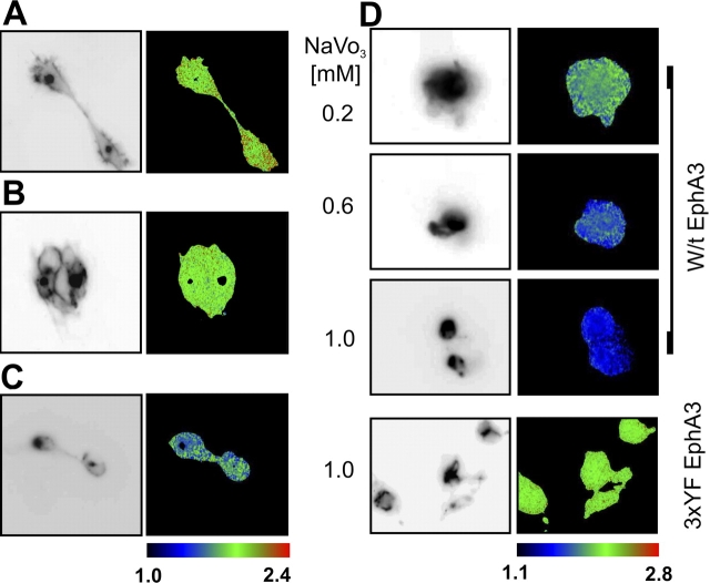
Eph receptors and their cell membrane-bound ephrin ligands regulate cell positioning and thereby establish or stabilize patterns of cellular organization. Although it is recognized that ephrin clustering is essential for Eph function, mechanisms that relay information of ephrin density into cell biological responses are poorly understood. We demonstrate by confocal time-lapse and fluorescence resonance energy transfer microscopy that within minutes of binding ephrin-A5-coated beads, EphA3 receptors assemble into large clusters. While remaining positioned around the site of ephrin contact, Eph clusters exceed the size of the interacting ephrin surface severalfold. EphA3 mutants with compromised ephrin-binding capacity, which alone are incapable of cluster formation or phosphorylation, are recruited effectively and become phosphorylated when coexpressed with a functional receptor. Our findings reveal consecutive initiation of ephrin-facilitated Eph clustering and cluster propagation, the latter of which is independent of ephrin contacts and cytosolic Eph signaling functions but involves direct Eph-Eph interactions.


Figures

Figure 1.
Figure 1.
The dynamics of ephrin-A5 clustering and internalization. (A) Confocal time-lapse microscopy of ephrin binding and clustering. Preclustered Alexa ephrin-A5Fc was added to EphA3/293 cells and confocal microscopic images of live cells during a 60 min time course were taken every minute starting 5 min before ephrin addition. Arrowheads highlight prominent ephrin cluster. A corresponding Video 1 is available at http://www.jcb.org/cgi/content/full/jcb.200312001/DC1. (B) EphA3/293 cells were treated with preclustered Alexa ephrin-A5Fc in the absence (I) or in the presence (II) of “Fc block,” or with nonclustered Alexa ephrin-A5Fc (III). After 30-min incubation, fixed cells were analyzed by confocal fluorescence microscopy. (C) Before stimulation with preclustered Alexa ephrin-A5Fc, the lysosomal compartment of EphA3/293 cells was stained with Lysotracker green. Alexa ephrin-A5Fc internalization was monitored sequentially at two excitation wavelengths. Resulting green (Lysotracker) and red (Alexa Fluor 546) images were merged. Bars, 20 μm.
Figure 2.
Figure 2.
Ephrin-A5 induced assembly of EphA3 signaling clusters. (A–C) EphA3-GFP transfected HEK293cells, unstimulated (A) or stimulated (30 min) with soluble (B) or preclustered ephrin-A5Fc (C) were fixed, permeabilized (0.1% Triton-X 100), and incubated with CY3-PY72. The GFP fluorescence is shown as inverted gray scale (left). FRET between EphA3GFP and CY3-PY72 was detected by FLIM, and phase lifetime maps (right) are shown. “Blacked-out” areas within the lifetime maps mask regions that were excluded from FLIM analysis. They include areas within cells where high GFP fluorescence intensity resulted in saturated detection. (D) Pervanadate-induced Eph receptor phosphorylation. HEK293 cells expressing w/t EphA3GFP or 3YF EphA3GFP were incubated with pervanadate (30 min) at the indicated concentrations. Fixed cells were examined for FRET as described in A–C. Left, GFP fluorescence; right, GFP fluorescence lifetime phase maps. Tabulated color codes (dark blue to red) indicate GFP lifetimes in nanoseconds.
Figure 3.
Figure 3.
EphA3 signaling clusters extend beyond ephrin contact surfaces. (A) GFP ephrin-A2–expressing HEK293 cells were stimulated with Alexa EphA3-Fc–coated beads. The GFP ephrin-A2 and Alexa EphA3-Fc populations in contact were monitored by FLIM. Transmitted light (I), GFP fluorescence (II), phase lifetime maps (III), and the fraction (population) of ephrin-A2 in contact with Alexa EphA3-Fc (IV) are presented. Bar, 20 μm. (B) EphA3-GFP–expressing HEK293 cells were stimulated with ephrin-A5–coated beads and processed as described in Fig. 2. Images of transmitted light (I), GFP fluorescence (II), phase lifetime maps (III), and the fraction (population) of phosphorylated EphA3 (IV) are shown. Bar, 20 μm. (A and B) Tabulated color codes indicate GFP lifetimes in nanoseconds (III) and probabilities of phosphorylated EphA3GFP (IV). Arrowheads indicate the position of ephrin-coated beads.
Figure 4.
Figure 4.
Recruitment of EphA3 into signaling clusters is independent of direct ephrin contact. (A) Model of the complex between Eph and ephrin binding domains, illustrating amino acid substitutions that affect ephrin binding. The cartoon is derived from the crystal structure of the EphB2/ephrin-B2 binding domains (Himanen et al., 2001). EphA3 interfaces in the putative heterodimerization (D) and heterotetramerization sites (T) that are involved in ephrin-A5 interactions as assessed by mutagenesis analysis (Smith et al., 2003) are indicated by yellow/green shading. The positions of two ephrin-binding point mutations in nb-EphA3 are indicated by yellow arrowheads. (B) HEK293cells, transfected either with [Phe152-Leu] EphA3GFP or nb-EphA3-GFP alone, or in combination with unlabeled w/t EphA3 as indicated were stimulated with ephrin-A5Fc beads, and fixed cells were analyzed for FRET between the donor GFP and CY3-PY72 as described in Fig. 2. Transmitted light (left), GFP fluorescence (middle), and phase lifetime maps (right) are shown. (C) HEK293 cells were transfected, alone or in combination, with nb-EphA3-GFP or with untagged w/t EphA3, 3YFEphA3, or K653M EphA3 as indicated. After stimulation, protein A–bound EphA3 signaling complexes were analyzed with anti-GFP (α GFP, top) and antiphosphotyrosine (α PY, middle) antibodies. Total cell lysates were probed with anti-EphA3 antibodies to assess comparative expression levels (α EphA3, bottom). Irrelevant lanes have been removed from the Western blot. (D) HEK293 cells transfected, alone or in combination, with nb-EphA3-GFP and EphA3Δcyto. Protein A–bound complexes, recovered as in Fig. 4 C, were analyzed with anti-GFP (top) and anti-EphA3 antibodies (middle). Lysates were analyzed for EphA3 expression levels (bottom).

References

    1. Bastiaens, P.I., and R. Pepperkok. 2000. Observing proteins in their natural habitat: the living cell. Trends Biochem. Sci. 25:631–637. - PubMed
    1. Boyd, A.W., and M. Lackmann. 2001. Signals from Eph and ephrin proteins: a developmental tool kit. Sci STKE. 2001:RE20. - PubMed
    1. Boyd, A.W., L.D. Ward, I.P. Wicks, R.J. Simpson, E. Salvaris, A. Wilks, K. Welch, M. Loudovaris, S. Rockman, and I. Busmanis. 1992. Isolation and characterization of a novel receptor-type protein tyrosine kinase (hek) from a human pre-B cell line. J. Biol. Chem. 267:3262–3267. - PubMed
    1. Brown, A., P.A. Yates, P. Burrola, D. Ortuno, A. Vaidya, T.M. Jessell, S.L. Pfaff, D.D. O'Leary, and G. Lemke. 2000. Topographic mapping from the retina to the midbrain is controlled by relative but not absolute levels of EphA receptor signaling. Cell. 102:77–88. - PubMed
    1. Coulthard, M.G., J.D. Lickliter, N. Subanesan, K. Chen, G.C. Webb, A.J. Lowry, S. Koblar, C.D. Bottema, and A.W. Boyd. 2001. Characterization of the Epha1 receptor tyrosine kinase: expression in epithelial tissues. Growth Factors. 18:303–317. - PubMed

Publication types