. 2004 Mar 1;164(5):701-15.
doi: 10.1083/jcb.200312070.

Septin collar formation in budding yeast requires GTP binding and direct phosphorylation by the PAK, Cla4

Affiliations

Septin collar formation in budding yeast requires GTP binding and direct phosphorylation by the PAK, Cla4

Matthias Versele et al. J Cell Biol. .

Abstract

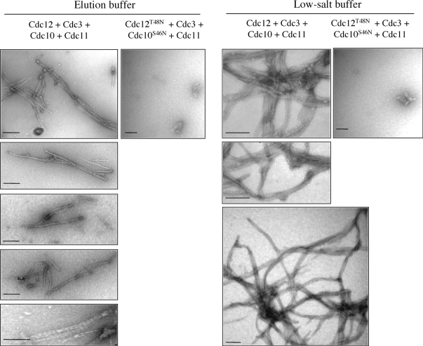
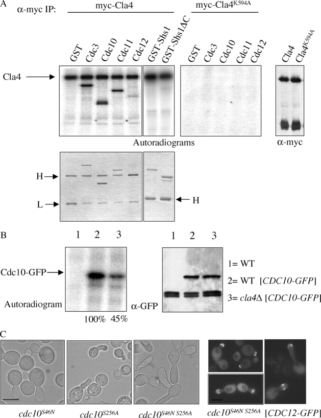
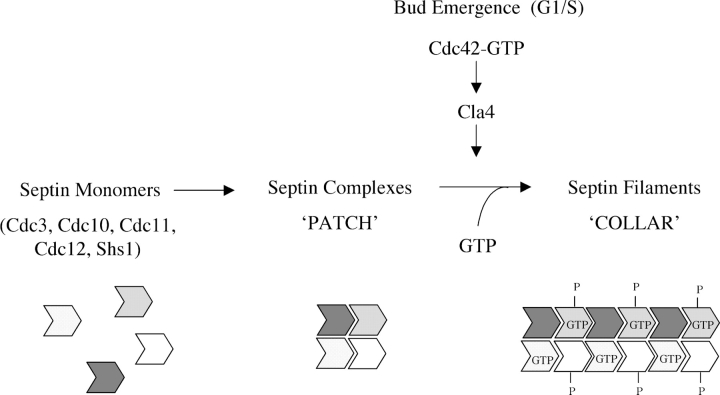
Assembly at the mother-bud neck of a filamentous collar containing five septins (Cdc3, Cdc10, Cdc11, Cdc12, and Shs1) is necessary for proper morphogenesis and cytokinesis. We show that Cdc10 and Cdc12 possess GTPase activity and appropriate mutations in conserved nucleotide-binding residues abrogate GTP binding and/or hydrolysis in vitro. In vivo, mutants unable to bind GTP prevent septin collar formation, whereas mutants that block GTP hydrolysis do not. GTP binding-defective Cdc10 and Cdc12 form soluble heteromeric complexes with other septins both in yeast and in bacteria; yet, unlike wild-type, mutant complexes do not bind GTP and do not assemble into filaments in vitro. Absence of a p21-activated protein kinase (Cla4) perturbs septin collar formation. This defect is greatly exacerbated when combined with GTP binding-defective septins; conversely, the septin collar assembly defect of such mutants is suppressed efficiently by CLA4 overexpression. Cla4 interacts directly with and phosphorylates certain septins in vitro and in vivo. Thus, septin collar formation may correspond to septin filament assembly, and requires both GTP binding and Cla4-mediated phosphorylation of septins.


Figures

Figure 1.
Figure 1.
Purified Cdc10 and Cdc12 bind and hydrolyze GTP. (A) The indicated septin preparations (1–5 μg) were analyzed by SDS-PAGE and stained with Coomassie dye. (B) GTPase activity was measured by phosphate release from γ[32P]GTP for Cdc3 (⋄), Cdc10 (♦), Cdc11 (X) and Cdc12-His6 (•), and His6-GST (*). (C) GTPase activity of Cdc12-His6 (•), Cdc12ΔC-His6 (○), Cdc12(S43V)-His6 (▴), and Cdc12(T48N)-His6 (▪), as in B. (D) GTPase activity of Cdc12ΔC-His6 (○), Cdc12ΔC(S43V)-His6 (Δ), and Cdc12ΔC(T48N)-His6 (□), as in B. (E) [32P]GTPγS binding by His6-tagged Cdc12 (•), Cdc12(S43V) (▴), and Cdc12(T48N) (▪). (F) [32P]GTPγS binding by His6-tagged Cdc12ΔC (○), Cdc12ΔC(S43V) (Δ), and Cdc12ΔC(T48N) (□). For B–F, representative data are shown for experiments that were each repeated two to three (or more) times with different septin preparations and varying substrate or ligand concentrations.
Figure 2.
Figure 2.
GTP binding-dependent functions of Cdc12 and Cdc10 overlap. (A) Wild-type (BY4741) transformed with empty vector (YCplac33), cdc12(S43V) (YMVB2), cdc12(T48N) (YMVB1), cdc12(T48N) (YMVB1) carrying an episomal vector overexpressing cdc12(T48N) (pMVB49), and cdc12(T48N) (YMVB3) carrying a CEN plasmid with CDC12 (pMVB39), were grown at 30 or 37°C for 16 h under selective conditions. Cells were stained with DAPI and examined by Nomarski optics (differential interference contrast) or fluorescence microscopy (DAPI). Bar, 5 μm. (B) Wild-type (YMVB25), cdc10(S46N) (YMVB6), cdc12(T48N) (YMVB24), cdc10(S46N) cdc12(T48N) (YMVB8), and cdc10(S41V) cdc12(S43V) (YMVB60) cells were grown in YPD to mid-exponential phase at 26, 30, or 37°C, and morphology of the cells was examined by bright-field microscopy. Bar, 5 μm.
Figure 3.
Figure 3.
Septin–septin interactions do not require GTP binding. (A) Wild-type (BY4741) or cdc12(T48N) cells (YMVB3) transformed with CEN plasmids containing Cdc3-GFP, Cdc11-GFP (pSB5), or Cdc10-GFP (pLA-10) were grown to mid-exponential phase at 30°C and shifted to 37°C for 8 h. Extracts were subjected to immunoprecipitation with anti-Cdc12ΔC antibody. The inputs and resulting immune complexes were solubilized in SDS-PAGE sample buffer and analyzed by immunoblotting. (B) Cdc12-His6 and Cdc12(T48N)-His6 (0.1 μM) were incubated with 0.1 mM GTP for 1 h, and their ability to bind the indicated proteins (immobilized as GST fusions) was assessed. Input represents 20% of the total amount of Cdc12-His6 and Cdc12(T48N)-His6 initially added in each binding reaction. (C) Septin complexes were purified on Ni2+-saturated NTA-agarose from E. coli cells coexpressing His6-Cdc12 and the other septins indicated. Eluates were resolved on SDS-PAGE, stained with Coomassie blue. (D) GTP binding to the His6Cdc12–Cdc3–Cdc10–Cdc11 complex (•), the His6Cdc12(T48N)–Cdc3–Cdc10(S46N)–Cdc11 complex (▪), and His6-GST (negative control; ▴), immobilized on Ni2+-NTA beads. Values represent the average (error bars indicate the range) for the results of a typical experiment, where each measurement was performed in duplicate.
Figure 3.
Figure 3.
Septin–septin interactions do not require GTP binding. (A) Wild-type (BY4741) or cdc12(T48N) cells (YMVB3) transformed with CEN plasmids containing Cdc3-GFP, Cdc11-GFP (pSB5), or Cdc10-GFP (pLA-10) were grown to mid-exponential phase at 30°C and shifted to 37°C for 8 h. Extracts were subjected to immunoprecipitation with anti-Cdc12ΔC antibody. The inputs and resulting immune complexes were solubilized in SDS-PAGE sample buffer and analyzed by immunoblotting. (B) Cdc12-His6 and Cdc12(T48N)-His6 (0.1 μM) were incubated with 0.1 mM GTP for 1 h, and their ability to bind the indicated proteins (immobilized as GST fusions) was assessed. Input represents 20% of the total amount of Cdc12-His6 and Cdc12(T48N)-His6 initially added in each binding reaction. (C) Septin complexes were purified on Ni2+-saturated NTA-agarose from E. coli cells coexpressing His6-Cdc12 and the other septins indicated. Eluates were resolved on SDS-PAGE, stained with Coomassie blue. (D) GTP binding to the His6Cdc12–Cdc3–Cdc10–Cdc11 complex (•), the His6Cdc12(T48N)–Cdc3–Cdc10(S46N)–Cdc11 complex (▪), and His6-GST (negative control; ▴), immobilized on Ni2+-NTA beads. Values represent the average (error bars indicate the range) for the results of a typical experiment, where each measurement was performed in duplicate.
Figure 4.
Figure 4.
Septin filament assembly in vitro requires GTP binding. Filament formation of the indicated complexes (see Fig. 3 C) in elution buffer or after dialysis against low salt buffer was visualized by negative-stain transmission EM. Bars, 200 nm.
Figure 5.
Figure 5.
GTP binding is necessary for septin collar assembly. (A) Wild-type cells (BY4741) carrying CDC12-GFP (pLP29), and cdc10(S46N) cdc12(T48N) (YMVB8) expressing cdc12(T48N)-GFP (pMVB91) were grown to mid-exponential phase on SCD(-His) at 26°C, synchronized with α-factor, released and shifted to 37°C, and samples were taken at the indicated times. (B) The same strains as in A were grown to mid-exponential phase on SCD(-His) at 26°C, synchronized with hydroxyurea, released and shifted to 37°C, and samples were taken at the indicated times. BF, bright-field microscopy; GFP, fluorescence microscopy. Bar, 5 μm.
Figure 6.
Figure 6.
Synergistic roles for Cla4 phosphorylation and GTP binding in septin collar assembly. (A) A cla4Δ mutant (YMVB12), cdc12(T48N) (YMVB3), cla4Δ cdc12(T48N) (YMVB14), cdc12(T48N) cdc10(S46N) (YMVB50), and cla4Δ cdc12(T48N) cdc10(S46N) (YMVB51) were grown to mid-exponential phase on YPD at 26, 30, or 37°C, and examined by bright-field microscopy. (B) The cdc12(T48N) cdc10(S46N) (YMVB50) double mutant carrying either empty vector (YCplac33) or CEN vectors expressing CLA4 (pMVB113), SKM1 (YCpUG-Myc-SKM1), STE20 (pCJ160), or kinase-dead CLA4(K495A) (pMVB112) under control of the GAL1-promoter were grown on SC-Ura+Raf at 26°C overnight, transferred to SC-Ura+Gal for 3 h, shifted to 37°C, and examined as in A at the indicated times. Bars, 5 μm.
Figure 7.
Figure 7.
Cla4 phosphorylates septins. (A) NH2-terminally myc-tagged Cla4 (pMVB113) or Cla4(K495A) (pMVB112) were expressed in cla4Δ cells (YMVB12) and recovered by immunoprecipitation, and equal amounts, as verified by immunoblotting (outer right panel), were used in immune complex kinase assays, with the indicated substrates. Cdc12-His6 was present, but not resolved from the heavy (H) and light (L) chains of the anti-Myc mAb. The gel containing GST-Shs1 and GST-Shs1ΔC was run longer to ensure separation of GST-Shs1 from Cla4. (B) Wild-type cells carrying empty vector (YCplac33) or YCp-CDC10-GFP, and an isogenic cla4Δ mutant (YMVB12) carrying YCp-CDC10-GFP, were labeled with [32P]H3PO4, lysed, and the resulting extracts were subjected to immunoprecipitation with anti-GFP antibody. The precipitates were resolved by SDS-PAGE and incorporation was quantified using a PhosphorImager. Equal recovery of Cdc10-GFP was confirmed by immunoblotting (right). (C) The following strains, cdc10(S46N) (YMVB6), cdc10(S256A) (YMVB53), cdc10(S46N S256A) (YMVB54), and cdc10(S46N S256A) expressing CDC12-GFP, were grown at 26°C to mid-exponential phase, shifted to 37°C for 4 h, and viewed by differential interference contrast (left) or fluorescence microscopy (right). Bar, 5 μm.
Figure 8.
Figure 8.
GTP binding and Cla4-mediated phosphorylation in filament assembly and septin collar formation. The diagram summarizes our results (see Discussion), but is not meant to imply when GTP binding occurs (during nascent synthesis and folding of each individual septin, or at a later time) or a regulatory role for GTP-for-GDP exchange in triggering filament assembly.

Similar articles

Cited by

References

    1. Adam, J.C., J.R. Pringle, and M. Peifer. 2000. Evidence for functional differentiation among Drosophila septins in cytokinesis and cellularization. Mol. Biol. Cell. 11:3123–3135. - PMC - PubMed
    1. Barral, Y., M. Parra, S. Bidlingmaier, and M. Snyder. 1999. Nim1-related kinases coordinate cell-cycle progression with the organization of the peripheral cytoskeleton in yeast. Genes Dev. 13:176–187. - PMC - PubMed
    1. Barral, Y., V. Mermali, M.S. Mooseker, and M. Snyder. 2000. Compartmentalization of the cell cortex by septins is required for maintenance of cell polarity in yeast. Mol. Cell. 5:841–851. - PubMed
    1. Boeke, J.D., J. Trueheart, G. Natsoulis, and G.R. Fink. 1987. 5-Flouroorotic acid as a selective agent in yeast molecular genetics. Methods Enzymol. 154:164–175. - PubMed
    1. Bouquin, N., Y. Barral, R. Courbeyrette, M. Blondel, M. Snyder, and C. Mann. 2000. Regulation of cytokinesis by the Elm1 protein kinase in Saccharomyces cerevisiae. J. Cell Sci. 113:1435–1445. - PubMed

Publication types

MeSH terms