. 2004 Mar 1;164(5):769-79.
doi: 10.1083/jcb.200307137.

Distinct roles for ADAM10 and ADAM17 in ectodomain shedding of six EGFR ligands

Affiliations

Distinct roles for ADAM10 and ADAM17 in ectodomain shedding of six EGFR ligands

Umut Sahin et al. J Cell Biol. .

Abstract

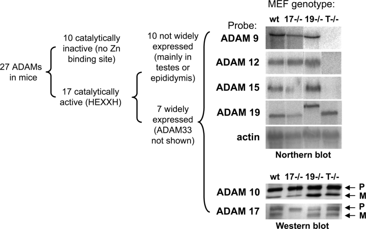
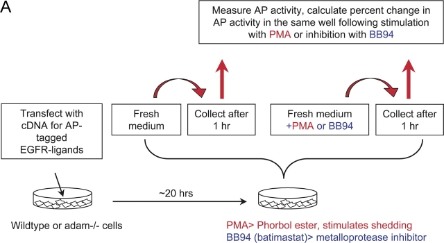
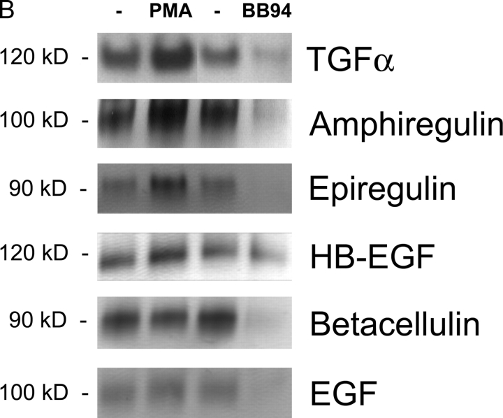
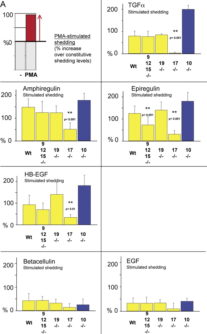
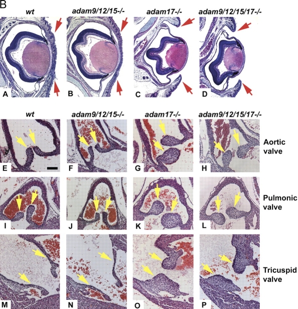
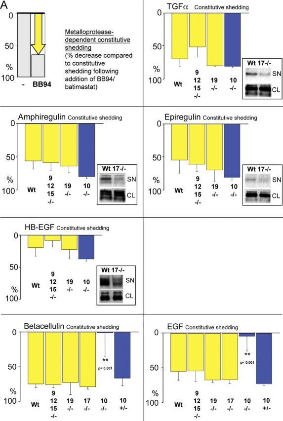
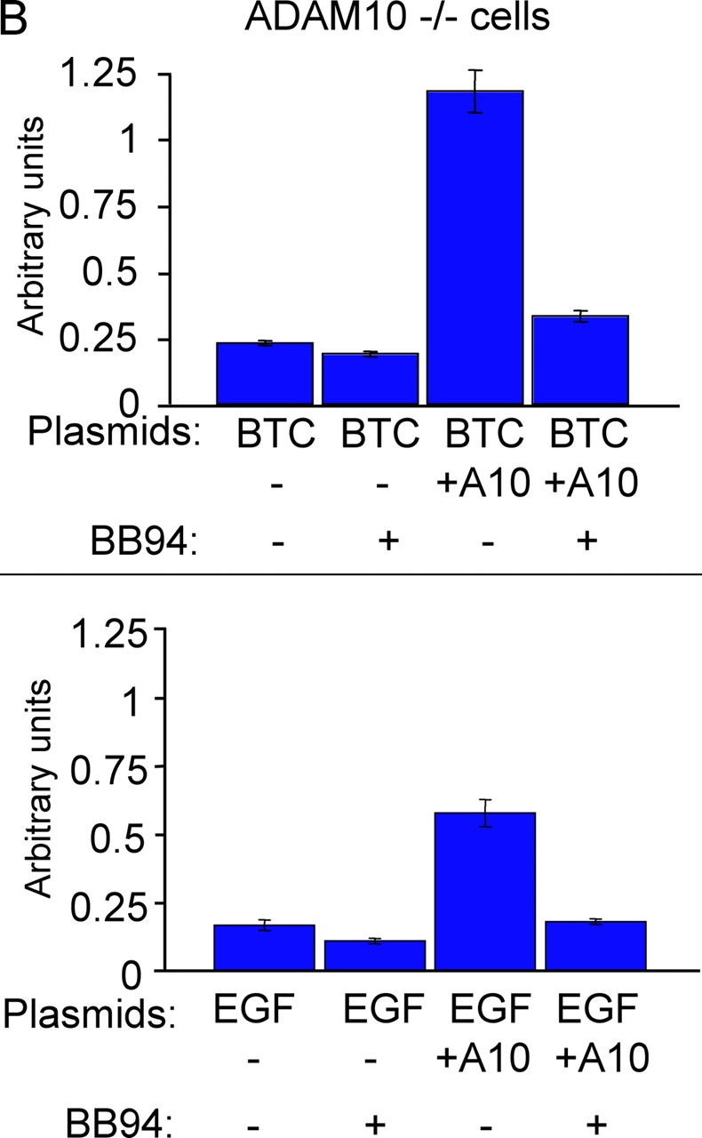
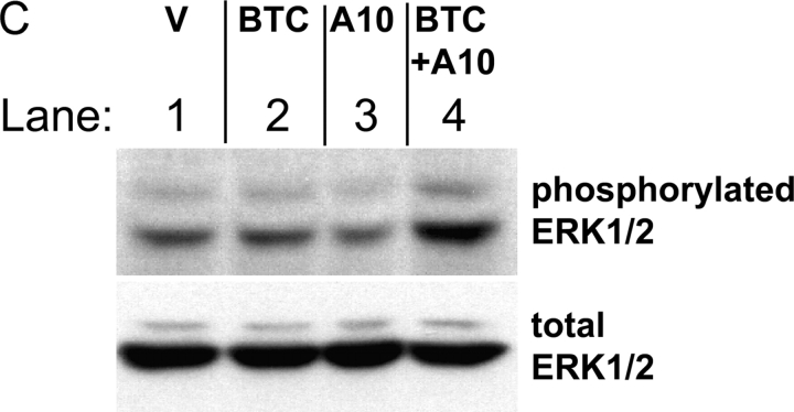
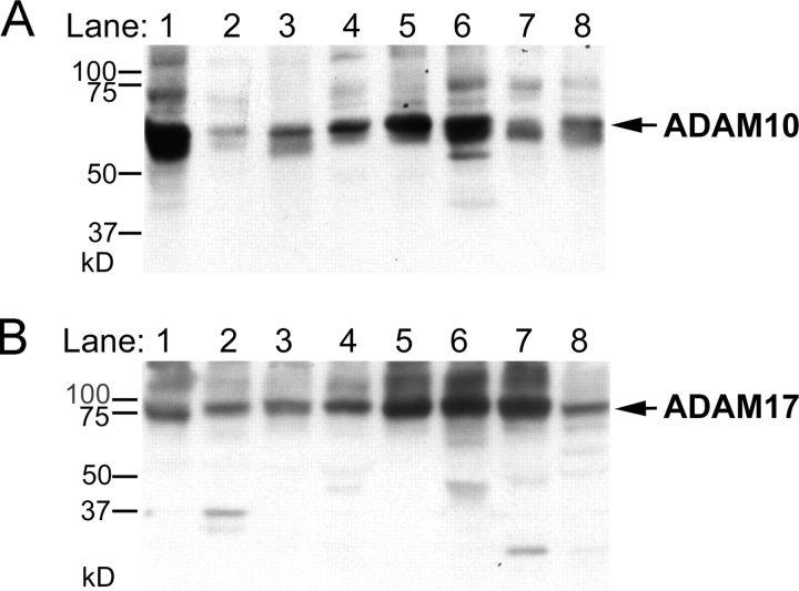
All ligands of the epidermal growth factor receptor (EGFR), which has important roles in development and disease, are released from the membrane by proteases. In several instances, ectodomain release is critical for activation of EGFR ligands, highlighting the importance of identifying EGFR ligand sheddases. Here, we uncovered the sheddases for six EGFR ligands using mouse embryonic cells lacking candidate-releasing enzymes (a disintegrin and metalloprotease [ADAM] 9, 10, 12, 15, 17, and 19). ADAM10 emerged as the main sheddase of EGF and betacellulin, and ADAM17 as the major convertase of epiregulin, transforming growth factor alpha, amphiregulin, and heparin-binding EGF-like growth factor in these cells. Analysis of adam9/12/15/17-/- knockout mice corroborated the essential role of adam17-/- in activating the EGFR in vivo. This comprehensive evaluation of EGFR ligand shedding in a defined experimental system demonstrates that ADAMs have critical roles in releasing all EGFR ligands tested here. Identification of EGFR ligand sheddases is a crucial step toward understanding the mechanism underlying ectodomain release, and has implications for designing novel inhibitors of EGFR-dependent tumors.


Figures

Figure 1.
Figure 1.
Expression of widely expressed and catalytically active ADAMs in MEFs. ADAMs are grouped by expression pattern and presence or absence of a catalytic site (HEXXH) in the metalloprotease domain. 27 ADAMs have been identified in mice, of which 10 lack an HEXXH sequence, and are presumably not catalytically active. Out of 17 ADAMs with an HEXXH sequence, 10 are mainly expressed in the testes or epididymis, or are not widely expressed (J.M. White, University of Virginia, Charlottesville, VA; http://www.people.virginia.edu/%7Ejw7g/Table_of_the_ADAMs.html). Six of the seven widely expressed HEXXH-containing ADAMs were included in this paper. The top right panel shows a Northern blot analysis of the expression of ADAMs 9, 12, 15, and 19 in primary MEFs. The ADAM19 mRNA in adam19 −/− cells is larger than in the other cells analyzed because the ADAM19 gene is disrupted by insertion of a secretory gene trap (Zhou et al., 2004). The bottom right panel is a Western blot depicting expression of ADAMs 10 and 17 in the primary embryonic fibroblasts used here. Both pro- and mature ADAM10 are expressed in all primary mEFs analyzed here, and pro- and mature ADAM17 are expressed in wild-type, adam19 −/−, and adam9/12/15 −/− cells. Note that the exon containing the Zn2+-binding catalytic site of ADAM17 is deleted in adam17 −/− cells (ADAM17ΔZn/ΔZn). This will most likely impair proper protein folding, resulting in retention of mutant ADAM17 in the ER by chaperones and subsequent degradation (Suzuki et al., 1998). wt, wild type; 17−/−, adam17 −/−; 19−/−, adam19 −/−; T−/−, adam9/12/15 −/− triple knockout; P, pro-form; M, mature.
Figure 2.
Figure 2.
Shedding of EGFR ligands in wild-type primary MEFs. (A) Diagram of a typical shedding experiment (see text for details). (B) Detection of shed AP-tagged EGFR ligands after renaturation in SDS gels (see Materials and methods for details). The left lane shows the AP-tagged forms of TGFα, amphiregulin, epiregulin, HB-EGF, betacellulin, and EGF released in 1 h into the supernatant of a single well each of transfected mEF under resting conditions. The next lane shows the EGFR ligands released in 1 h from the same well after addition of PMA, a phorbol ester that stimulates ectodomain shedding. The third lane shows EGFR ligands released from a separate well in 1 h under resting conditions, and the fourth lane shows the released EGFR ligands in that same well after addition of the hydroxamate-based metalloprotease inhibitor batimastat (BB94).
Figure 2.
Figure 2.
Shedding of EGFR ligands in wild-type primary MEFs. (A) Diagram of a typical shedding experiment (see text for details). (B) Detection of shed AP-tagged EGFR ligands after renaturation in SDS gels (see Materials and methods for details). The left lane shows the AP-tagged forms of TGFα, amphiregulin, epiregulin, HB-EGF, betacellulin, and EGF released in 1 h into the supernatant of a single well each of transfected mEF under resting conditions. The next lane shows the EGFR ligands released in 1 h from the same well after addition of PMA, a phorbol ester that stimulates ectodomain shedding. The third lane shows EGFR ligands released from a separate well in 1 h under resting conditions, and the fourth lane shows the released EGFR ligands in that same well after addition of the hydroxamate-based metalloprotease inhibitor batimastat (BB94).
Figure 3.
Figure 3.
PMA-stimulated shedding of EGFR ligands in adam −/− cells. (A) The first panel shows an explanatory diagram depicting how increases in ectodomain shedding of different EGFR ligands from a given well are presented in the remaining panels of this figure. Shedding stimulated by 20 ng/ml PMA for 1 h is calculated as the percent increase in AP activity over constitutive shedding in the same well for 1 h. The next panels only show the PMA-dependent increase in shedding over constitutive levels for each EGFR ligand and each adam −/− cell type. Data from primary mEFs (yellow bars) are compiled from separate experiments using cells from three or more litters for each adam −/− mouse line. Only adam10 −/− and control adam10 +/− cells were immortalized (blue bars). Overall, at least four separate wells were evaluated per EGFR ligand. The results indicate that ADAM17 is the major stimulated sheddase for TGFα, amphiregulin, epiregulin, and HB-EGF. ADAMs 9, 12, or 15 (or a combination of two or more of these ADAMs) also contribute to stimulated epiregulin shedding. On the other hand, the shedding of betacellulin and EGF is only weakly stimulated by PMA. Because the increase in stimulated shedding is small, no statistically significant differences in stimulated shedding of betacellulin or EGF was seen in adam −/− cells compared with wild-type controls. (B) Histological analysis of sectioned hematoxylin and eosin–stained eyes and eyelids (A–D), aortic valves (E–H), pulmonic valves (I–L), and tricuspid valves (M–P) of newborn wild-type (A, E, I, and M), adam9/12/15 −/− (B, F, J, and N), adam17 −/− (C, G, K, and O), and adam9/12/15/17 −/− (D, H, L, and P) mice. Eyelids of wild-type and adam9/12/15 −/− mice are closed at birth (A and B), whereas those of adam17 −/− and adam9/12/15/17 −/− mice are open (C and D). The aortic, pulmonic, and tricuspid valves of adam9/12/15 −/− mice (F, J, and N) resemble those of wild-type mice (E, I, and M, respectively), whereas these valves are thickened and misshapen in adam17 −/− (G, K, and C) and adam9/12/15/17 −/− quadruple knockout mice (H, L, and P). The valve defects in adam9/12/15/17 −/− quadruple knockout mice, which also include thickened and misshapen mitral valves (not depicted), are comparable to those in adam17 −/− mice. Eyelids in A–D marked by red arrows, heart valves in E–P marked by yellow arrows. Bar (E–P), 100 μm.
Figure 3.
Figure 3.
PMA-stimulated shedding of EGFR ligands in adam −/− cells. (A) The first panel shows an explanatory diagram depicting how increases in ectodomain shedding of different EGFR ligands from a given well are presented in the remaining panels of this figure. Shedding stimulated by 20 ng/ml PMA for 1 h is calculated as the percent increase in AP activity over constitutive shedding in the same well for 1 h. The next panels only show the PMA-dependent increase in shedding over constitutive levels for each EGFR ligand and each adam −/− cell type. Data from primary mEFs (yellow bars) are compiled from separate experiments using cells from three or more litters for each adam −/− mouse line. Only adam10 −/− and control adam10 +/− cells were immortalized (blue bars). Overall, at least four separate wells were evaluated per EGFR ligand. The results indicate that ADAM17 is the major stimulated sheddase for TGFα, amphiregulin, epiregulin, and HB-EGF. ADAMs 9, 12, or 15 (or a combination of two or more of these ADAMs) also contribute to stimulated epiregulin shedding. On the other hand, the shedding of betacellulin and EGF is only weakly stimulated by PMA. Because the increase in stimulated shedding is small, no statistically significant differences in stimulated shedding of betacellulin or EGF was seen in adam −/− cells compared with wild-type controls. (B) Histological analysis of sectioned hematoxylin and eosin–stained eyes and eyelids (A–D), aortic valves (E–H), pulmonic valves (I–L), and tricuspid valves (M–P) of newborn wild-type (A, E, I, and M), adam9/12/15 −/− (B, F, J, and N), adam17 −/− (C, G, K, and O), and adam9/12/15/17 −/− (D, H, L, and P) mice. Eyelids of wild-type and adam9/12/15 −/− mice are closed at birth (A and B), whereas those of adam17 −/− and adam9/12/15/17 −/− mice are open (C and D). The aortic, pulmonic, and tricuspid valves of adam9/12/15 −/− mice (F, J, and N) resemble those of wild-type mice (E, I, and M, respectively), whereas these valves are thickened and misshapen in adam17 −/− (G, K, and C) and adam9/12/15/17 −/− quadruple knockout mice (H, L, and P). The valve defects in adam9/12/15/17 −/− quadruple knockout mice, which also include thickened and misshapen mitral valves (not depicted), are comparable to those in adam17 −/− mice. Eyelids in A–D marked by red arrows, heart valves in E–P marked by yellow arrows. Bar (E–P), 100 μm.
Figure 4.
Figure 4.
Batimastat-sensitive constitutive shedding of EGFR ligands in adam −/− cells. (A) A diagram indicating how the batimastat-sensitive component of ectodomain shedding of different EGFR ligands from a given well of resting cells was determined. Shedding of EGFR ligands in 1 h from resting cells is used as a reference to determine the percentage of batimastat-sensitive constitutive shedding (percent decrease after addition of batimastat). The next panels show the batimastat-sensitive decrease in shedding of each EGFR ligand in each adam −/− cell type. As in Fig. 3, separate experiments were performed with cells from three or more litters for each adam −/− mouse line. At least six separate wells were analyzed for adam10 −/− and control adam10 +/− cells, which were immortalized (blue bars). Several wells were evaluated in each experiment for each lot of cells and for each EGFR ligand. The results show that ADAMs 9, 10, 12, 15, and 19 are not essential for the batimastat-sensitive constitutive shedding of TGFα, amphiregulin, epiregulin, and HB-EGF. The absolute levels of constitutive release of each EGFR ligand were comparable between wild-type, adam9/12/15 −/−, and adam19 −/− cells (not depicted). However, the levels of constitutive shedding of TGFα, amphiregulin, epiregulin, and HB-EGF were significantly reduced in adam17 −/− cells compared with wild-type controls (see inset gel figures; SN, supernatant; CL, cell lysate), indicating that ADAM17 is also the major constitutive sheddase for these ligands. Data on the batimastat-sensitive shedding in adam17 −/− cells are not included in the graph because addition of batimastat further decreases the small amount of constitutive shedding in adam17 −/− cells. Thus, another metalloprotease besides ADAM17 apparently makes a very minor contribution to shedding of these four substrates. The batimastat-sensitive shedding of betacellulin and EGF was very similar in adam9/12/15 −/− , adam19 −/− , adam17 −/−, and adam10 +/− mEFs compared with wild-type controls. Although the absolute levels of constitutive shedding from these cells were also very similar (not depicted), constitutive shedding of betacellulin from adam10 −/− cells was decreased by 87.5%, whereas shedding of EGF was decreased by 49.7% compared with adam10 +/− cells. In the absence of ADAM10, the remaining small amount of constitutive shedding was not inhibitable by batimastat. (B) Batimastat-sensitive shedding of betacellulin (BTC) and EGF from adam10 −/− cells can be rescued by cotransfection with wild-type ADAM10 (A10; each bar represents the results from six tissue culture wells). These results confirm that the defect in EGF and betacellulin shedding in adam10 / cells is indeed due to the absence of ADAM10. (C) Constitutive phosphorylation of ERK1/2 in adam10 −/− cells transfected with the pcDNA3 vector (V, lane 1) is not increased by transfection with BTC (lane 2) or ADAM10 (lane 3). However, ERK1/2 phosphorylation is increased when BTC and ADAM10 are cotransfected (lane 4), demonstrating that ADAM10 is critical for BTC-dependent EGFR signaling in these cells. The bottom panel shows the same blot reprobed with antibodies against total ERK1/2 to confirm equal loading in all lanes.
Figure 4.
Figure 4.
Batimastat-sensitive constitutive shedding of EGFR ligands in adam −/− cells. (A) A diagram indicating how the batimastat-sensitive component of ectodomain shedding of different EGFR ligands from a given well of resting cells was determined. Shedding of EGFR ligands in 1 h from resting cells is used as a reference to determine the percentage of batimastat-sensitive constitutive shedding (percent decrease after addition of batimastat). The next panels show the batimastat-sensitive decrease in shedding of each EGFR ligand in each adam −/− cell type. As in Fig. 3, separate experiments were performed with cells from three or more litters for each adam −/− mouse line. At least six separate wells were analyzed for adam10 −/− and control adam10 +/− cells, which were immortalized (blue bars). Several wells were evaluated in each experiment for each lot of cells and for each EGFR ligand. The results show that ADAMs 9, 10, 12, 15, and 19 are not essential for the batimastat-sensitive constitutive shedding of TGFα, amphiregulin, epiregulin, and HB-EGF. The absolute levels of constitutive release of each EGFR ligand were comparable between wild-type, adam9/12/15 −/−, and adam19 −/− cells (not depicted). However, the levels of constitutive shedding of TGFα, amphiregulin, epiregulin, and HB-EGF were significantly reduced in adam17 −/− cells compared with wild-type controls (see inset gel figures; SN, supernatant; CL, cell lysate), indicating that ADAM17 is also the major constitutive sheddase for these ligands. Data on the batimastat-sensitive shedding in adam17 −/− cells are not included in the graph because addition of batimastat further decreases the small amount of constitutive shedding in adam17 −/− cells. Thus, another metalloprotease besides ADAM17 apparently makes a very minor contribution to shedding of these four substrates. The batimastat-sensitive shedding of betacellulin and EGF was very similar in adam9/12/15 −/− , adam19 −/− , adam17 −/−, and adam10 +/− mEFs compared with wild-type controls. Although the absolute levels of constitutive shedding from these cells were also very similar (not depicted), constitutive shedding of betacellulin from adam10 −/− cells was decreased by 87.5%, whereas shedding of EGF was decreased by 49.7% compared with adam10 +/− cells. In the absence of ADAM10, the remaining small amount of constitutive shedding was not inhibitable by batimastat. (B) Batimastat-sensitive shedding of betacellulin (BTC) and EGF from adam10 −/− cells can be rescued by cotransfection with wild-type ADAM10 (A10; each bar represents the results from six tissue culture wells). These results confirm that the defect in EGF and betacellulin shedding in adam10 / cells is indeed due to the absence of ADAM10. (C) Constitutive phosphorylation of ERK1/2 in adam10 −/− cells transfected with the pcDNA3 vector (V, lane 1) is not increased by transfection with BTC (lane 2) or ADAM10 (lane 3). However, ERK1/2 phosphorylation is increased when BTC and ADAM10 are cotransfected (lane 4), demonstrating that ADAM10 is critical for BTC-dependent EGFR signaling in these cells. The bottom panel shows the same blot reprobed with antibodies against total ERK1/2 to confirm equal loading in all lanes.
Figure 5.
Figure 5.
Evaluation of ADAM10 and ADAM17 protein levels in different mouse tissues. Western blots of mouse tissue extracts were probed with a polyclonal antiserum against ADAM10 (A) or ADAM17 (B). Equal amounts of Con A–enriched glycoproteins from the following tissues were loaded per lane: brain (lane 1), skeletal muscle (lane 2), kidney (lane 3), heart (lane 4), lung (lane 5), spleen (lane 6), testis (lane 7), and liver (lane 8). The arrow indicates the position of ADAM10 in A, and of ADAM17 in B.

References

    1. Asakura, M., M. Kitakaze, S. Takashima, Y. Liao, F. Ishikura, T. Yoshinaka, H. Ohmoto, K. Node, K. Yoshino, H. Ishiguro, et al. 2002. Cardiac hypertrophy is inhibited by antagonism of ADAM12 processing of HB-EGF: metalloproteinase inhibitors as a new therapy. Nat. Med. 8:35–40. - PubMed
    1. Black, R.A., and J.M. White. 1998. ADAMs: focus on the protease domain. Curr. Opin. Cell Biol. 10:654–659. - PubMed
    1. Borrell-Pages, M., F. Rojo, J. Albanell, J. Baselga, and J. Arribas. 2003. TACE is required for the activation of the EGFR by TGF-α in tumors. EMBO J. 22:1114–1124. - PMC - PubMed
    1. Brachmann, R., P.B. Lindquist, M. Nagashima, W. Kohr, T. Lipari, M. Napier, and R. Derynck. 1989. Transmembrane TGF-α precursors activate EGF/TGF-α receptors. Cell. 56:691–700. - PubMed
    1. Chesneau, V., D. Becherer, Y. Zheng, H. Erdjument-Bromage, P. Tempst, and C.P. Blobel. 2003. Catalytic properties of ADAM19. J. Biol. Chem. 278:22331–22340. - PubMed

Publication types

MeSH terms

Substances