. 2004 Mar 1;199(5):617-27.
doi: 10.1084/jem.20031935.

Mapping of a functional recombination motif that defines isotype specificity for mu-->gamma3 switch recombination implicates NF-kappaB p50 as the isotype-specific switching factor

Affiliations

Mapping of a functional recombination motif that defines isotype specificity for mu-->gamma3 switch recombination implicates NF-kappaB p50 as the isotype-specific switching factor

Amy L Kenter et al. J Exp Med. .

Abstract

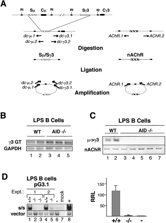
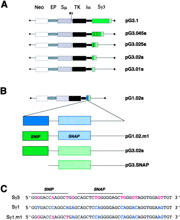
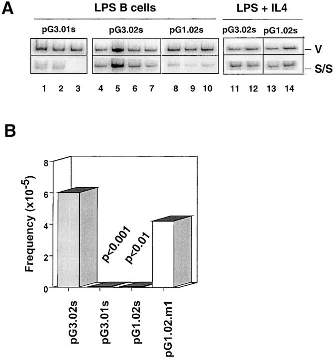
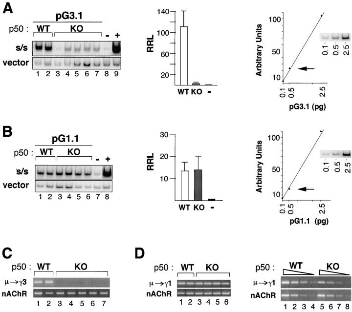
Ig class switch recombination (CSR) requires expression of activation-induced deaminase (AID) and production of germline transcripts to target S regions for recombination. However, the mechanism of CSR remains unclear. Here we show that an extrachromosomal S plasmid assay is AID dependent and that a single consensus repeat is both necessary and sufficient for isotype-specific CSR. Transfected switch substrates specific for mu-->gamma3 and mu-->gamma1 are stimulated to switch with lipopolysaccharide (LPS) alone or LPS and interleukin-4, respectively. An Sgamma3/Sgamma1 substrate containing only three Sgamma3-associated nucleotides reconstituted LPS responsiveness and permitted mapping of a functional recombination motif specific for mu-->gamma3 CSR. This functional recombination motif colocalized with a binding site for NF-kappaB p50, and p50 binding to this site was previously established. We show a p50 requirement for plasmid-based mu-->gamma3 CSR using p50-deficient B cells. Switch junctions from p50-deficient B cells showed decreased lengths of microhomology between Smu and Sgamma3 relative to wild-type cells, indicating a function for p50 in the mechanics of CSR. We note a striking parallel between the affects of p50 and Msh2 deficiency on Smu/Sgamma3 junctions. The data suggest that p50 may be the isotype-specific factor in mu-->gamma3 CSR and epistatic with Msh2.


Figures

Figure 1.
Figure 1.
Switch plasmid-based μ→γ3 CSR in LPS B cells is AID dependent. (A) A diagram of the DC-PCR strategy for endogenous loci is shown. A portion of the IgH locus is diagramed before and after μ→γ3 recombination. EcoRI sites (RI) flank the 5′ and 3′ ends of the Sμ and Sγ3 regions, respectively, and are preserved after CSR. After digestion with EcoRI, the DNA is ligated. Nested primer sets specific for sites at the 5′ end of Sμ (dc-μ.1, dc-μ.2) and the 3′ end of Sγ3 (dc-γ3.1, dc-γ3.2) amplify the region spanning the circle joint and yield a specific S/S DC-PCR product. The positions and orientations of the primer sets are shown before and after ligation. The nAChR gene serves as an internal control for digestion and ligation. (B–D) B cells from AID+/+ and AID−/− spleens were activated in culture with LPS for 3 (B) or 5 d (C) or were activated with LPS for 3 d and then transfected with pG3.1 and grown in LPS culture for an additional 3 d (D). (B) RT-PCR for detection of GAPDH and the γ3 germline transcript (γ3 GT) was performed on RNA of LPS-activated B cells from AID+/+ (lanes 1 and 2) or AID−/− (lanes 3–5) mice. (C) DC-PCR was used to detect endogenous μ→γ3 switching in DNA from LPS-activated AID+/+ or AID−/− B cells (top). The nAchR gene was used as a positive control (bottom). (D) LPS activated B cells from AID+/+ or AID−/− mice were transfected with the pG3.1 plasmid and used in a plasmid-based DC-PCR assay. The RRL for pG3.1 is the ratio of the radioactivity associated with the 180-bp (S/S) fragment to that of the 510-bp (vector) fragment. Intact pG3.1 (0.5 pg) demonstrated to be in the linear range of detection in the DC-PCR assay (not depicted) was used as a negative control (lane 6), and its RRL was set to 1 (right). The RRL values for the AID+/+ and AID−/− transfections were normalized to the negative control. In the right panel AID+/+ and AID−/− samples are designated +/+ and −/−, respectively. The intact negative control is indicated as −. The average RRLs from two independent experiments for pG3.1 is plotted (right), and SDs are shown.
Figure 2.
Figure 2.
Structure of switch plasmids related to pG3.1. (A) Intact pG3.1 contains a neomycin-resistance gene (neo), the Ig μ intronic enhancer (E), an IgH variable region promoter (P), a thymidine kinase (TK) gene, the promoter for α gts (), and the Sμ and Sγ3 regions. PG3.01s is identical to pG3.1 except for the deletion of the Sγ3 region and retention two nonconsensus tandem repeats from the extreme 3′ end of genomic Sγ3 (35). The plasmids pG3.02s, pG3.025s, and pG3.045s contain 1, 2, and 6 consensus Sγ3 tandem repeats, respectively. The nonconsensus and consensus tandem repeats located in Sγ3 are indicated as green hatched and solid green boxes, respectively. (B) PG1.02s contains an Sγ1 consensus repeat, which is depicted by the solid blue box. The Sγ1 consensus repeat is expanded to show two subsections, SNIP (solid) and SNAP (speckled), separated by a short and long spacer, as indicated. Sγ1 and Sγ3 DNA sequences are shown in blue and green, respectively. PG1.02.m1 contains a chimeric repeat including the Sγ1 repeat backbone and an Sγ3 SNIP site. PG3.SNAP contains a 30-bp truncated Sγ3 consensus repeat centered on the SNAP site. (C) The DNA sequence for Sγ3 and Sγ1 consensus repeats are shown. In the chimeric Sγ1.m1 repeat, the Sγ1 and Sγ3 SNIP sites differ at three nucleotide positions.
Figure 3.
Figure 3.
Switch substrates require a single Sγ consensus repeat to support CSR. (A) Switch plasmids, as indicated, were transfected into 1.B4.B6 cells and analyzed for CSR using the plasmid-based DC-PCR assay (left). The RRLs are the results of five to six transfections from at least two independent experiments, and SDs are shown (right). The RRL is calculated for each plasmid as the ratio of radioactivity associated with the 180-bp S/S fragment to that of the 510-bp vector-associated fragment. The RRLs are not normalized. (B) Switch plasmids were transfected into LPS-activated B cells and analyzed by plasmid-based DC-PCR using either SacI or SacI and BglI digestion. (C) The switching activity of pG3.1, pG3.02s, pG3.SNAP, and pG3.01s were compared in LPS-activated B cells using the bacterial transformation assay. DNA recovered from nuclei of the transfected cells was untreated or digested with EcoRI and then transformed into bacteria. S/S recombinant frequency was as follows: pG3.1 (17/41,540); pG3.02s (10/110,060); pG3.01s (0/231,800); and pG3.SNAP (6/570,400). Switch frequency was obtained by dividing the number of S/S recombinant transformants by the total number of transformants and multiplying by 105. Results are summarized from at least three to six transfections from two to three independent experiments. p-values, derived by χ2 analysis, are positioned above the histograms and indicate the confidence level that the plasmid switch frequency in the pG3.02s was significantly different from that obtained from pG3.01s and pG3.SNAP.
Figure 4.
Figure 4.
Substitution of three nucleotides in the Sγ1 SNIP site leads to reconstitution of switching in LPS B cells for the pG1.02s.m1 plasmid. (A) The plasmids, pG3.01s, pG3.02s, and pG1.02s, were transfected into B cells activated with LPS in the presence or absence of IL-4 and then harvested and analyzed by DC-PCR. The 510-bp vector (V) and the 180-bp S/S PCR fragments are shown. (B) The plasmids, pG3.01s, pG3.02s, pG1.02s, and pG1.02s.m1, were transfected into B cells activated with LPS then harvested and analyzed using the bacterial transformation assay. S/S recombinant frequency was as follows: pG3.02s (10/110,060); pG3.01s (0/231,800); pG1.02s (0/138,800); and pG1.02.m1 (9/212,080). Switch frequency was obtained by dividing the number of S/S transformants by the total number of transformants and multiplying by 105. Results are summarized from at least three to six transfections from two to three independent experiments. p-values are positioned above the histograms. Values of p were derived by χ2 analysis.
Figure 5.
Figure 5.
Plasmid-based μ→γ3 CSR is abolished in NF-kB p50-deficient B cells. (A and B) B cells from NF-κB p50 WT and knockout (KO) spleens were T cell depleted, activated with LPS or LPS + αδdex + IL-4 + IL-5 + TGFβ for 3 d, then transfected with pG3.1 (A, left) or pG1 (B, left), respectively, grown in culture for an additional 3 d, and then used in a plasmid-based DC-PCR assay. The PCR products representing the switched plasmid (S/S) and the vector backbone are shown from independent transfected samples. The average RRLs from two independent experiments for pG3.1 (A) and pG1 (B) is plotted (middle), and SDs are shown. Standard curves (fivefold dilutions) for vector DC-PCR products are shown (right). The arrow indicates the concentration of plasmid (0.5 pg) used in the DC-PCR assays shown here. (C) Endogenous DC-PCR assays for μ→γ3 CSR in LPS-activated B cells from p50 WT (lanes 1 and 2) and KO (lanes 3–7) are shown. The nAChR locus is used as a control for sample loading and ligation. (D) Endogenous DC-PCR assays for μ→γ3 CSR in LPS + αδdex + IL-4 + IL-5 + TGFβ activated B cells are shown for p50 WT (lanes 1 and 2) and KO (lanes 3–6) in the left panel. Fourfold serial dilution of representative DC-PCR samples are shown in the right panel for WT (lanes 1–4) and KO (lanes 5–8). The nAChR locus is used as a control for sample loading and ligation.

Similar articles

Cited by

References

    1. Stavnezer, J. 2000. Molecular processes that regulate class switching. Curr. Top. Microbiol. Immunol. 245:127–168. - PubMed
    1. Manis, J.P., M. Tian, and F.W. Alt. 2002. Mechanism and control of class-switch recombination. Trends Immunol. 23:31–39. - PubMed
    1. Muramatsu, M., K. Kinoshita, S. Fagarasan, S. Yamada, Y. Shinkai, and T. Honjo. 2000. Class switch recombination and hypermutation require activation-induced cytidine deaminase (AID), a potential RNA editing enzyme. Cell. 102:553–563. - PubMed
    1. Muramatsu, M., V.S. Sankaranand, S. Anant, M. Sugai, K. Kinoshita, N.O. Davidson, and T. Honjo. 1999. Specific expression of activation-induced cytidine deaminase (AID), a novel member of the RNA-editing deaminase family in germinal center B cells. J. Biol. Chem. 274:18470–18476. - PubMed
    1. Revy, P., T. Muto, Y. Levy, F. Geissmann, A. Plebani, O. Sanal, N. Catalan, M. Forveille, R. Dufourcq-Labelouse, A. Gennery, et al. 2000. Activation-induced cytidine deaminase (AID) deficiency causes the autosomal recessive form of the Hyper-IgM syndrome (HIGM2). Cell. 102:565–575. - PubMed

Publication types

MeSH terms