. 2004 Mar;24(6):2286-95.
doi: 10.1128/MCB.24.6.2286-2295.2004.

Replication stalling at Friedreich's ataxia (GAA)n repeats in vivo

Affiliations

Replication stalling at Friedreich's ataxia (GAA)n repeats in vivo

Maria M Krasilnikova et al. Mol Cell Biol. 2004 Mar.

Abstract

Friedreich's ataxia (GAA)n repeats of various lengths were cloned into a Saccharymyces cerevisiae plasmid, and their effects on DNA replication were analyzed using two-dimensional electrophoresis of replication intermediates. We found that premutation- and disease-size repeats stalled the replication fork progression in vivo, while normal-size repeats did not affect replication. Remarkably, the observed threshold repeat length for replication stalling in yeast (approximately 40 repeats) closely matched the threshold length for repeat expansion in humans. Further, replication stalling was strikingly orientation dependent, being pronounced only when the repeat's homopurine strand served as the lagging strand template. Finally, it appeared that length polymorphism of the (GAA)n. (TTC)n repeat in both expansions and contractions drastically increases in the repeat's orientation that is responsible for the replication stalling. These data represent the first direct proof of the effects of (GAA)n repeats on DNA replication in vivo. We believe that repeat-caused replication attenuation in vivo is due to triplex formation. The apparent link between the replication stalling and length polymorphism of the repeat points to a new model for the repeat expansion.


Figures

FIG. 1.
FIG. 1.
Strategy for cloning long (GAA)n · (TTC)n repeats (see the text for details).
FIG. 2.
FIG. 2.
Electrophoretic analysis of replication intermediates in yeast. (A) Structure of the BglI-linearized pYES derivatives containing (GAA)n · (TTC)n repeats. In this linear depiction, the 2μm replication origin is roughly in the middle of the plasmid. (GAA)n · (TTC)n repeats (black box) were cloned into the BsgI site, being replicated by left-to-right replication fork. (B) Schematic representation for the two-dimensional neutral/neutral electrophoretic analysis. Cleavage of replication intermediates with restriction enzymes BsaI and BglI generates Y-shaped DNA molecules, and the size of a Y increases with replication progression. Replication blockage at a repeat leads to the accumulation of replication intermediates of a given size and shape, as shown in bold (right panel). Separation by two-dimensional agarose electrophoresis reveals a Y-arc (left panel). Partial replication blockage by a repeat should result in the appearance of a bulge (black circle) on the replication arc. Complete replication blockage should lead to the appearance of a spike. Arrowheads in the right panel point to the portion of the Y-arc analyzed by phosphorimaging.
FIG. 3.
FIG. 3.
Electrophoretic analysis of replication intermediates for pYES derivatives carrying various (GAA)n · (TTC)n repeats. (A) Primary electrophoretic data. Plasmids were named according to the repetitive sequence in the lagging strand template. Arrows show replication stall sites. (B) Quantitative analysis of Y-arcs for plasmids carrying (GAA)n · (TTC)n repeats. The analyzed portion of the Y-arc is marked by arrowheads in Fig. 1B. Peaks on densitograms correspond to bulges on the Y-arcs shown in Fig. 2A.
FIG. 4.
FIG. 4.
Replication of the pYES-GAA228 plasmid maintained under conditions of transcriptional repression (Glu) or activation (Gal). Arrows show replication stall sites.
FIG. 5.
FIG. 5.
(GAA)n repeats do not affect transcription in yeast. Plasmids were the same that were used in replication studies. Transcription from the GAL1 promoter was analyzed by Northern hybridization with the radiolabeled PvuII-XhoI probe, corresponding to the +78-to-+190 part of the GAL1 transcript, immediately upstream of the repeat. A molecular weight ladder is marked by arrows.
FIG. 6.
FIG. 6.
Relative copy numbers of plasmids containing various (GAA)n · (TTC)n repeats as determined by the combination of dot blot and Southern hybridization assays. Plasmids are named according to sequences of the lagging strand template. The horizontal line reflects the copy number of a control plasmid, carrying a nonrepetitive sequence, used for normalization.
FIG. 7.
FIG. 7.
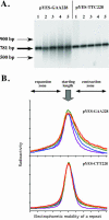
Length polymorphism of the (GAA)228 · (TTC)228 repeat in the two orientations relative to the origin. (A) Experimental Southern hybridization data. Plasmids are named according to sequences of the lagging strand template. Numbers 1 to 5 correspond to five consecutive rounds of cultivation. (B) Quantitative analysis of the Southern hybridization data using a PhosphorImager. Purple, blue, green, yellow, and red lines correspond to cultivation rounds one through five, respectively.
FIG. 8.
FIG. 8.
A model for replication blockage by (GAA)n · (TTC)n repeats leading to their expansions. (A) A portion of the lagging strand template with a (GAA)n run folds back to form a stable triplex that stalls the leading DNA polymerase. (B) Slow unraveling of this triplex could be accompanied by polymerase dissociation and misalignment of the newly synthesized and template DNA strands. (C) Resumption of DNA synthesis upon triplex dismantling would then lead to repeat expansions or contractions. Red line, homopurine strand; green line, homopyrimidine strand; black line, flanking DNA. Arrows depict Okazaki fragments. Yellow circles represent leading and lagging DNA polymerases. Brown circles symbolize genome guardians, such as DNA helicases, SSB proteins, etc.

Similar articles

Cited by

References

    1. Bidichandani, S. I., T. Ashizawa, and P. I. Patel. 1998. The GAA triplet-repeat expansion in Friedreich ataxia interferes with transcription and may be associated with an unusual DNA structure. Am. J. Hum. Genet. 62:111-121. - PMC - PubMed
    1. Bowater, R. P., and R. D. Wells. 2001. The intrinsically unstable life of DNA triplet repeats associated with human hereditary disorders. Prog. Nucleic Acid Res. Mol. Biol. 66:159-202. - PubMed
    1. Brewer, B. J., and W. L. Fangman. 1987. The localization of replication origins on ARS plasmids in S. cerevisiae. Cell 51:463-471. - PubMed
    1. Campuzano, V., L. Montermini, Y. Lutz, L. Cova, C. Hindelag, S. Jiralerspong, Y. Trottier, S. J. Kish, B. Faucheux, P. Trouillas, F. J. Authier, A. Durr, J.-L. Mandel, A. L. Vescovi, M. Pandolfo, and M. Koenig. 1997. Frataxin is reduced in Friedreich ataxia patients and is associated with mitochondrial membranes. Hum. Mol. Genet. 6:1771-1780. - PubMed
    1. Campuzano, V., L. Montermini, M. D. Molto, L. Pianese, M. Cossee, F. Cavalcanti, E. Monros, F. Rodius, F. Duclos, A. Monticelli, F. Zara, J. Canizares, H. Koutnikova, S. I. Bidichandari, C. Gellera, A. Brice, P. Trouillas, G. De Michele, A. Filla, R. De Frutos, F. Palau, P. I. Patel, S. Di Donato, J.-L. Mandel, S. Cocozza, M. Koenig, and M. Pandolfo. 1996. Friedreich's ataxia: autosomal recessive disease caused by an intronic GAA triplet repeat expansion. Science 271:1423-1427. - PubMed

Publication types

Substances