. 2004 Mar;24(6):2332-43.
doi: 10.1128/MCB.24.6.2332-2343.2004.

Inhibition of the Jun N-terminal protein kinase pathway by SHIP-1, a lipid phosphatase that interacts with the adaptor molecule Dok-3

Affiliations

Inhibition of the Jun N-terminal protein kinase pathway by SHIP-1, a lipid phosphatase that interacts with the adaptor molecule Dok-3

Jeffrey D Robson et al. Mol Cell Biol. 2004 Mar.

Abstract

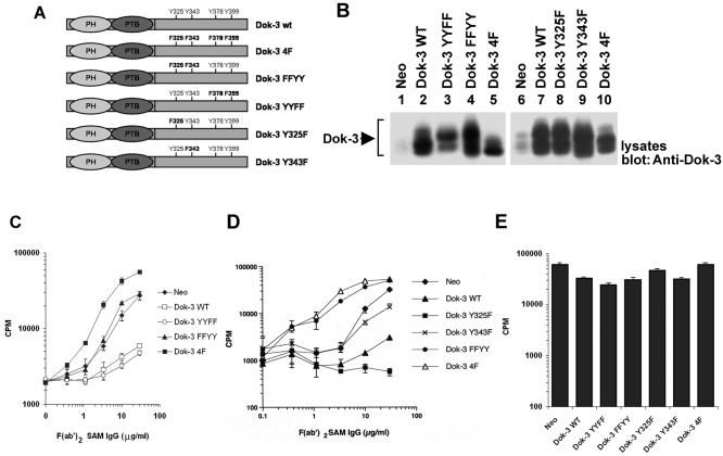
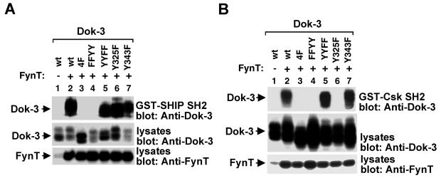
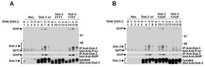
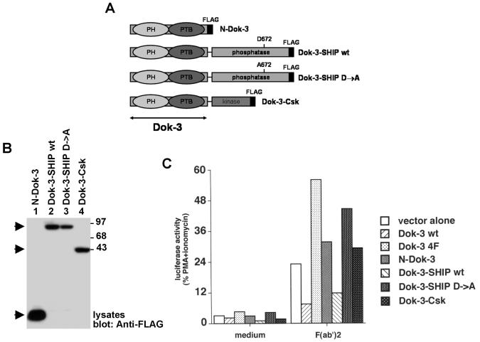
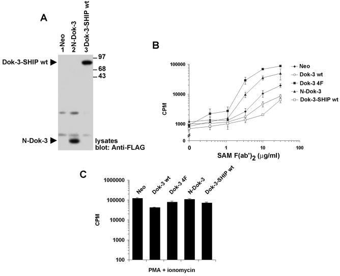
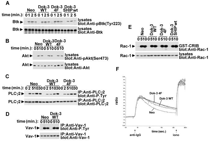
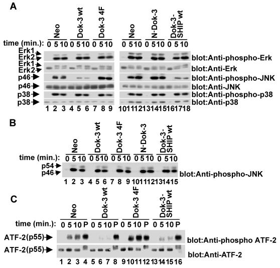
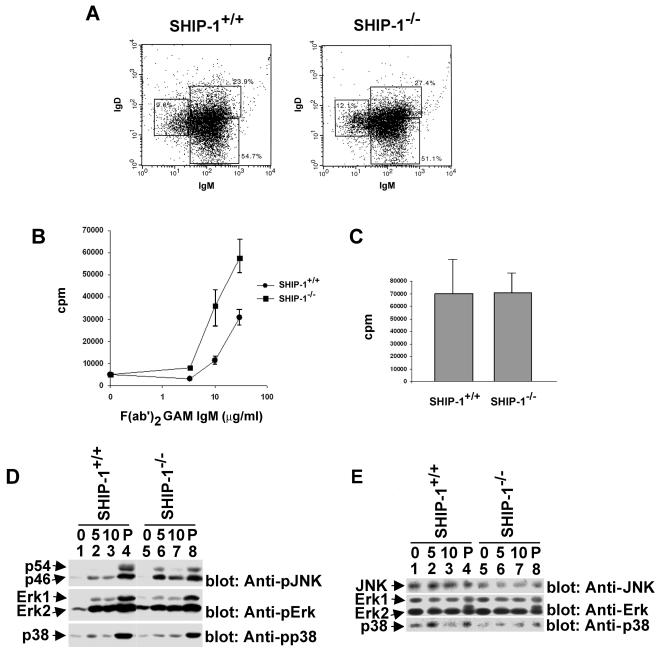
Dok-3 is a Dok-related adaptor expressed in B cells and macrophages. Previously, we reported that Dok-3 is an inhibitor of B-cell activation in A20 B cells and that it associates with SHIP-1, a 5' inositol-specific lipid phosphatase, as well as Csk, a negative regulator of Src kinases. Here, we demonstrate that Dok-3 suppresses B-cell activation by way of its interaction with SHIP-1, rather than Csk. Our biochemical analyses showed that the Dok-3-SHIP-1 complex acts by selectively inhibiting the B-cell receptor (BCR)-evoked activation of the Jun N-terminal protein kinase (JNK) cascade without affecting overall protein tyrosine phosphorylation or activation of previously described SHIP-1 targets like Btk and Akt/PKB. Studies of B cells derived from SHIP-1-deficient mice showed that BCR-triggered activation of JNK is enhanced in the absence of SHIP-1, implying that the Dok-3-SHIP-1 complex (or a related mechanism) is a physiological negative regulator of the JNK cascade in normal B cells. Together, these data elucidate the mechanism by which Dok-3 inhibits B-cell activation. Furthermore, they provide evidence that SHIP-1 can be a negative regulator of JNK signaling in B cells.


Figures

FIG. 1.
FIG. 1.
Tyrosines 325 and 343 are required for the inhibitory effect of Dok-3 in A20 B cells. (A) Primary structures of the Dok-3 mutants created for this study. The positions of the PH and PTB domains, as well as of the four carboxyl-terminal tyrosines, are highlighted. (B) Expression of the various Dok-3 polypeptides in A20 B cells. The expression of Dok-3 in pools of three independent transfectants for each of the Dok-3-encoding constructs was measured by immunoblotting of equivalent amounts of total cell proteins with anti-Dok-3. (C) Impact of Dok-3 YYFF and Dok-3 FFYY on BCR-induced IL-2 secretion. Pools of three transfectants were activated for 24 h with the indicated amounts of F(ab′)2 fragments of SAM IgG. IL-2 release was determined in a bioassay using the IL-2-dependent T-cell line HT-2. Assays were done in triplicate, and average values with standard deviations are shown. (D) Effect of Dok-3 Y325F and Dok-3 Y343F on BCR-triggered IL-2 production. The experiment was conducted as outlined for panel C, except that other transfectants were tested. (E) Influence of Dok-3 polypeptides on responsiveness to PMA plus ionomycin. Cells were stimulated for 24 h with PMA and ionomycin. IL-2 secretion was subsequently measured as described for panel C.
FIG. 2.
FIG. 2.
Differential binding of the SHIP-1 or Csk SH2 domain to tyrosine-phosphorylated Dok-3. Cos-1 cells were transfected with the indicated dok-3 cDNAs in the presence or absence of FynT Y528F. Equivalent amounts of total cell proteins were then incubated with either GST-SHIP-1 SH2 domains (A) or GST-Csk SH2 domains (B). After several washes, the association with Dok-3 was detected by immunoblotting with anti-Dok-3 (top blocks). The expressions of Dok-3 (middle blocks) and FynT (bottom blocks) were verified by immunoblotting of total cell lysates with anti-Dok-3 and anti-FynT, respectively.
FIG. 3.
FIG. 3.
Tyrosines 325 and 343 are required for the association between Dok-3 and SHIP-1 in B cells. Pools of three independent transfectants of the indicated A20 derivatives were stimulated for various periods of time with F(ab′)2 fragments of SAM IgG. Following lysis, Dok-3 was immunoprecipitated from equivalent quantities of total cell proteins and probed by immunoblotting with anti-phosphotyrosine (Anti-P.tyr; top blocks) or anti-SHIP-1 (middle blocks). The presence of Dok-3 was confirmed by immunoblotting of equivalent amounts of total cell lysates with anti-Dok-3 (bottom blocks). (A) Comparison of Dok-3 FFYY and Dok-3 YYFF. Note that a small amount of Dok-3-associated SHIP-1 could be seen upon longer autoradiographic exposures in cells expressing the neo gene or Dok-3 FFYY (data not shown). This is presumably due to an interaction between endogenous Dok-3 and SHIP-1. (B) Comparison of Dok-3 Y325F and Dok-3 Y343F.
FIG. 4.
FIG. 4.
The catalytic activity of SHIP-1, but not Csk, is sufficient to mediate the inhibitory impact of Dok-3 on IL-2 promoter activation. (A) Schematic representation of the chimeric proteins used for this experiment (see text for details). All chimeras contained a FLAG epitope tag at the carboxyl terminus. (B) Expression of Dok-3 chimeras in transiently transfected A20 cells. Cells were transfected with the indicated cDNAs in the presence of an IL-2 promoter-luciferase reporter. After 40 h, expression of the chimeric proteins was revealed by immunoblotting of equivalent amounts of total cell proteins with anti-FLAG. (C) Luciferase assays. Transfected cells were stimulated or not for 6 h with F(ab′)2 fragments of SAM IgG. Luciferase was then measured, as described in Materials and Methods. Values are presented as percentages of the luciferase activity induced by stimulation with PMA plus ionomycin. Similar conclusions were reached when values were calculated as the increase in induction (n-fold) over unstimulated controls (data not shown).
FIG. 5.
FIG. 5.
The catalytic region of SHIP-1 is sufficient to mediate the inhibitory effect of Dok-3 on BCR-triggered cytokine production. Pools of stable A20 transfectants expressing Dok-3-SHIP-1 or N-Dok-3 were tested. (A) Expression levels. The expression levels of the chimeric proteins were ascertained by immunoblotting of equivalent quantities of cell proteins with anti-FLAG. (B) BCR-induced IL-2 secretion. Antigen receptor-triggered IL-2 secretion was tested as outlined for Fig. 1C. Assays were done in triplicate and average values with standard deviations are shown. (C) Responsiveness to PMA plus ionomycin. Cells were tested as detailed for Fig. 1E. Assays were done in triplicate, and average values with standard deviations are shown.
FIG. 6.
FIG. 6.
Lack of influence of Dok-3 on multiple signaling pathways in A20 B cells. A20 derivatives were stimulated for the indicated periods of time with F(ab′)2 fragments of SAM IgG. (A) BCR-induced Btk activation. Lysates were probed by immunoblotting with phosphospecific anti-Btk antibodies (anti-pY223) (upper block). The abundance of Btk was verified by reprobing of the immunoblot membrane with anti-Btk (lower block). (B) BCR-triggered Akt/PKB activation. Lysates were probed by immunoblotting with phosphospecific anti-Akt antibodies (anti-pS473) (upper block). The abundance of Akt was verified by reprobing of the immunoblot membrane with anti-Akt (lower block). (C) BCR-induced PLC-γ2 tyrosine phosphorylation. PLC-γ2 was immunoprecipitated from cell lysates and probed by immunoblotting with anti-phosphotyrosine (upper block). The abundance of PLC-γ2 was verified by reprobing of the membrane with anti-PLC-γ2. (D) BCR-elicited Vav-1 tyrosine phosphorylation. Vav-1 was immunoprecipitated from cell lysates and probed by immunoblotting with anti-phosphotyrosine (upper block). The abundance of Vav-1 was verified by reprobing of the membrane with anti-Vav-1. (E) BCR-induced Rac-1 activation. Activated Rac-1 was recovered from cell lysates by using a GST fusion protein encompassing the Rac-1-binding region of PAK (GST-CRIB). Associated Rac-1 was detected by immunoblotting with anti-Rac-1. The abundance of Rac-1 in the A20 derivatives was verified by immunoblotting of total cell lysates with anti-Rac-1. (F) BCR-evoked calcium fluxes. Cells were stimulated and calcium fluxes were monitored, as outlined in Materials and Methods.
FIG. 7.
FIG. 7.
Selective inhibition of the JNK cascade by the Dok-3-SHIP-1 complex. (A) Effect of Dok-3 polypeptides on BCR-induced MAPK activation. Pools of A20 derivatives expressing the indicated polypeptides were stimulated for various periods of time with F(ab′)2 fragments of SAM IgG (3 μg/ml). After lysis, the activation of Erk-1 and Erk-2 (top blocks), JNK (third blocks from top), and p38 (fifth blocks) was assayed by immunoblotting of equivalent amounts of total cell proteins with antibodies specific for the activated forms of MAPKs. Levels of expression of MAPKs were confirmed by immunoblotting with anti-Erk (second blocks), anti-JNK (fourth blocks), and anti-p38 (sixth blocks). (B) Impact of Dok-3 polypeptides on PMA-induced activation of JNK. The experiment was conducted as detailed for panel A, except that cells were stimulated with PMA (100 nM). (C) Effect of Dok-3 polypeptides on activation of transcription factor ATF-2. Pooled cell lines were stimulated for the indicated times with F(ab′)2 fragments of SAM IgG (3 μg/ml) or PMA (100 nM; lanes P). The activation of ATF-2 was assayed by immunoblotting of equivalent quantities of cellular proteins with anti-phospho-ATF-2 (pT69/T71).
FIG. 8.
FIG. 8.
SHIP-1 is required for JNK regulation in ex vivo mouse B cells. B cells were purified from SHIP-1+/+ and SHIP-1−/− mice and then propagated for 60 h in LPS-containing culture medium as detailed in Materials and Methods. (A) Flow cytometry. After propagation in LPS-containing medium, B-cell populations were identified by flow cytometry using antibodies against surface IgM and surface IgD. Percentages of cells in the various populations are shown. (B) BCR-induced proliferation. LPS-propagated cells were stimulated for 48 h with the indicated concentrations of F(ab′)2 fragments of GAM IgM. Proliferation was then assessed by measuring tritiated thymidine incorporation. Assays were done in triplicate. Average values with standard deviations are shown. (C) LPS-induced proliferation. The experiment was conducted as detailed for panel B, except that cells were stimulated with LPS (20 μg/ml). Assays were done in triplicate, and average values with standard deviations are shown. (D) BCR-triggered activation of MAPKs. Cells were stimulated and lysates were analyzed as detailed for Fig. 7. (E) Levels of MAPKs in ex vivo mouse B cells. The abundance of the various MAPKs in the cells used in panel D was assessed by immunoblotting of cell lysates with the indicated antibodies.

References

    1. Abraham, N., M. C. Miceli, J. R. Parnes, and A. Veillette. 1991. Enhancement of T-cell responsiveness by the lymphocyte-specific tyrosine protein kinase p56lck. Nature 350:62-66. - PubMed
    1. Aman, M. J., T. D. Lamkin, H. Okada, T. Kurosaki, and K. S. Ravichandran. 1998. The inositol phosphatase SHIP inhibits Akt/PKB activation in B cells. J. Biol. Chem. 273:33922-33928. - PubMed
    1. Benschop, R. J., and J. C. Cambier. 1999. B cell development: signal transduction by antigen receptors and their surrogates. Curr. Opin. Immunol. 11:143-151. - PubMed
    1. Bolland, S., R. N. Pearse, T. Kurosaki, and J. V. Ravetch. 1998. SHIP modulates immune receptor responses by regulating membrane association of Btk. Immunity 8:509-516. - PubMed
    1. Brauweiler, A., I. Tamir, J. Dal Porto, R. J. Benschop, C. D. Helgason, R. K. Humphries, J. H. Freed, and J. C. Cambier. 2000. Differential regulation of B cell development, activation, and death by the src homology 2 domain-containing 5′ inositol phosphatase (SHIP). J. Exp. Med. 191:1545-1554. - PMC - PubMed

Publication types

MeSH terms

Substances