. 2004 Mar;24(6):2444-54.
doi: 10.1128/MCB.24.6.2444-2454.2004.

Elongation inhibition by DRB sensitivity-inducing factor is regulated by the A20 promoter via a novel negative element and NF-kappaB

Affiliations

Elongation inhibition by DRB sensitivity-inducing factor is regulated by the A20 promoter via a novel negative element and NF-kappaB

Elena Ainbinder et al. Mol Cell Biol. 2004 Mar.

Abstract

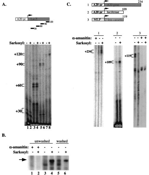
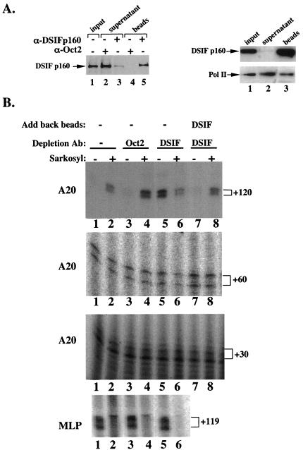
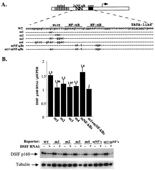
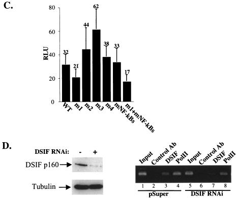
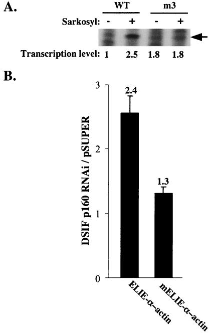
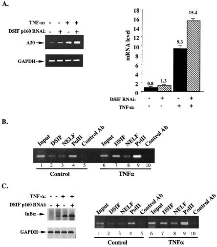
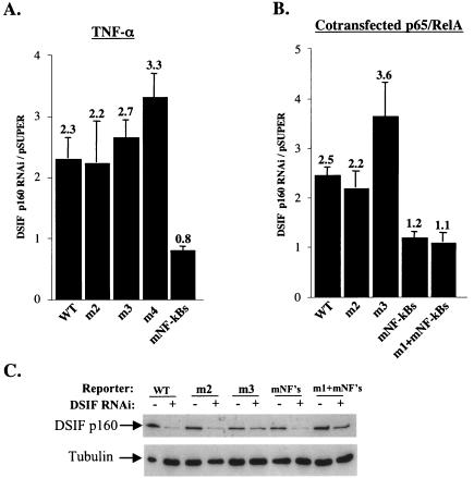
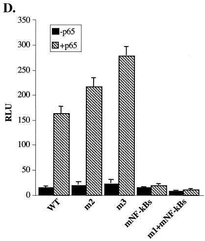
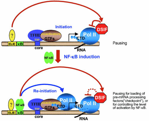
A20 is an immediate-early NF-kappaB target gene. Prior to NF-kappaB stimulation, the A20 promoter is bound by the polymerase II machinery to allow rapid transcription activation. Here we show that the basal A20 transcription is repressed at the level of elongation in a promoter-specific fashion. Immunodepletion in vitro and RNA interference in cultured cells suggest that the basal elongation inhibition is conferred by DRB sensitivity-inducing factor (DSIF). We have identified a negative upstream promoter element called ELIE that controls DSIF activity. Remarkably, following NF-kappaB stimulation, inhibition of the A20 promoter by DSIF persists, but it is now regulated by NF-kappaB rather than ELIE. Similar regulation by DSIF is shown for another NF-kappaB-responsive gene, the IkappaBalpha gene. These findings reveal an intimate and dynamic relationship between DSIF inhibition of elongation and promoter-bound transcription factors. The potential significance of the differential regulation of DSIF activity by cis-acting elements is discussed.


Figures

FIG. 1.
FIG. 1.
(A) Transcription directed by the A20 promoter is inhibited at the level of elongation. In vitro transcription reactions were performed with nuclear extract prepared from nonstimulated Jurkat cells and analyzed by primer extension assay with primers corresponding to the +30, +60, +90, and +120 positions relative to the A20 transcription start site (as depicted in the upper panel). In lanes 2, 4, 6, and 8 Sarkosyl (0.2%) was added exactly 1.5 min after the addition of nucleotides (initiation). (B) In vitro transcription with immobilized template. A DNA fragment composed of the A20 promoter and 120 nucleotides of downstream sequence was immobilized on magnetic beads and incubated with nuclear extract. After addition of nucleotides (including [32P]UTP) Sarkosyl was added as indicated and either washed with transcription buffer 20 s later (lanes 5 and 6) or left untreated (lanes 1 to 4). Transcription from the washed template was then resumed with the addition of transcription buffer and unlabeled nucleotides. α-Amanitin (5 μM) was added to the reaction mixtures in the indicated lanes to verify that the synthesized transcript was Pol II specific. (C) A20 elongation inhibition is promoter driven and promoter specific. Shown are in vitro transcription reactions analyzed either by run-on assay (subpanels 1 and 3) or by primer extension (subpanel 2) with A20 promoter with its native downstream fragment (subpanel 1) or A20 promoter fused to the luciferase gene (subpanel 2) as the template. MLP U-less reporter was used as a control (subpanel 3). (The structure of the templates is shown schematically in the upper panel.) α-Amanitin (5 μM) was added where indicated to verify Pol II transcription. Sarkosyl (0.2%) was added exactly 1.5 min after the addition of nucleotides to the indicated reaction mixtures.
FIG. 2.
FIG. 2.
Inhibition of transcription elongation directed by the A20 promoter is mediated by DSIF. (A) Left panel: immunoblot analysis of Jurkat cell nuclear extract immunodepleted of DSIF p160 by using anti-DSIF p160 antibody. Anti-Oct-2 antibodies were used as the specificity control for the immunodepletion. Ten percent of precipitated proteins was loaded in lanes 4 and 5. Right panel: analysis of Pol II in DSIF-depleted extract. Shown are the results of immunoblot analysis of nuclear extract that was either untreated (lane 1) or immunodepleted of DSIF (lane 2) by using anti-DSIF p160 antibody and anti-Pol II antibody (large subunit). The beads (lane 3) represent the immunoprecipitated complex. (B) Transcription activity of the DSIF p160-depleted extract. Shown are the results of in vitro transcription reactions with either A20-lacZ or MLP U-less templates and Jurkat cell nuclear extract that was untreated (lanes 1 and 2) or immunodepleted with the indicated antibodies. The A20 transcripts were analyzed by primer extension assay with primers corresponding to +30, +60, and +120 relative to the transcription start site. The MLP transcript was analyzed by the run-on assay. In lanes 2, 4, 6, and 8 Sarkosyl (0.2%) was added 1.5 min after the addition of nucleotides. In lanes 7 and 8 the beads containing immunoprecipitated and washed DSIF were added back to the DSIF-depleted extract.
FIG. 3.
FIG. 3.
In unstimulated cells DSIF inhibits the A20 transcription and is regulated by a specific upstream negative element. (A) A20 promoter mutants. The schematic of the A20 promoter is shown at the top. The DNA sequence of the region in the A20 promoter that was subjected to mutations is expanded in the top (WT) row, and only the substituted nucleotides are denoted. The two NF-κB binding sites, the TATA-like sequence, and the putative ELIE are indicated. (B) The effect of DSIF p160 knockdown on the transcription activity of the A20 promoter. 293T cells were transfected with a plasmid expressing DSIF p160 RNAi or the empty expression plasmid pSuper together with a luciferase reporter gene controlled by A20 promoter variants shown in panel A. Promoter activity is presented as the ratio between the luciferase reporter activity in the presence of DSIF RNAi and its activity in the presence of the RNAi parental vector (pSuper). Luciferase activities were normalized to the activity of cotransfected RSV promoter-driven Renilla reporter luciferase that was used to correct for differences in transfection efficiencies. The data represent the means and standard deviations of seven transfection experiments, each done independently in duplicate. The bottom panel shows results of an immunoblot analysis of a representative transfection experiment using anti-DSIF p160 antibody and antitubulin, which served as a control for protein loading. (C) The effect of the mutations on the basal activity of the A20 promoter is presented as relative luciferase units (RLU). (D) Effect of DSIF RNAi on the endogenous A20 promoter occupancy by DSIF and Pol II. 293T cells were transfected with a plasmid expressing DSIF p160 RNAi or the empty expression plasmid pSuper. Forty-eight hours later ChIP assays were performed on these cells with DSIF, Pol II, and control antibodies. The bound A20 promoter region was analyzed by PCR (right panel). The left panel shows results of immunoblot analysis of DSIF and tubulin of these cells. WT, wild type; Ab, antibody.
FIG. 3.
FIG. 3.
In unstimulated cells DSIF inhibits the A20 transcription and is regulated by a specific upstream negative element. (A) A20 promoter mutants. The schematic of the A20 promoter is shown at the top. The DNA sequence of the region in the A20 promoter that was subjected to mutations is expanded in the top (WT) row, and only the substituted nucleotides are denoted. The two NF-κB binding sites, the TATA-like sequence, and the putative ELIE are indicated. (B) The effect of DSIF p160 knockdown on the transcription activity of the A20 promoter. 293T cells were transfected with a plasmid expressing DSIF p160 RNAi or the empty expression plasmid pSuper together with a luciferase reporter gene controlled by A20 promoter variants shown in panel A. Promoter activity is presented as the ratio between the luciferase reporter activity in the presence of DSIF RNAi and its activity in the presence of the RNAi parental vector (pSuper). Luciferase activities were normalized to the activity of cotransfected RSV promoter-driven Renilla reporter luciferase that was used to correct for differences in transfection efficiencies. The data represent the means and standard deviations of seven transfection experiments, each done independently in duplicate. The bottom panel shows results of an immunoblot analysis of a representative transfection experiment using anti-DSIF p160 antibody and antitubulin, which served as a control for protein loading. (C) The effect of the mutations on the basal activity of the A20 promoter is presented as relative luciferase units (RLU). (D) Effect of DSIF RNAi on the endogenous A20 promoter occupancy by DSIF and Pol II. 293T cells were transfected with a plasmid expressing DSIF p160 RNAi or the empty expression plasmid pSuper. Forty-eight hours later ChIP assays were performed on these cells with DSIF, Pol II, and control antibodies. The bound A20 promoter region was analyzed by PCR (right panel). The left panel shows results of immunoblot analysis of DSIF and tubulin of these cells. WT, wild type; Ab, antibody.
FIG. 4.
FIG. 4.
Analysis of ELIE. (A) In vitro transcription reactions analyzed by run-on assay using wild-type or m3 A20 promoter variants (Fig. 3). The correctly initiated transcript (shown by arrow) was quantified and is presented as a ratio to the activity of the wild-type (WT) A20 promoter in the absence of Sarkosyl. (B) Small DNA fragments derived from the A20 promoter containing either full-length ELIE or ELIE with 3 nucleotides deleted (ELIE-α-actin and mELIE-α-actin, respectively) were cloned in front of a minimal α-actin core promoter driving a luciferase reporter gene. These reporter plasmids we cotransfected into 293T cells along with either the plasmid expressing DSIF p160 RNAi or the empty expression plasmid pSuper, and 48 h later luciferase activity was measured and normalized to the activity of cotransfected RSV-Renilla luciferase and is presented as relative luciferase units. The data represent the means and standard deviations of five transfection experiments done independently in duplicate.
FIG. 5.
FIG. 5.
DSIF controls the transcription of the endogenous A20 gene in unstimulated and TNF-α-stimulated cells. (A) 293T cells transfected with either pSuper or DSIF p160 RNAi plasmids were left untreated or treated with TNF-α for 1 h. Total RNA prepared from these cells was used either for standard RT-PCR (left panel) or quantitative real-time PCR (right panel) analysis of A20 mRNA level. For a control, analysis of GAPDH mRNA level was performed as well. (B) ChIP assay using soluble chromatin extract from control or TNF-α-treated Jurkat cells and antibodies directed against DSIF p160, NELF-A, and Pol II. The precipitated DNAs were used for PCR amplifications with primers spanning the core and promoter-proximal regions of the A20 gene. (C) The endogenous IκBα gene was analyzed similarly to the A2O gene by RT-PCR (left panel) and by chromatin immunoprecipitation (right panel) as described for panels A and B. Ab, antibody.
FIG. 6.
FIG. 6.
DSIF inhibitory activity in NF-κB-stimulated A20 promoter is mediated by NF-κB. (A) 293T cells were cotransfected with various A20 promoter reporters and either pSuper or DSIF p160 RNAi, and 48 h posttransfection cells were treated with TNF-α for 4.5 h. The effect of DSIF p160 depletion is presented as the ratio of the relative luciferase activity in the presence of DSIF RNAi to the activity in the presence of the RNAi parental vector (pSuper). (B) 293T cells were cotransfected with various A20 promoter reporters, p65-RelA expression vector, and either pSuper or DSIF p160 RNAi, and the effect of DSIF p160 depletion on the A20 promoter variants stimulated by NF-κB (p65-RelA) is presented as in panel A. (C) Immunoblot analysis of DSIF and tubulin from a representative transfection experiment. (D) Responsiveness of the various A20 promoter mutants to NF-κB (p65-RelA). Shown is the relative luciferase activity (luciferase units divided by the activity of cotransfected RSV promoter-driven Renilla reporter luciferase) of the transfection experiment described for panel B. The data represent the means and standard deviations of three to seven transfection experiments done independently in duplicate. WT, wild type.
FIG. 6.
FIG. 6.
DSIF inhibitory activity in NF-κB-stimulated A20 promoter is mediated by NF-κB. (A) 293T cells were cotransfected with various A20 promoter reporters and either pSuper or DSIF p160 RNAi, and 48 h posttransfection cells were treated with TNF-α for 4.5 h. The effect of DSIF p160 depletion is presented as the ratio of the relative luciferase activity in the presence of DSIF RNAi to the activity in the presence of the RNAi parental vector (pSuper). (B) 293T cells were cotransfected with various A20 promoter reporters, p65-RelA expression vector, and either pSuper or DSIF p160 RNAi, and the effect of DSIF p160 depletion on the A20 promoter variants stimulated by NF-κB (p65-RelA) is presented as in panel A. (C) Immunoblot analysis of DSIF and tubulin from a representative transfection experiment. (D) Responsiveness of the various A20 promoter mutants to NF-κB (p65-RelA). Shown is the relative luciferase activity (luciferase units divided by the activity of cotransfected RSV promoter-driven Renilla reporter luciferase) of the transfection experiment described for panel B. The data represent the means and standard deviations of three to seven transfection experiments done independently in duplicate. WT, wild type.
FIG. 7.
FIG. 7.
Model explaining the differential role of DSIF inhibition of elongation in nonstimulated and stimulated cells. In unstimulated cells (upper panel), the promoters of rapidly induced NF-κB target genes, like A20, are bound by the entire Pol II transcription apparatus, but the basal activity is low since this complex is capable of initiation but not reinitiation (1). Under these conditions, a factor that binds ELIE on the A20 promoter inhibits the basal transcription at the level of elongation via DSIF. This effect is required to keep the basal activity of the prebound transcription complex much lower until NF-κB is stimulated. After NF-κB is induced, it enhances the reinitiation rate but, on the other hand, is responsible for inhibition of elongation by DSIF. This inhibition might serve to load the elongating polymerase with pre-mRNA processing factors in order to ensure that each transcript that has been initiated will be completely processed into mature and translatable mRNA. Alternatively, this inhibition may be used to control the level of activation of the A20 gene by NF-κB, which might be important under certain physiological conditions. CTD, C-terminal domain; GTFs, general transcription factors; TBP, TATA binding protein.

Similar articles

Cited by

References

    1. Ainbinder, E., M. Revach, O. Wolstein, S. Moshonov, and R. Dikstein. 2002. The mechanism of rapid transcriptional induction of tumor necrosis factor alpha-responsive genes by NF-κB. Mol. Cell. Biol. 22:6354-6362. - PMC - PubMed
    1. Barboric, M., R. M. Nissen, S. Kanazawa, N. Jabrane-Ferrat, and B. M. Peterlin. 2001. NF-κB binds P-TEFb to stimulate transcriptional elongation by RNA polymerase II. Mol. Cell 8:327-337. - PubMed
    1. Bourgeois, C. F., Y. K. Kim, M. J. Churcher, M. J. West, and J. Karn. 2002. Spt5 cooperates with human immunodeficiency virus type 1 Tat by preventing premature RNA release at terminator sequences. Mol. Cell. Biol. 22:1079-1093. - PMC - PubMed
    1. Brummelkamp, T. R., R. Bernards, and R. Agami. 2002. A system for stable expression of short interfering RNAs in mammalian cells. Science 296:550-555. - PubMed
    1. Conaway, J. W., A. Shilatifard, A. Dvir, and R. C. Conaway. 2000. Control of elongation by RNA polymerase II. Trends Biochem. Sci. 25:375-380. - PubMed

Publication types

MeSH terms

LinkOut - more resources