. 2004 Mar;24(6):2526-35.
doi: 10.1128/MCB.24.6.2526-2535.2004.

Np95 is a histone-binding protein endowed with ubiquitin ligase activity

Affiliations

Np95 is a histone-binding protein endowed with ubiquitin ligase activity

Elisabetta Citterio et al. Mol Cell Biol. 2004 Mar.

Abstract

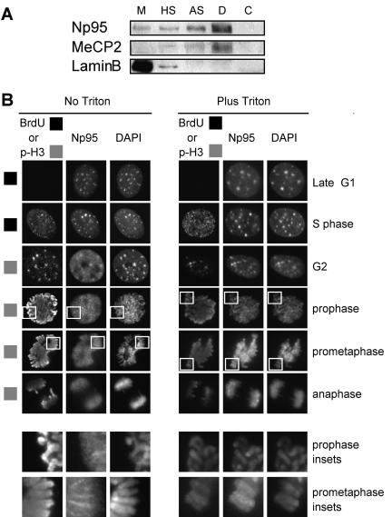
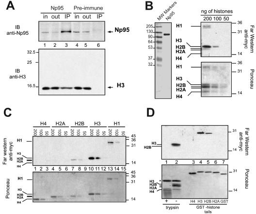
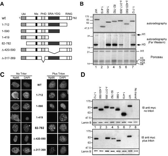
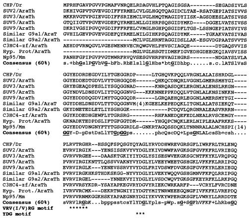
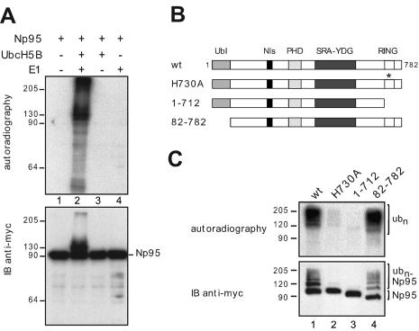
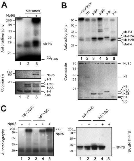
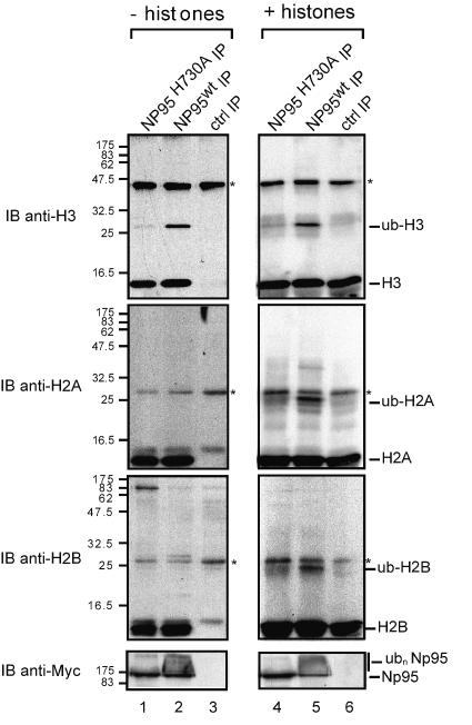
Np95 is an important determinant in cell cycle progression. Its expression is tightly regulated and becomes detectable shortly before the entry of cells into S phase. Accordingly, Np95 is absolutely required for the G1/S transition. Its continued expression throughout the S/G2/M phases further suggests additional roles. Indeed, Np95 has been implicated in DNA damage response. Here, we show that Np95 is tightly bound to chromatin in vivo and that it binds to histones in vivo and in vitro. The binding to histones is direct and shows a remarkable preference for histone H3 and its N-terminal tail. A novel protein domain, the SRA-YDG domain, contained in Np95 is indispensable both for the interaction with histones and for chromatin binding in vivo. Np95 contains a RING finger. We show that this domain confers E3 ubiquitin ligase activity on Np95, which is specific for core histones, in vitro. Finally, Np95 shows specific E3 activity for histone H3 when the endogenous core octamer, coimmunoprecipitating with Np95, is used as a substrate. Histone ubiquitination is an important determinant in the regulation of chromatin structure and gene transcription. Thus, the demonstration that Np95 is a chromatin-associated ubiquitin ligase suggests possible molecular mechanisms for its action as a cell cycle regulator.


Figures

FIG. 1.
FIG. 1.
Np95 is a chromatin-associated protein. (A) Biochemical fractionation of NIH 3T3 cell extracts. Fractions, prepared as described in Materials and Methods, are as follows: C, cytoplasmic; D, DNase I; AS, ammonium sulfate; HS, high salt (2 M NaCl); M, nuclear matrix. Fractions were immunoblotted with the antibodies indicated on the left. Anti-MeCP2 and anti-lamin B antibodies were used as controls to verify the purity of the fractions. (B) Triton-resistant association of Np95 with chromatin. NIH 3T3 cells were grown on coverslips and serum starved for 48 h. Ten percent FBS was then added to stimulate reentry into the cell cycle. After 16 h, 10 μM BrdU was added and the mixture was incubated for 10 min to identify cells in S phase. Cells were then harvested 16 and 23 h after FBS addition. Before fixation, half of the coverslips were treated with 0.5% Triton X-100 (Plus Triton) to remove soluble proteins or mock treated (No Triton). Cells were then stained with anti-Np95 antibody, together with either anti-BrdU (for cells harvested at 16 h [marked with black squares]) or anti-anti-pH3 (specific for histone H3 phosphorylated at serine 10) antibody to reveal cells in G2/M (for cells harvested at 23 h [marked with grey squares]). Counterstaining was done with DAPI. Representative pictures of the indicated phases of the cell cycle are shown. The insets (last two rows, corresponding to magnifications of the areas indicated in the prophase and prometaphase panels) show details of the distribution of Np95 on the mitotic chromosomes.
FIG. 2.
FIG. 2.
Np95 interacts with histones in vivo and in vitro. (A) Endogenous Np95 coimmunoprecipitates with histone H3. Total extracts from asynchronously growing NIH 3T3 cells were immunoprecipitated with either anti-Np95 or preimmune serum (indicated at the top). The immunoprecipitates (IP) were washed under stringent conditions and immunoblotted (IB) with anti-Np95 (top) or anti-H3 (bottom) serum. A fraction (10%) of the starting material (in, input) and of the supernatant of the immunoprecipitates (out, output) is also shown. (B) Np95 interacts with H3, H2B, and H1. (Left) Coomassie staining of the purified myc-His6-Np95 (Np95) protein used in all subsequent far-Western assays and enzymatic assays (details are given in Materials and Methods). (Right) The indicated amounts (shown at the top) of a mixture of commercially available core histones and H1 were detected in a far-Western assay with purified Np95, followed by immunostaining with anti-myc. Ponceau staining of the blot is also shown (bottom). (C) Np95 interacts with individual histones. Recombinantly produced histones, in various amounts (indicated at the top), were detected in a far-Western assay with pure Np95 as in panel B. Ponceau staining of the blot is also shown (bottom). (D) Np95 interacts with histone tails. Four micrograms of polynucleosomes treated with trypsin for 30 min (lane 1) or mock treated (lane 2) or 1 μg of GST fusion proteins of each of the core histone tails (lanes 3, 4, 5, and 6, also indicated at the bottom), or of GST (lane 7) was detected in a far-Western assay with pure Np95 as in panel B. Ponceau staining of the blot is also shown (bottom). The asterisk indicates a band corresponding to the trypsin inhibitor used to terminate the reaction in the first two lanes. In all panels, where appropriate, the positions of histones are indicated and molecular mass (MW) markers are shown (sizes are in kilodaltons).
FIG. 3.
FIG. 3.
The SRA-YDG domain is essential for the interaction of Np95 with histones in vitro and with chromatin in vivo. (A) Schematic representation of wild-type (wt) and mutant Np95 proteins. Nls, putative nuclear localization signal; PHD, PHD domain; SRA-YDG, SRA-YDG domain; RING, RING finger domain. All constructs were myc-His6 tagged. (B) Interaction of wild-type (WT) and various mutant proteins with histones in vitro. (Top) The Np95 constructs indicated in panel A were in vitro translated in the presence of [35S]methionine. The autoradiogram shows the labeled reaction products. (Middle and bottom) One microgram of a mixture of commercially available core histones and H1 was detected in a far-Western assay with 35S-labeled mutant Np95 proteins (as at the top), followed by autoradiography. Ponceau staining of the blot is also shown (bottom). The middle part represents an assembly of individual strips. (C) The SRA-YDG domain is necessary for interaction of Np95 with chromatin in vivo. NIH 3T3 cells were grown on coverslips and transfected with 2 μg of each of the myc-His6-tagged Np95 constructs indicated in panel A. Before fixation, cells were either treated with 0.5% Triton X-100 (Plus Triton) or mock treated (No Triton). The cells were then stained with anti-myc (Np95) and counterstained with DAPI. Representative pictures are shown. (D) NIH 3T3 cells were transfected (transf.) with the indicated constructs as in panel C. Cells were then either treated with 0.5% Triton X-100 or mock treated in situ (see Materials and Methods). Lysates of Triton-extracted whole cells were then obtained and analyzed in an immunoblot (IB) assay with anti-myc. An antibody against lamin B was used as a control. In all panels, where appropriate, the positions of histones are indicated and molecular mass markers are shown (sizes are in kilodaltons).
FIG. 4.
FIG. 4.
Alignment of SRA-YDG domains. Shown are alignments of various SRA-YDG domains and the consensus sequence of the domain (obtained at a plurality of 60%) based on the domain architecture, named the SET and RING finger-associated domain, present in the SMART database (accession no. SM00466). The domain is also described in detail in reference with its 13 conserved G's (underlined in the alignment) and the VRV(I/V)RG and YDG motifs (indicated by asterisks in the alignment). Abbreviations: Dr, Deinococcus radiodurans; AraTh, Arabidopsis thaliana; Mm, Mus musculus. The sequences shown are as follows: CHP (conserved hypothetical protein), accession no. Q9RU61; SUV2, accession no. AAK28967; SUV3, accession no. Q9C5P4 and Q9SSL7; SUV5, accession no. O82175; SUV9, accession no. AAK28974; Similar G9a1, accession no. AAD26896; Similar G9a2, accession no. AAD15582; C3HC4-zf, accession no. AAC28190; Hyp. Prot (hypothetical protein), accession no. AAC95167; Np95, accession no. Q9Z1H6.
FIG. 5.
FIG. 5.
Np95 has E2-dependent Ub ligase activity in vitro. (A) In vitro ubiquitination assay. Purified Np95 (as in Fig. 2B; see also Materials and Methods) was incubated with ATP, radioactively labeled Ub, and the indicated combinations (+/−) of mammalian E1 and recombinant E2 (UbcH5B) enzymes and assayed for Ub ligase activity. High-molecular-weight Ub conjugates were detected by autoradiography (top). Np95 was detected by immunoblotting (IB) with anti-myc (bottom). (B) Schematic representation of wild-type (wt) and mutant Np95 proteins. All constructs were myc-His6 tagged. Domains are as in Fig. 3A. (C) The RING finger domain of Np95 is required for Ub ligation. Wild-type and mutant Np95 proteins were incubated in an in vitro ubiquitination reaction mixture containing 32P-labeled Ub, E1, and UbcH5B enzymes, followed by autoradiography (top). Ub conjugates (ubn) are indicated. Np95 proteins and their Ub conjugates (ubn-Np95) were detected by immunoblotting with anti-myc (bottom). In all panels, where appropriate, the positions of Np95 and its Ub conjugates (ubn-Np95) are indicated and molecular mass markers are shown (sizes are in kilodaltons).
FIG. 6.
FIG. 6.
Np95 is an E3 ligase for core histones in vitro. (A) Np95 ubiquitinates core histones in vitro. Ubiquitination reaction mixtures containing 4 μg of histones purified from calf thymus, 32P-labeled Ub (32P-ub), E1, and UbcH5B were prepared with (+) or without (−) purified Np95. Ubiquitinated histones (ub-Hs) were detected by autoradiography (top). (B) Purified Np95 was incubated with the indicated substrate (1 μg of H1 purified from calf thymus or recombinant H2A, H2B, H3, or H4) or no substrate (− substrate) in a ubiquitination assay, followed by autoradiography (top). In both panels A and B, Coomassie staining of the top parts shows Np95 and the substrates present in the reaction mixtures (bottom parts). (C) NF-Y is not a substrate for Np95. NF-YABC or NF-YBC complexes were incubated in ubiquitination reaction mixtures in the presence (+) or absence (−) of Np95 as indicated. High-molecular-weight products of Np95 self-ubiquitination (ubn-Np95) were detected by autoradiography (top). The NF-YB subunit was detected by immunoblotting (IB) with anti-NF-YB (bottom). Note the lack of a gel-retarded product in the anti-NF-YB blot, which would be indicative of Ub conjugates. In all panels, where appropriate, the positions of individual proteins or of their Ub conjugates (ubn-) are indicated and molecular mass markers are shown (sizes are in kilodaltons).
FIG. 7.
FIG. 7.
Np95 promotes ubiquitination of interacting histone H3 ex vivo. Phoenix cells were transfected with myc-tagged Np95wt, Np95H730A, or the vector alone (control [ctrl]), and equivalent amounts of cell lysates were immunoprecipitated with anti-myc (see Materials and Methods). Immunoprecipitates (IP) were assayed for ubiquitination activity in the presence of Ub, E1, and UbcH5B in the absence (lanes 1 to 3) or presence (lanes 4 to 6) of histones purified from calf thymus. Detection in an immunoblot (IB) assay was done with the antibodies indicated on the left. In all panels, where appropriate, the positions of individual proteins or of their Ub conjugates are indicated and molecular mass markers are shown (sizes are in kilodaltons). Asterisks in all panels indicate nonspecific bands.

References

    1. Baarends, W. M., J. W. Hoogerbrugge, H. P. Roest, M. Ooms, J. Vreeburg, J. H. Hoeijmakers, and J. A. Grootegoed. 1999. Histone ubiquitination and chromatin remodeling in mouse spermatogenesis. Dev. Biol. 207:322-333. - PubMed
    1. Baumbusch, L. O., T. Thorstensen, V. Krauss, A. Fischer, K. Naumann, R. Assalkhou, I. Schulz, G. Reuter, and R. B. Aalen. 2001. The Arabidopsis thaliana genome contains at least 29 active genes encoding SET domain proteins that can be assigned to four evolutionarily conserved classes. Nucleic Acids Res. 29:4319-4333. - PMC - PubMed
    1. Bonapace, I. M., L. Latella, R. Papait, F. Nicassio, A. Sacco, M. Muto, M. Crescenzi, and P. P. Di Fiore. 2002. Np95 is regulated by E1A during mitotic reactivation of terminally differentiated cells and is essential for S phase entry. J. Cell Biol. 157:909-914. - PMC - PubMed
    1. Briggs, S. D., T. Xiao, Z. W. Sun, J. A. Caldwell, J. Shabanowitz, D. F. Hunt, C. D. Allis, and B. D. Strahl. 2002. Gene silencing: trans-histone regulatory pathway in chromatin. Nature 418:498. - PubMed
    1. Caretti, G., M. C. Motta, and R. Mantovani. 1999. NF-Y associates with H3-H4 tetramers and octamers by multiple mechanisms. Mol. Cell. Biol. 19:8591-8603. - PMC - PubMed

Publication types

MeSH terms