. 2004 Mar;24(6):2546-59.
doi: 10.1128/MCB.24.6.2546-2559.2004.

Transforming growth factor beta-mediated transcriptional repression of c-myc is dependent on direct binding of Smad3 to a novel repressive Smad binding element

Affiliations

Transforming growth factor beta-mediated transcriptional repression of c-myc is dependent on direct binding of Smad3 to a novel repressive Smad binding element

Joshua P Frederick et al. Mol Cell Biol. 2004 Mar.

Abstract

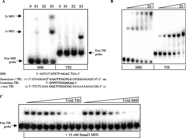
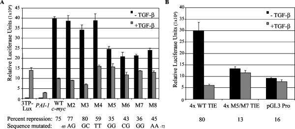
Smad proteins are the most well-characterized intracellular effectors of the transforming growth factor beta (TGF-beta) signal. The ability of the Smads to act as transcriptional activators via TGF-beta-induced recruitment to Smad binding elements (SBE) within the promoters of TGF-beta target genes has been firmly established. However, the elucidation of the molecular mechanisms involved in TGF-beta-mediated transcriptional repression are only recently being uncovered. The proto-oncogene c-myc is repressed by TGF-beta, and this repression is required for the manifestation of the TGF-beta cytostatic program in specific cell types. We have shown that Smad3 is required for both TGF-beta-induced repression of c-myc and subsequent growth arrest in keratinocytes. The transcriptional repression of c-myc is dependent on direct Smad3 binding to a novel Smad binding site, termed a repressive Smad binding element (RSBE), within the TGF-beta inhibitory element (TIE) of the c-myc promoter. The c-myc TIE is a composite element, comprised of an overlapping RSBE and a consensus E2F site, that is capable of binding at least Smad3, Smad4, E2F-4, and p107. The RSBE is distinct from the previously defined SBE and may partially dictate, in conjunction with the promoter context of the overlapping E2F site, whether the Smad3-containing complex actively represses, as opposed to transactivates, the c-myc promoter.


Figures

FIG. 1.
FIG. 1.
Targeted deletion of Smad3 results in elimination of TGF-β growth inhibitory effects and elevated levels of c-Myc. (A) The rate of DNA synthesis of primary keratinocytes derived from wild-type (+/+) and Smad3ex1/ex1 homozygous null (−/−) murine keratinocytes treated with vehicle or 100 pM TGF-β was measured by [3H]thymidine incorporation as described in Materials and Methods. The difference of the measured amount of incorporated [3H]thymidine of cells treated with vehicle minus that of TGF-β-treated cells was divided by the vehicle control counts per minute and plotted as the percent growth inhibition. The presented data are representative of values obtained from three individual lines of each genotype. (B) Murine colon epithelium was isolated from wild-type (+/+) and Smad3ex1/ex1 homozygous null (−/−) 3- to 5-month-old littermates, and Western analysis for c-Myc and actin, as a loading control, of 30 μg of protein from the epithelium lysate are depicted. Brackets indicate two individual sets of wild-type and Smad3ex1/ex1 homozygous null littermates (litter A and litter B).
FIG. 2.
FIG. 2.
TGF-β-mediated repression of c-myc transcriptional initiation is reduced in the absence of functional Smad3. (A) BALB/MK cells or primary keratinocytes derived from wild-type and Smad3ex1/ex1 homozygous null littermates were treated with 400 nM TGF-β or vehicle for 1 h. RPAs of 10 μg of BALB/MK and 15 μg of primary keratinocyte total RNA were performed as described in Materials and Methods. The depicted RPA is representative of three independent experiments. (B) Densitometric analysis of a phosphorimager scan of the RPA depicted in panel A was used to quantify mRNA levels. Values for c-myc band intensities were normalized to β-actin levels, which were quantified with a lighter phosphorimage exposure to obtain more accurate assessments of the relatively high β-actin intensities. The repression was calculated by dividing the quantified, normalized value of c-myc in the absence of TGF-β (control) by corresponding levels in the presence of TGF-β. (C) Total RNA was isolated from control HaCaT and S3 DN HaCaT cells treated with 100 pM TGF-β for 0, 2, 4, and 8 h, as indicated. RPAs of 5 μg of total RNA per condition were performed as described in Materials and Methods to determine mRNA levels of c-myc, PAI-1, L32, and GAPDH. PAI-1 levels were assessed as a positive control, as this gene is known to be transactivated by TGF-β through a mechanism that is partially dependent on Smad3 recruitment to promoter-contained SBEs. L32 and GAPDH mRNA levels served as loading control indicators. The depicted RPA is representative of three independent experiments.
FIG. 3.
FIG. 3.
Smad3 and Smad4 cooverexpression represses endogenous c-myc expression. (A) BALB/MK cells either were treated with 400 nM TGF-β or vehicle for 60 min or were infected with recombinant adenoviruses (Ad) carrying empty CMV vector (V) or vector containing cDNA for Smad2 (2), Smad3 (3), and/or Smad4 (4) at a total multiplicity of infection of 100 for 12 h. RPA of 15 μg of total RNA isolated from these cells is shown. Results are representative of three experiments. Immunoblot analysis of whole-cell lysates from the infected cells with anti-Smad1/2/3 (sc-7960; Santa Cruz) is shown in the middle panel. The blot was reprobed with anti-Smad4 (sc-7966; Santa Cruz), and these results are depicted in the lower panel. (B) Densitometric analysis of a phosphorimager scan of the RPA in panel A was completed, and repression of normalized, quantified values of band intensities was calculated as described in the legend for Fig. 2B and graphed, with the exception that the value derived from empty vector adenovirus-infected cells was used as the control level.
FIG. 4.
FIG. 4.
The MH1 domain of Smad3 specifically and directly binds to the c-myc TIE with a relative affinity similar to that of the SBE. (A) A 15 nM concentration of recombinant Smad1 (S1), Smad2 (S2), or Smad3 (S3) MH1 domain was individually incubated with radiolabeled SBE and c-myc TIE-containing probes, and an EMSA was performed as described in Materials and Methods. Two Smad3 MH1 complexes shifted the 16-bp SBE probe due to the incorporated palindromic double site, as depicted in bold. One molecule of the MH1 domain bound to one molecule of SBE is represented by 1x MH1, whereas two molecules of the MH1 domain bound to one molecule of DNA are represented by 2x MH1. The employed 30-bp c-myc TIE probe, equivalent to nucleotides of the c-myc promoter −92 to −63 relative to the P2 transcriptional start site, is depicted below. Sequence encompassing the first identified TIE within the rat stromelysin promoter as well as the composition of the subsequently proposed “consensus” TIE are listed below. Nucleotides in bold represent invariance, whereas lowercase letters signify preferred nucleotides. (B) The relative affinities of the Smad3 MH1-SBE and Smad3-TIE interactions were determined by EMSA, in which increasing concentrations of Smad3 MH1 recombinant protein (S3) were incubated with a fixed amount of radiolabeled SBE or c-myc TIE probe. Threefold serial dilutions of protein, from 556 to 2.29 nM, were used as indicated. The free or unbound probes are the lowest bands depicted within their respective lanes. (C) The similarity of the relative affinities of Smad3 MH1 for SBE and c-myc TIE probes was confirmed by a DNA cold competitor EMSA as described in Materials and Methods. Fifteen nanomolar Smad3 MH1 domain recombinant protein was incubated with radiolabeled c-myc TIE and increasing amounts of cold competitor DNA. The EMSA samples incubated with unlabeled c-myc TIE oligonucleotide are shown on the left, whereas those with unlabeled SBE are on the right. Threefold dilutions of DNA, from 700 to 0.320 nM, were used as indicated. All the data depicted are representative of at least three independent experiments.
FIG. 5.
FIG. 5.
The c-myc TIE contains a novel Smad binding site, or RSBE, maximally comprised of 5′-TTGGCGGGAA-3′. (A) DNA cold competitor EMSAs similar to that described in the legend for Fig. 4C were performed to map the Smad3 binding sequence. A 15 nM concentration of recombinant Smad3 MH1 domain was incubated with radiolabeled, wild-type c-myc TIE probe and a 1.365 μM concentration of the indicated unlabeled TIE competitor. Complexes of Smad3 MH1 bound to the radiolabeled probe (S3 MH1) and unbound, free probe are indicated by arrows. The sequences for the wild-type (WT) c-myc TIE and various, scanning TIE mutants (M1 to M10 and M“SBE” or MS TIE) are listed below. The mutated nucleotides are underlined, and the sequence with homology to a consensus TIE is depicted in bold. The top panel demonstrates an equal amount of employed cold competitor DNA. The shown EMSA is representative of five similar experiments. (B) The underlined guanines 5′-GGCGG-3′ (−79 to −75 relative to the P2 transcriptional start site) within the c-myc TIE were shown to be important in mediating Smad3 binding by methylation interference assay and are boxed. The 30-bp, wild-type c-myc TIE oligonucleotide was radiolabeled on the 5′ end of the sense strand of DNA, methylated with DMS, and then incubated with recombinant Smad3 MH1 domain. The methylated DNA-protein mix was separated by PAGE, and free, or unbound, probe as well as protein-shifted probe was isolated by electroelution. The free (outer two lanes) and Smad3 MH1-bound (middle lane) probes were then piperidine cleaved and separated by denaturing PAGE. Nucleotide −86 relative to the c-myc P2 transcriptional start site is shown at the bottom and −62 is at the top. (C) The binding affinity of recombinant Smad3 MH1 domain (S3) to wild-type c-myc TIE (WT TIE) probe compared to M“SBE” TIE probe was assessed by EMSA. M“SBE” TIE represents a c-myc TIE sequence in which the putative SBE sequence, 5′-GGCT-3′ (−84 to −81) was mutated to TTAA. Threefold serial dilutions of recombinant Smad3 MH1, from 1.668 to 2.29 mM, were incubated with WT TIE and M“SBE” TIE probes as indicated. The presented EMSA is representative of four independent experiments. (D) A 300-μg aliquot of whole-cell lysate (WCL) obtained from sonicated HaCaT cells treated with vehicle or TGF-β for 1 h was incubated with equimolar amounts of biotinylated double-strand oligonucleotides (B-DNA). The SBE sequence used is listed in Fig. 4A, and the composition of the wild-type and mutated c-myc TIE oligonucleotides (WT, MS, and M3 to M6) is depicted in panel A. DNA-bound protein complexes were isolated by subsequent incubation with streptavidin-linked agarose beads, centrifugation, and washing. DNA-bound protein was then separated by sodium dodecyl sulfate-PAGE and analyzed by Western blotting for Smad3 and Smad4. (E) The DNAP experiment shown is exactly the same as that depicted in panel D with the exception that 300 μg of nuclear extract (NE) was used in place of whole-cell lysate. The shown DNAPs are representative of three independent experiments.
FIG. 6.
FIG. 6.
The RSBE characterized by Smad-DNA interaction studies within the c-myc TIE is required and sufficient for TGF-β-mediated transcriptional repression. (A) HaCaT cells were transiently transfected with equal amounts of the indicated promoter-driven luciferase reporters and cultured in the absence and presence of 100 pM TGF-β for 24 h. Cells were then harvested, and levels of luciferase activity were measured by a luminometer. 3TP-lux and PAI-1 are promoter reporters that are transactivated by TGF-β in part through recruitment of Smad3 to contained SBEs. Nucleotides −279 to +337 relative to the P2 transcriptional start site of the c-myc promoter were subcloned into a reporter vector, and mutations were created (M2 to M8) corresponding to those utilized in the characterization of the protein-DNA interaction. The sequence that was mutated within the reporter constructs (−85 to −72) is listed below. The percent of TGF-β-mediated repression was calculated as the measured luciferase activity from exponentially growing cells (−TGF-β) (control) divided by the difference of the measured activity of TGF-β-treated cells (+TGF-β) from that of the control, and this value is listed below the indicated reporter construct. (B) HaCaTs were transiently transfected as for panel A with luciferase reporter constructs containing four copies of sequence (−87 to −72) encompassing the wild-type c-myc TIE (4x WT TIE) or corresponding M5/M7 mutant TIE (4x M5/M7 TIE), subcloned upstream of an SV40 promoter-driven reporter vector. The empty vector, pGL3 Pro, was also transfected to serve as a negative control. Percent repression was calculated as for panel A and is listed below the indicated construct. The presented data are representative of three independent experiments.
FIG. 7.
FIG. 7.
The extended c-myc TIE is comprised of a closely overlapping RSBE and E2F site. (A) An E2F-4/DP-1 heterodimer binding site within the c-myc TIE was mapped as described in the legend for Fig. 5A with the exception that 150 nM of an equimolar mixture of recombinant E2F-4/DP-1 DBDs was employed in place of Smad3 MH1. The extended c-myc TIE is depicted below, with the sequence with homology to a consensus TIE in bold, the RSBE in italics, and the E2F site underlined. (B) A DNAP was performed with whole-cell lysate as described in the legend for Fig. 5A, and immunoblots were probed for p107 and E2F-4. The data presented are representative of three independent experiments.

Similar articles

Cited by

References

    1. Abdollah, S., M. Macias-Silva, T. Tsukazaki, H. Hayashi, L. Attisano, and J. L. Wrana. 1997. TβRI phosphorylation of Smad2 on Ser465 and Ser467 is required for Smad2-Smad4 complex formation and signaling. J. Biol. Chem. 272:27678-27685. - PubMed
    1. Akiyoshi, S., H. Inoue, J. Hanai, K. Kusanagi, N. Nemoto, K. Miyazono, and M. Kawabata. 1999. c-Ski acts as a transcriptional co-repressor in transforming growth factor-beta signaling through interaction with smads. J. Biol. Chem. 274:35269-35277. - PubMed
    1. Alexandrow, M. G., M. Kawabata, M. Aakre, and H. L. Moses. 1995. Overexpression of the c-Myc oncoprotein blocks the growth-inhibitory response but is required for the mitogenic effects of transforming growth factor beta 1. Proc. Natl. Acad. Sci. USA 92:3239-3243. - PMC - PubMed
    1. Attisano, L., and J. L. Wrana. 2000. Smads as transcriptional co-modulators. Curr. Opin. Cell Biol. 12:235-243. - PubMed
    1. Baker, J. C., and R. M. Harland. 1997. From receptor to nucleus: the Smad pathway. Curr. Opin. Genet. Dev. 7:467-473. - PubMed

Publication types

MeSH terms

LinkOut - more resources