. 2004 Mar 9;101(10):3581-6.
doi: 10.1073/pnas.0400373101. Epub 2004 Mar 1.

Systemic delivery of human microdystrophin to regenerating mouse dystrophic muscle by muscle progenitor cells

Affiliations

Systemic delivery of human microdystrophin to regenerating mouse dystrophic muscle by muscle progenitor cells

Estanislao Bachrach et al. Proc Natl Acad Sci U S A. .

Abstract

Cell-based therapy for Duchenne muscular dystrophy patients and mdx mice has proven to be a safe but ineffective form of treatment. Recently, a group of cells called muscle side population (SP) cells have been isolated based on their ability to efflux the DNA-binding dye Hoechst. To understand the potential of skeletal muscle SP cells to serve as precursors for muscle, SP cells from the two mice strains mdx(5cv) and C57BL/6N were isolated, transduced, and transplanted. Under coculture conditions with myogenic cells, some cells within the SP cell population can give rise to early Pax7-positive satellite cells and other later stage myogenic cells. Transduced SP cells were transplanted via the tail vein and were shown to successfully deliver enhanced GFP and human microdystrophin to the skeletal muscle of nonirradiated mdx(5cv) mice, thus demonstrating their ability to travel through the capillaries and enter into damaged muscle. These results demonstrate that i.v. delivery of genes via SP cells is possible and that these SP cells are capable of recapitulating the myogenic lineage. Because this approach shows definitive engraftment by using autologous transplantation of noninjured recipients, our data may have substantial implications for therapy of muscular dystrophy.


Figures

Fig. 1.
Fig. 1.
FACS analysis of C57BL/6N SP cells and mdx5cv SP cells. (A) Hoechst 33342 and propidium iodide profiles of SP and MP cells from skeletal muscle of mdx5cv and C57BL/6N wild type mice. (B) Inhibition of Hoechst 33342 dye efflux by verapamil for mdx5cv and C57BL/6N SP cells.
Fig. 2.
Fig. 2.
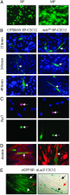
eGFP-lentiviral transduction of muscle SP and MP cells and coculture with C2C12 cells. (A) eGFP expression by direct fluorescence microscopy of l-MSCV-eGFP-infected-SP and l-MSCV-eGFP-infected-MP cells. Results from the mdx5cv transduction are presented, but no differences were found between the mdx5cv and C57BL/6N mice. mdx5cv and C57BL/6N eGFP-SP cells were cocultured with differentiated C2C12-derived myotubes (B) or undifferentiated C2C12-derived myocytes (C and D) C2C12 cells. (B) A time course of coculture with nuclei stained with 4′,6-diamidino-2-phenylindole (DAPI) (blue) and eGFP in green. The mdx5cv SP cells were found aligned at 12 h and fused into multinucleated myotubes as early as 24 h (eGFP-SP arrow). The C57BL/6N SP cells were not found aligned until 24 h and did not fused into myotubes until 48 h postcoculture (eGFP-SP arrowhead). (C) Coculture of mdx5cv and C57BL/6N eGFP-SP cells with C2C12 in proliferation media before confluence. Staining was done with Pax7 (Texas red) and DAPI (blue). Equal numbers of Pax7-eGFP-SP-derived cells were detected in both strains (arrows). (D) Four days after addition of confluence fusion medium, the cultures were stained with desmin (Texas red) and DAPI (blue) Desmin-positive-eGFP-SP-derived myotubes were detected in both strains (arrows). (E) Coculture of eGFP-SP cells with nLacZ marked C2C12 myogenic cells. Equal numbers of muscle eGFP-SP cells and nLacZ-C2C12 cells were cocultured. Multinucleated myotubes, which expressed eGFP but not nLacZ, were seen (black and white arrows), as well as multinucleated myotubes expressing both eGFP and nLacZ (black and white arrowheads). SP-derived cells not fused into multinucleated myofibers were also observed (red arrows).
Fig. 3.
Fig. 3.
Culture of single myofibers from transplanted animals. (A) Light microscopy of an example of single myofiber culture. (Aa) Day 1, satellite cells (arrow) migrate to the periphery of the fiber. (Ab) Week 1, in culture satellite cells (arrow) differentiate into myoblasts (arrowhead). (Ac) Myocytes are detected during the first week in culture (arrow). (Ad) Days 10-15, new multinucleated myotubes appear (arrow). (B) Culture of single myofibers from eGFP-SP transplanted mice. (Ba) Day 1, culture of an eGFP single myofiber from a transplanted mdx5cv. (Bb) Day 2, detection of an eGFP multinucleated (DAPI in blue) myofiber (arrowhead) and an eGFP-Pax7-positive (Texas red)-SP-derived cell (arrow). (Bc) Day 5, detection of an eGFP-desmin-positive (Texas red)-SP derived myocyte (arrow) and a desmin-positive only (Texas red) myocyte (arrowhead). (Bd) Day 10, presence of an eGFP-demin-positive (Texas red)-SP-derived multinucleated (DAPI in blue) myotube (arrow). (C) Culture of single myofibers from eGFP-SP-transplanted mice. (Ca and Cb) Day 15, a myogenin-positive (Texas red plus DAPI) also expressing eGFP in a multinucleated myotube (arrow). (Cc and Cd) Day 15, desmin-positive myofibers (Texas red), which also express eGFP (arrow). Pictures are at low magnification (×20), except Ac and Bc (×40.)
Fig. 4.
Fig. 4.
Transduction and transplantation of muscle SP and MP cells with microdystrophin-expressing lentivirus. (A) Detection of a 137-kDa band (human microdystrophin) by immunoblot analysis of l-MSCV-μdys-infected-SP and l-MSCV-μdys-infected-MP cells extracts. (B) After sorting, 15,000 mdx5cv muscle SP or MP cells were transduced with microdystrophin lentiviral vectors and then intravenously transplanted into C57BL/6N or mdx5cv mice. Detection of mouse (m) dystrophin-positive revertant fibers (FITC stained green) and human (h) dystrophin (merge FITC and Texas red) in four different skeletal muscles from μdys-SP mdx5cv-transplanted mice with an anti-mouse dystrophin (which also recognizes human dystrophin) and a specific anti-human dystrophin antibody 4 weeks after transplantation, respectively. Nuclei were stained with DAPI (blue). (Magnification, ×20.)

References

    1. Hoffman, E. P., Brown, R. H., Jr., & Kunkel, L. M. (1987) Cell 51, 919-928. - PubMed
    1. Cox, G. A., Cole, N. M., Matsumura, K., Phelps, S. F., Hauschka, S. D., Campbell, K. P., Faulkner, J. A. & Chamberlain, J. S. (1993) Nature 364, 725-729. - PubMed
    1. Tremblay, J. P., Malouin, F., Roy, R., Huard, J., Bouchard, J. P., Satoh, A. & Richards, C. L. (1993) Cell Transplant. 2, 99-112. - PubMed
    1. Mendell, J. R., Kissel, J. T., Amato, A. A., King, W., Signore, L., Prior, T. W., Sahenk, Z., Benson, S., McAndrew, P. E., Rice, R., et al. (1995) N. Engl. J. Med. 333, 832-838. - PubMed
    1. Gussoni, E., Blau, H. M. & Kunkel, L. M. (1997) Nat. Med. 3, 970-977. - PubMed

Publication types

MeSH terms

LinkOut - more resources