. 2004 Mar 9;101(10):3569-74.
doi: 10.1073/pnas.0304987101. Epub 2004 Mar 1.

Isolation and characterization of a digoxin transporter and its rat homologue expressed in the kidney

Affiliations

Isolation and characterization of a digoxin transporter and its rat homologue expressed in the kidney

Tsuyoshi Mikkaichi et al. Proc Natl Acad Sci U S A. .

Abstract

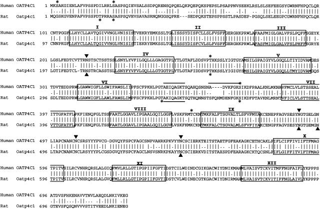
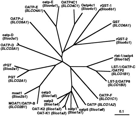
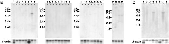
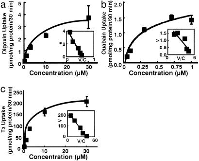
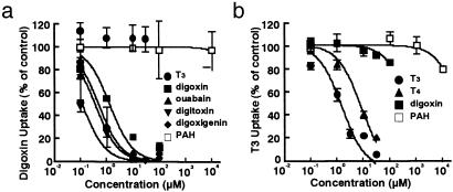
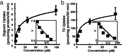
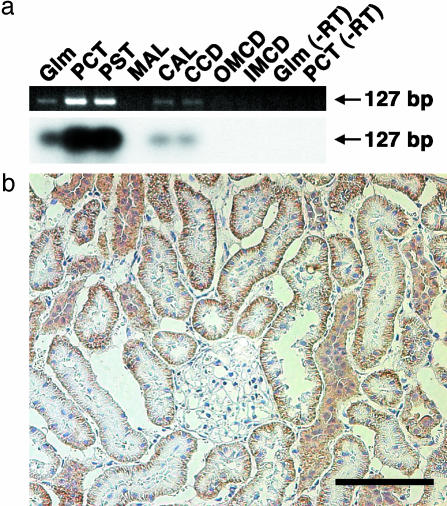
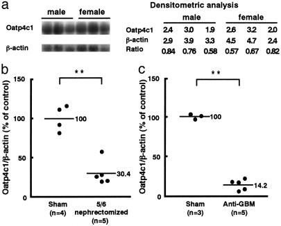
Digoxin, which is one of the most commonly prescribed drugs for the treatment of heart failure, is mainly eliminated from the circulation by the kidney. P-glycoprotein is well characterized as a digoxin pump at the apical membrane of the nephron. However, little is known about the transport mechanism at the basolateral membrane. We have isolated an organic anion transporter (OATP4C1) from human kidney. Human OATP4C1 is the first member of the organic anion transporting polypeptide (OATP) family expressed in human kidney. The isolated cDNA encodes a polypeptide of 724 aa with 12 transmembrane domains. The genomic organization consists of 13 exons located on chromosome 5q21. Its rat counterpart, Oatp4c1, is also isolated from rat kidney. Human OATP4C1 transports cardiac glycosides (digoxin, K(m) = 7.8 microM and ouabain, K(m) = 0.38 microM), thyroid hormone (triiodothyronine, K(m) = 5.9 microM and thyroxine), cAMP, and methotrexate in a sodium-independent manner. Rat Oatp4c1 also transports digoxin (K(m) = 8.0 microM) and triiodothyronine (K(m) = 1.9 microM). Immunohistochemical analysis reveals that rat Oatp4c1 protein is localized at the basolateral membrane of the proximal tubule cell in the kidney. These data suggest that human OATP4C1/rat Oatp4c1 might be a first step of the transport pathway of digoxin and various compounds into urine in the kidney.


Figures

Fig. 1.
Fig. 1.
Alignment of deduced amino acid sequence of human OATP4C1 and rat Oatp4c1. The two sequences are aligned with single letter notation by inserted gaps (-) to achieve maximum homology. The 12 transmembrane domains (boxes) were assigned on the basis of hydrophobicity analysis. Potential N-glycosylation sites (filled triangles) and possible phosphorylation sites (asterisks) are indicated. Possible ATP-binding sites (bar) are also indicated.
Fig. 2.
Fig. 2.
Phylogenetic relationship among human OATP4C1, rat Oatp4c1, and the OATP family. Branch lengths are drawn to scale.
Fig. 3.
Fig. 3.
(a) Human Northern blot analysis. Lanes are as follows: 1, heart; 2, brain; 3, placenta; 4, lung; 5, liver; 6, skeletal muscle; 7, kidney; 8, pancreas; 9, spleen; 10, thymus; 11, prostate; 12, testis; 13, ovary; 14, small intestine; 15, colon; 16, peripheral blood leukocyte; 17, stomach; 18, thyroid; 19, spinal cord; 20, lymph node; 21, trachea; 22, adrenal gland; 23, bone marrow; 24, fetal brain; 25, fetal lung; 26, fetal liver; 27, fetal kidney. (b) Rat Northern blot analysis. Lanes are as follows: 1, heart; 2, brain; 3, spleen; 4, lung; 5, liver; 6, skeletal muscle; 7, kidney; 8, testis.
Fig. 4.
Fig. 4.
Kinetics of human OATP4C1-mediated digoxin (a), ouabain (b), and T3 (c) uptake into transfected MDCK cells. (Inset) Eadie-Hofstee plot. V, velocity; V/C, velocity per concentration of substrate.
Fig. 5.
Fig. 5.
Dose-dependent inhibition by various compounds on [3H]digoxin (a) or [125I]T3 (b) uptake via human OATP4C1. The OATP4C1-mediated 0.1 μM [3H]digoxin or 0.1 μM [125I]T3 uptake was determined in the absence or presence of unlabeled compounds for 30 min.
Fig. 6.
Fig. 6.
Rat Oatp4c1-mediated transport in transfected MDCK cell. Kinetics of rat Oatp4c1-mediated digoxin (a) and T3 (b) uptake into transfected MDCK cells. (Inset) Eadie-Hofstee plot. V, velocity; V/C, velocity per concentration of substrate.
Fig. 7.
Fig. 7.
(a) RT-PCR along rat dissected nephron segment. The PCR product amplified was electrophoresed and stained with ethidium bromide (Upper). Subsequently, gels were transferred and hybridized with [32P]dCTP-labeled rat Oatp4c1 cDNA (Lower). Glm, glomerulus; PCT, proximal convoluted tubule; PST, proximal straight tubule; MAL, medullary thick ascending limb; CAL, cortical thick ascending limb; CCD, cortical collecting duct; OMCD, outer medullary collecting duct; IMCD, inner medullary collecting duct. (b) Immunohistochemistry of rat Oatp4c1 in rat kidney. The rat Oatp4c1 immunoreactivity was significantly detected at the basolateral membrane of the proximal tubule cell. (Scale bar = 100 μm.)
Fig. 8.
Fig. 8.
Northern blot analysis of the rat Oatp4c1 mRNA (total RNA 20 μg per lane) in the intact male and female Sprague-Dawley rat kidneys (a), male 5/6 nephrectomized rat kidneys (b), and male anti-GBM Ab-injected WKY rat kidneys (c). **, P < 0.01, 5/6 nephrectomized rat or anti-GBM injected rat vs. sham control.

References

    1. The Digitalis Investigation Group (1997) N. Engl. J. Med. 336, 525-533. - PubMed
    1. Koren, G. (1987) Clin. Pharmacokinet. 13, 334-343. - PubMed
    1. Inui, K., Masuda, S. & Saito, H. (2000) Kidney Int. 58, 944-958. - PubMed
    1. Dresser, M. J., Leabman, M. K. & Giacomini, K. M. (2001) J. Pharm. Sci. 90, 397-421. - PubMed
    1. Woodland, C., Ito, S. & Koren, G. (1998) Ther. Drug Monit. 20, 134-138. - PubMed

Publication types

MeSH terms

Associated data

LinkOut - more resources