. 2004 Mar 9;101(10):3414-9.
doi: 10.1073/pnas.0305892101. Epub 2004 Mar 1.

Solution structure of dengue virus capsid protein reveals another fold

Affiliations

Solution structure of dengue virus capsid protein reveals another fold

Lixin Ma et al. Proc Natl Acad Sci U S A. .

Abstract

Dengue virus is responsible for approximately 50-100 million infections, resulting in nearly 24,000 deaths annually. The capsid (C) protein of dengue virus is essential for specific encapsidation of the RNA genome, but little structural information on the C protein is available. We report the solution structure of the 200-residue homodimer of dengue 2 C protein. The structure provides, to our knowledge, the first 3D picture of a flavivirus C protein and identifies a fold that includes a large dimerization surface contributed by two pairs of helices, one of which has characteristics of a coiled-coil. NMR structure determination involved a secondary structure sorting approach to facilitate assignment of the intersubunit nuclear Overhauser effect interactions. The dimer of dengue C protein has an unusually high net charge, and the structure reveals an asymmetric distribution of basic residues over the surface of the protein. Nearly half of the basic residues lie along one face of the dimer. In contrast, the conserved hydrophobic region forms an extensive apolar surface at a dimer interface on the opposite side of the molecule. We propose a model for the interaction of dengue C protein with RNA and the viral membrane that is based on the asymmetric charge distribution of the protein and is consistent with previously reported results.


Figures

Fig. 1.
Fig. 1.
A multiple sequence alignment of flavivirus C proteins reproduced from multalign (30). Residues with high similarity (>50%) are red, and the conserved residues are highlighted. The secondary structure is indicated at the top. Helices are V26-L35, K45-T58, A63-W69, and K74-N96. The conserved hydrophobic region of flaviviruses is shaded gray. DEN2, dengue type 2; DEN1, dengue type 1; DEN3, dengue type 3, DEN4, dengue type 4; KUN, Kunjin; WNV, West Nile virus; MVE, Murray Valley encephalitis; JEV, Japanese encephalitis; SLE, St. Louis encephalitis; YFV, yellow fever; TBE; LIV, louping ill; LAN, Langat; POW, Powassan virus.
Fig. 2.
Fig. 2.
Structure of DEN2C, residues 21–100. Labels for one of the two dimer subunits are designated with prime (′). Helix α1 is blue, α2 is green, α3 is yellow, and α4 is magenta. (A) Stereoview of backbone tracing for set of 20 refined solution structures prepared by using insight ii (Molecular Simulation). (B) Ribbon diagram of DEN2C dimer and separated monomer. (C) A 90° rotation of B about a horizontal axis. (D) As in C, rotated 90° about a vertical axis. Figs. 2, 3, 4 were generated by using molscript (31) and raster3d (32).
Fig. 3.
Fig. 3.
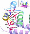
Close-up view of the environment of W69 and hydrophobic interactions (orange) of the monomer core. R68 is shown in indigo. (Inset) The context of the full dimer.
Fig. 4.
Fig. 4.
(A) Cross-eye stereoview of the side-chain interactions that form the hydrophobic α4–α4′ interface along the buried edge of the helix pair. (B) Cross-eye stereoview of the dimer interface formed by the α2–α2′ helix pair (green), and interactions between α2 and α4′ (magenta), which are equivalent to α2′ and α4. The primed subunit is in front (α2′) and on the left (α4′). Ionic interactions involving the single acidic residue, E87 (yellow), and K45 (dark blue) and R55′ (cyan) are indicated with dotted lines. Residues L46, F53, and L57 (purple) from both subunits (top of view) are exposed to solvent and form a large continuous hydrophobic surface. Buried residues F56 and F84 (purple) form dimer contacts.
Fig. 5.
Fig. 5.
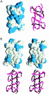
Solvent-accessible surfaces colored by residue type for the DEN2C dimer. Lysine and arginine are blue, and glutamic acid is red (not visible in these views). A main-chain tracing in the same orientation is shown with the surface representation. (A) Charged surface of the α4–α4′ helix pair. (B) Cross-eyed stereoview of the apolar surface formed by the α2–α2′ helix pair. Generated by using grasp (33).
Fig. 6.
Fig. 6.
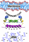
Model for molecular interactions between structural components of flavivirus. The viral membrane is shown on top near the hydrophobic cleft, and the viral RNA is shown on the bottom near the positively charged surface of DEN2C.

Similar articles

Cited by

References

    1. Gubler, D. J. (2002) Trends Microbiol. 10, 100-103. - PubMed
    1. Lindenbach, B. D. & Rice, C. M. (2001) in Fields Virology, eds. Knipe, D. M. & Howley, P. M. (Lippincott, Williams & Wilkins, Philadelphia), pp. 991-1041.
    1. Kuhn, R. J., Zhang, W., Rossmann, M. G., Pletnev, S. V., Corver, J., Lenches, E., Jones, C. T., Mukhopadhyay, S., Chipman, P. R., Strauss, E. G., et al. (2002) Cell 108, 717-725. - PMC - PubMed
    1. Rey, F. A., Heinz, F. X., Mandl, C., Kunz, C. & Harrison, S. C. (1995) Nature 375, 291-298. - PubMed
    1. Zhang, W., Chipman, P. R., Corver, J., Johnson, P. R., Zhang, Y., Mukhopadhyay, S., Baker, T. S., Strauss, J. H., Rossmann, M. G. & Kuhn, R. J. (2003) Nat. Struct. Biol., 10, 907-912. - PMC - PubMed

Publication types

MeSH terms

Associated data

LinkOut - more resources