. 2004 Mar;186(6):1658-66.
doi: 10.1128/JB.186.6.1658-1666.2004.

The Legionella pneumophila PilT homologue DotB exhibits ATPase activity that is critical for intracellular growth

Affiliations

The Legionella pneumophila PilT homologue DotB exhibits ATPase activity that is critical for intracellular growth

Jessica A Sexton et al. J Bacteriol. 2004 Mar.

Abstract

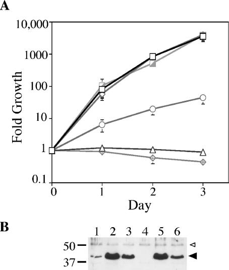
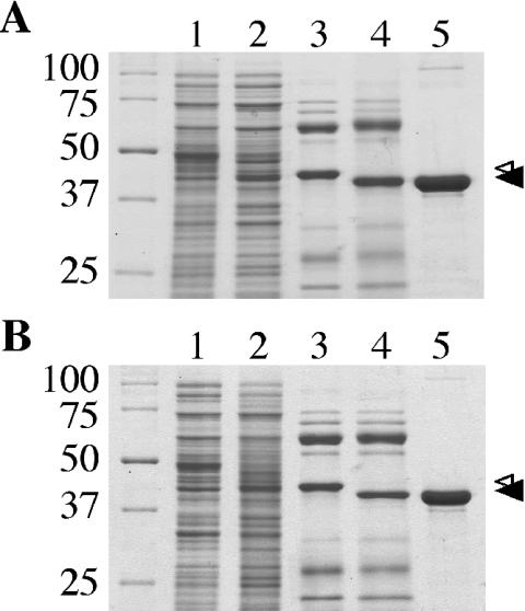
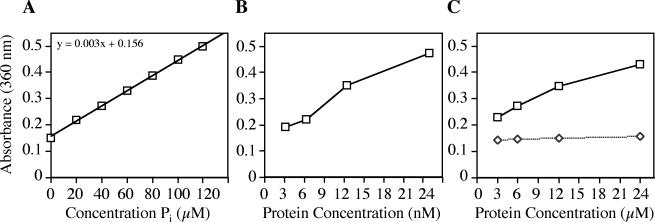
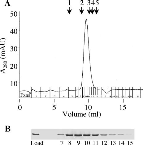
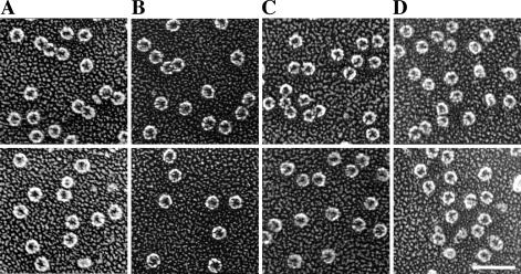
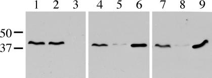
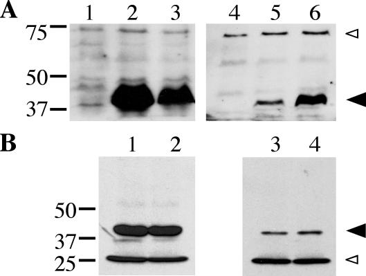
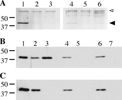
The ability of Legionella pneumophila to grow and cause disease in the host is completely dependent on a type IV secretion system known as the Dot/Icm complex. This membrane-spanning apparatus translocates effector molecules into host cells in a process that is poorly understood but that is known to require the putative ATPase DotB. One possible role for DotB is suggested by its similarity to the PilT family of proteins, which mediate pilus retraction. To better understand the molecular behavior of DotB, we have purified the protein and shown that it forms stable homohexameric rings and hydrolyzes ATP with a specific activity of 6.4 nmol of ATP/min/mg of protein. ATPase activity is critical to the function of DotB, as alteration of the conserved Walker box lysine residue resulted in a mutant protein, DotB K162Q, which failed to bind or hydrolyze ATP and which could not complement a DeltadotB strain for intracellular growth in macrophages. Consistent with the ability of DotB to interact with itself, the dotBK162Q allele exhibited transdominance over wild-type dotB, providing the first example of such a mutation in L. pneumophila. Finally, the DotB K162Q mutant protein had a significantly enhanced membrane localization in L. pneumophila compared to wild-type DotB, suggesting a relationship between nucleotide binding and membrane association. These results are consistent with a model in which DotB cycles between the cytoplasm and the Dot/Icm complex at the membrane, where it hydrolyzes nucleotides to provide energy to the complex.


Figures

FIG. 1.
FIG. 1.
Complementation of a dotB mutant for intracellular growth and transdominance of DotB K162Q in wild-type L. pneumophila. (A) Intracellular growth in U937 monocytes. L. pneumophila ΔdotB strain JV918 containing empty vector pJB908 (triangles), the DotB complementing clone pJB1153 (hatched squares), and the DotB K162Q clone pJB1568 (diamonds) was assayed for growth in U937 cells over time. In addition, L. pneumophila wild-type strain Lp02 containing empty vector pJB908 (open squares), the DotB complementing clone pJB1153 (open diamonds), and the DotB K162Q clone pJB1568 (circles) was also assayed for growth in U937 cells. (B) Strains used in panel A were examined by Western blotting for relative DotB levels at the time of infection. Lane 1, Lp02(pJB908); lane 2, Lp02(pJB1153); lane 3, Lp02(pJB1568); lane 4, JV918(pJB908); lane 5, JV918 (pJB1153); lane 6, JV918(pJB1568). The molecular masses of relevant markers (in kilodaltons) are on the left. Large arrowhead, DotB or DotB K162Q band; small arrowhead, cross-reacting band that serves to confirm overall sample equivalence.
FIG. 2.
FIG. 2.
Purification of DotB and DotB K162Q from E. coli. (A) Coomassie-stained SDS-PAGE gel showing wild-type DotB purification steps. Lane 1, cell extract from E. coli C600 containing plasmid pJB1572, no induction; lane 2, cell extract from C600(pJB1572), IPTG induced; lane 3, eluate from Ni-NTA-agarose after incubation with His-DotB lysate; lane 4, Ni-NTA eluate after exposure to thrombin; lane 5, purified DotB after three additional separation procedures. (B) Coomassie-stained SDS-PAGE gel showing DotB K162Q purification steps. Lanes are as for panel A, except that DotB K162Q was expressed with C600(pJB2442). (A and B) Small arrowhead, His-DotB or His-DotB K162Q bands; large arrowhead, bands representing DotB or DotB K162Q with the His tags removed. Standards of known molecular masses (kilodaltons) are on the left.
FIG. 3.
FIG. 3.
ATPase activity of purified DotB. ATPase activity was determined by measuring the amount of free phosphate in the reaction by a colorimetric assay (EnzChek; Molecular Probes). (A) Standard curve showing absorbance at 360 nm as a function of the concentration of phosphate. (B) ATPase activity of the positive control apyrase. (C) ATPase activity of DotB (squares) and DotB K162Q (diamonds). The specific activities of apyrase and DotB were calculated to be 3.0 μmol/min/mg and 6.4 nmol/min/mg, respectively. Each point represents three replicates.
FIG. 4.
FIG. 4.
DotB is a hexamer. (A) Superose 12 elution profile for purified wild-type DotB. Milli-absorbance units (mAU) are shown as a function of elution volume. DotB eluted as a single peak corresponding to a molecular mass of 259 kDa. Arrows show elution peaks for standards of known molecular masses (Amersham Biosciences): 1, thyroglobulin (669 kDa); 2, ferritin (440 kDa); 3, catalase (232 kDa); 4, aldolase (158 kDa); 5, BSA (67 kDa). (B) Coomassie-stained SDS-PAGE gel demonstrating that the quantities of DotB in elution fractions (Fxns) 7 to 15 correlate with the single protein elution peak. The elution profile is representative of three experiments and is identical to that for DotB K162Q (data not shown).
FIG. 5.
FIG. 5.
Purified DotB and DotB K162Q proteins form hexameric rings ∼10 nm in diameter. (A to C) Wild-type DotB was visualized in the absence of added nucleotides (A), revealing the putative apo form, as well as in the presence of 1 mM ADP (B) or 1 mM ATPγS (C). (D) DotB K162Q was also visualized in the putative apoprotein (shown here) and nucleotide-bound (not shown) forms. Scale bar, 50 nm. Images are representative of several thousand molecules visualized for each condition.
FIG. 6.
FIG. 6.
Detection of mixed DotB/DotB K162Q multimers. Lysate from L. pneumophila Flag-DotB strain JV3079 containing one of three plasmids was subjected to Ni-NTA-agarose. Strains were JV3079 with empty vector pJB908 (lanes 1 to 3), JV3079 with the His-DotB complementing clone pJB1192 (lanes 4 to 6), and JV3079 with the His-DotB K162Q clone pJB2444 (lanes 7 to 9). For each strain, three samples were taken: total protein (lanes 1, 4, and 7), Ni-NTA-agarose flowthrough (lanes 2, 5, and 8), and Ni-NTA-agarose eluate (lanes 3, 6, and 9). Samples were separated via SDS-PAGE, blotted onto a polyvinylidene fluoride membrane, and used in a Flag Western blot to detect the presence of Flag-DotB. The molecular masses of relevant markers (in kilodaltons) are on the left.
FIG. 7.
FIG. 7.
Subcellular localization of DotB and DotB K162Q. (A) Total protein (lanes 1 to 3) and total membrane protein (lanes 4 to 6) fractions were taken from three L. pneumophila strains and were subjected to DotB Western blotting. Strains included the ΔdotB strain JV918 with empty vector pJB908 (lanes 1 and 4), JV918 with the DotB complementing clone pJB1153 (lanes 2 and 5), and JV918 with the DotB K162Q clone pJB1568 (lanes 3 and 6). The molecular masses of relevant markers (in kilodaltons) are on the left. (B) Total protein (lanes 1 and 2) and total membrane protein (lanes 3 and 4) fractions were taken from two E. coli strains and were used in DotB Western blotting. Strains included XL1-Blue with the DotB complementing clone pJB1153 (lanes 1 and 3) and XL1-Blue with the DotB K162Q clone pJB1565 (lanes 2 and 4). (A and B) Large arrowhead, DotB; small arrowhead, cross-reacting band that confirms overall sample equivalence. The molecular masses of relevant markers (in kilodaltons) are on the left. Results are representative of several experiments.
FIG. 8.
FIG. 8.
Nucleotide binding capacity of DotB and DotB K162Q. (A) Coomassie-stained SDS-PAGE gel showing elution fractions from DotB (lanes 1 to 3) and DotB K162Q (lanes 4 to 6) ATP binding reactions. Purified protein was incubated with ATP-agarose (lanes 1 and 4), AMP-agarose (lanes 2 and 5), or ATP-agarose in the presence of 10 mM competing free nucleotides (lanes 3 and 6), washed, and eluted. The molecular masses of relevant markers (in kilodaltons) are on the left. Large arrowhead, DotB; small arrowhead, BSA added to reactions to prevent nonspecific binding. (B) Western blot of fractions from binding reactions using L. pneumophila lysate containing wild-type DotB (ΔdotB strain JV918 with DotB complementing clone pJB1153). Whole-cell lysate (lane 1) was incubated with ATP-agarose (lanes 2 and 3), AMP-agarose (lanes 4 and 5), or ATP-agarose in the presence of 10 mM competing free nucleotide (lanes 6 and 7), washed, and eluted. Flowthrough (lanes 2, 4, and 6) and elution (lanes 3, 5, and 7) fractions were collected and subjected to DotB Western blotting. (C) Western blot of fractions from binding reactions using L. pneumophila lysate containing DotB K162Q (ΔdotB strain JV918 with DotB K162Q clone pJB1568). Whole-cell lysate (lane 1) was incubated with ATP-agarose (lanes 2 and 3), AMP-agarose (lanes 4 and 5), or ATP-agarose in the presence of 10 mM competing free nucleotides (lanes 6 and 7), washed, and eluted. Flowthrough (lanes 2, 4, and 6) and elution (lanes 3, 5, and 7) fractions were collected and subjected to DotB Western blotting. The molecular masses of relevant markers (in kilodaltons) are on the left.

References

    1. Ball, G., V. Chapon-Herve, S. Bleves, G. Michel, and M. Bally. 1999. Assembly of XcpR in the cytoplasmic membrane is required for extracellular protein secretion in Pseudomonas aeruginosa. J. Bacteriol. 181:382-388. - PMC - PubMed
    1. Berger, K. H., and R. R. Isberg. 1993. Two distinct defects in intracellular growth complemented by a single genetic locus in Legionella pneumophila. Mol. Microbiol. 7:7-19. - PubMed
    1. Bhattacharjee, M. K., S. C. Kachlany, D. H. Fine, and D. H. Figurski. 2001. Nonspecific adherence and fibril biogenesis by Actinobacillus actinomycetemcomitans: TadA protein is an ATPase. J. Bacteriol. 183:5927-5936. - PMC - PubMed
    1. Christie, P. J., and J. P. Vogel. 2000. Bacterial type IV secretion: conjugation systems adapted to deliver effector molecules to host cells. Trends Microbiol. 8:354-360. - PMC - PubMed
    1. Christie, P. J., J. E. Ward, Jr., M. P. Gordon, and E. W. Nester. 1989. A gene required for transfer of T-DNA to plants encodes an ATPase with autophosphorylating activity. Proc. Natl. Acad. Sci. USA 86:9677-9681. - PMC - PubMed

Publication types

MeSH terms

LinkOut - more resources