. 2004 Mar;186(6):1737-46.
doi: 10.1128/JB.186.6.1737-1746.2003.

Sustained photoevolution of molecular hydrogen in a mutant of Synechocystis sp. strain PCC 6803 deficient in the type I NADPH-dehydrogenase complex

Affiliations

Sustained photoevolution of molecular hydrogen in a mutant of Synechocystis sp. strain PCC 6803 deficient in the type I NADPH-dehydrogenase complex

Laurent Cournac et al. J Bacteriol. 2004 Mar.

Abstract

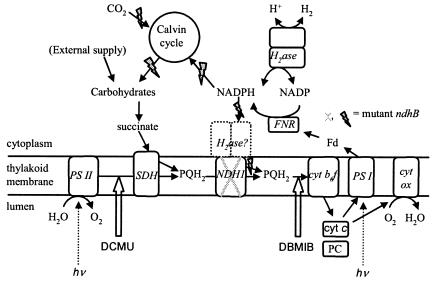
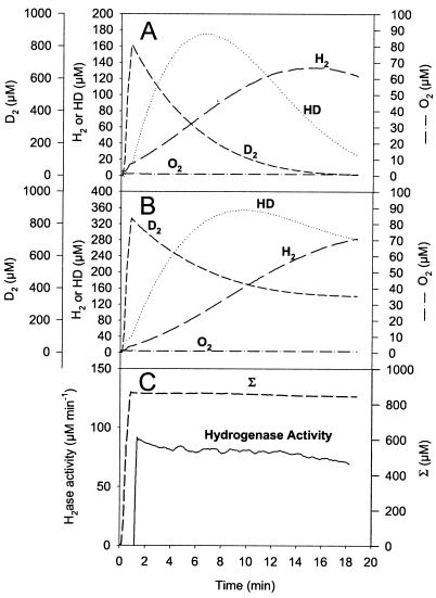
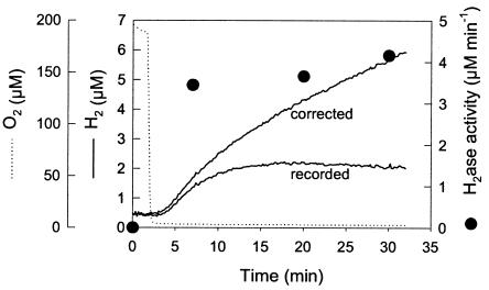
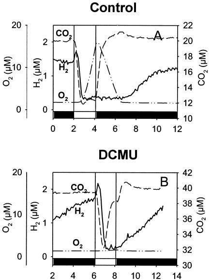
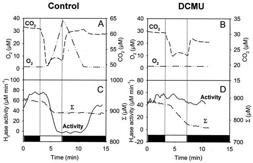
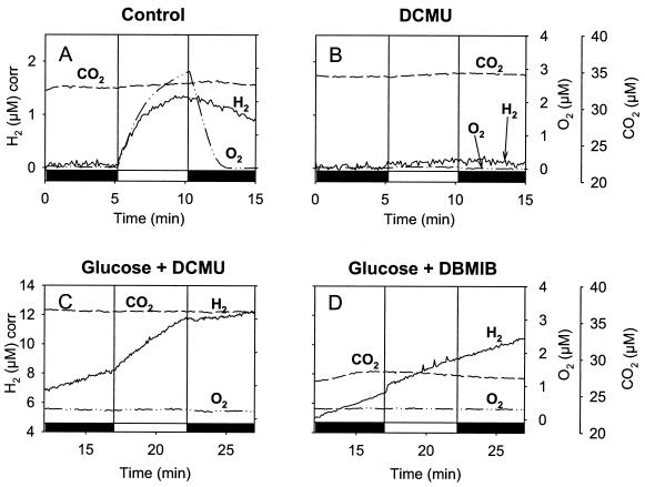
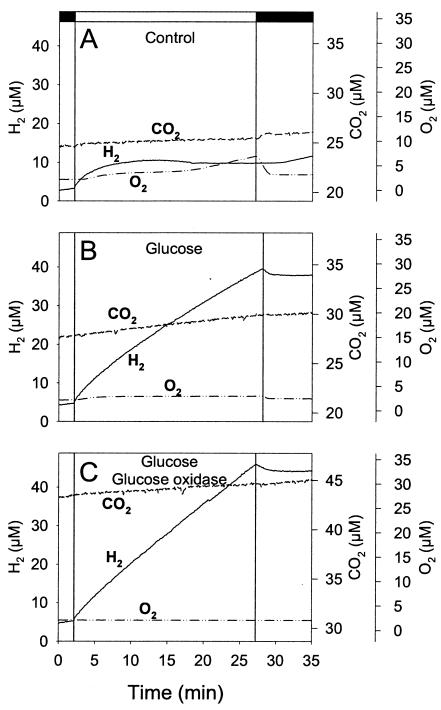
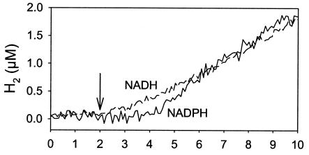
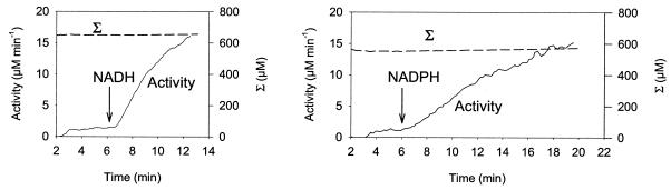
The interaction between hydrogen metabolism, respiration, and photosynthesis was studied in vivo in whole cells of Synechocystis sp. strain PCC 6803 by continuously monitoring the changes in gas concentrations (H2, CO2, and O2) with an online mass spectrometer. The in vivo activity of the bidirectional [NiFe]hydrogenase [H2:NAD(P) oxidoreductase], encoded by the hoxEFUYH genes, was also measured independently by the proton-deuterium (H-D) exchange reaction in the presence of D2. This technique allowed us to demonstrate that the hydrogenase was insensitive to light, was reversibly inactivated by O2, and could be quickly reactivated by NADH or NADPH (+H2). H2 was evolved by cells incubated anaerobically in the dark, after an adaptation period. This dark H2 evolution was enhanced by exogenously added glucose and resulted from the oxidation of NAD(P)H produced by fermentation reactions. Upon illumination, a short (less than 30-s) burst of H2 output was observed, followed by rapid H2 uptake and a concomitant decrease in CO2 concentration in the cyanobacterial cell suspension. Uptake of both H2 and CO2 was linked to photosynthetic electron transport in the thylakoids. In the ndhB mutant M55, which is defective in the type I NADPH-dehydrogenase complex (NDH-1) and produces only low amounts of O2 in the light, H2 uptake was negligible during dark-to-light transitions, allowing several minutes of continuous H2 production. A sustained rate of photoevolution of H2 corresponding to 6 micro mol of H2 mg of chlorophyll(-1) h(-1) or 2 ml of H2 liter(-1) h(-1) was observed over a longer time period in the presence of glucose and was slightly enhanced by the addition of the O2 scavenger glucose oxidase. By the use of the inhibitors DCMU [3-(3,4-dichlorophenyl)-1,1-dimethylurea] and DBMIB (2,5-dibromo-3-methyl-6-isopropyl-p-benzoquinone), it was shown that two pathways of electron supply for H2 production operate in M55, namely photolysis of water at the level of photosystem II and carbohydrate-mediated reduction of the plastoquinone pool.


Figures

FIG. 1.
FIG. 1.
Proposed electron transfer pathways in the light involved in H2 production and uptake in Synechocystis sp. strain PCC 6803. Abbreviations: H2ase, hydrogenase; PS I, photosystem I; PS II, photosystem II; cyt, cytochrome; cyt b6f, cytochrome b6f complex; cyt ox, cytochrome aa3 oxidase; SDH, succinate dehydrogenase; NDH-1, type I NAD(P)H-dehydrogenase; PQ, plastoquinone; PC, plastocyanin; FNR, ferredoxin-NADP+ reductase; Fd, ferredoxin. Electron transfers are represented by solid arrows; DCMU and DBMIB inhibition sites are indicated by thick white arrows. Enzyme names are in italics. Grey flash arrows indicate pathways which are impaired in the ndhB mutant M55.
FIG. 2.
FIG. 2.
H2 and HD production in exchange with D2 uptake catalyzed by Desulfovibrio fructosovorans [NiFe]hydrogenase. (A) Real concentrations of the hydrogen species present in the vessel (D2 concentration reaches zero). (B) Gas concentration changes corrected for consumption by the apparatus (shown is the equivalence between D2 uptake and H2 plus HD production). (C) Hydrogenase activity, calcu-lated as formula image , is expressed in nanomoles per milliliter per minute (or micromolar per minute). The sum (Σ) of D2 plus H2 plus HD concentrations is calculated as described in Materials and Methods. Σ remains constant since only the exchange reaction between hydrogen isotopes and protons of the medium is taking place here. Vexch and hydrogenase activity are then confounded.
FIG. 3.
FIG. 3.
Anaerobic H2 production in darkness. A cell suspension of Synechocystis sp. strain PCC 6803 (10 μg of Chl ml−1) in 35 mM HEPES buffer, pH 7.2, was placed in the measuring chamber of the online mass spectrometer, and catalase (500 U), glucose (5 mM), and glucose oxidase (30 U) were added to make the medium totally anaerobic. The concentrations of H2 (straight line, “recorded”) and O2 (dotted line) were recorded every 10 s at mass peaks 2 and 32, respectively. The curve labeled “corrected” shows H2 evolution in the medium corrected for the consumption by the apparatus and thus directly reflects H2 production by the cells. For the determination of hydrogenase activity by the H-D exchange reaction (•), the cells were preincubated in darkness under the same conditions as those described above for the indicated periods of time and then sparged with D2 and hydrogenase activity, based on the rates of H2 and HD formation, was calculated as described in Materials and Methods and in the legend to Fig. 2C.
FIG. 4.
FIG. 4.
Mass spectrometric measurements of H2, O2, and CO2 exchange during dark-light-dark transitions in WT Synechocystis cells adapted to dark anaerobic conditions. Shown are effects of the PS II inhibitor DCMU. (A) Transitory H2 output and H2 uptake at the onset of light. Cells of Synechocystis sp. strain PCC 6803 (10 μg of Chl ml−1) were preincubated anaerobically in darkness in the measuring chamber of the mass spectrometer, and H2 production was allowed to proceed in the dark. The cells were then illuminated briefly (2 min), and the concomitant changes in concentrations of dissolved gases H2, O2, and CO2 were measured every 4 s. (B) Same conditions as for panel A but in the presence of 75 μM DCMU. Dark periods are represented on the x axis by black bars, and the light period is represented by a white bar.
FIG. 5.
FIG. 5.
Mass spectrometric measurements of O2 and CO2 exchange and H-D exchange during dark-light-dark transitions in WT Synechocystis cells adapted to dark anaerobic conditions in the presence of D2. Shown are effects of the PS II inhibitor DCMU. (A and B) Time course of O2 and CO2 concentration changes. (C and D) Time course of hydrogenase activity measured by the H-D exchange reaction and of the sum (Σ) of D2 plus H2 plus HD concentrations, corrected for the apparatus consumption, determined in the presence of 400 μM D2 without further addition (A and C) or in the presence of 75 μM DCMU (B and D). Dark periods are represented on the x axis by black bars, and the light period is represented by a white bar.
FIG. 6.
FIG. 6.
H2 and O2 concentration changes in darkness and during a 5-min illumination period in the Synechocystis ndhB mutant M55. A culture of M55 (optical density at 730 nm = 1.5) was adapted to dark anaerobic conditions (A), in the presence of DCMU (75 μM) (B), in the presence of glucose (10 mM) and DCMU (75 μM) (C), or in the presence of glucose and DBMIB (20 μM) (D) in a closed reaction vessel connected to a mass spectrometer. The concentrations of H2, O2, and CO2 in the medium were continuously recorded. Dark periods are represented on the x axis by black bars, and the light period is represented by a white bar.
FIG. 7.
FIG. 7.
Mass spectrometric measurements of H2, O2, and CO2 exchange in the Synechocystis ndhB mutant M55 in darkness and during a 25-min illumination period. Cells were adapted for 1 h under anaerobiosis in the dark in a closed vessel connected to a mass spectrometer, and the concentrations of H2, O2, and CO2 in the medium were continuously monitored. (A) Control, (B) with 10 mM glucose, and (C) with 10 mM glucose plus glucose oxidase plus catalase. Conditions were the same as for Fig. 6 except for the duration of the illumination period. Dark periods are represented on the x axis by black bars, and the light period is represented by a white bar.
FIG. 8.
FIG. 8.
H2 evolution catalyzed by a Synechocystis cell extract with NADH or NADPH as the electron donor. The extract (200 μg of Chl) was gassed with argon, the chamber was closed, and O2 (monitored at mass peak 32) was removed by the addition of catalase, glucose, and glucose oxidase. NADH (1 mM) (dashed line) or NADPH (1 mM) (solid line) was added at 2 min (arrow).
FIG. 9.
FIG. 9.
Activation by NADH (left) or NADPH (right) of the hydrogenase activity in cell extracts of WT Synechocystis sp. strain PCC 6803 at pH 6.0. NADH or NADPH (0.2 mM) was added after the extract (73 μg of Chl) saturated with D2 was made anaerobic as described in the legend to Fig. 3. Two minutes after the O2 concentration had reached zero, the concentration changes in D2, HD, H2, and O2 were measured at mass peaks 4, 3, 2, and 32, respectively. Hydrogenase activity and the corrected total concentration of hydrogen species (Σ) were calculated as described in Materials and Methods and depicted in Fig. 2.

References

    1. Abdel-Basset, R., and K. P. Bader. 1998. Physiological analyses of the hydrogen gas exchange in cyanobacteria. J. Photochem. Photobiol. B Biol. 43:146-151.
    1. Appel, J., S. Phunpruch, K. Steinmuller, and R. Schulz. 2000. The bidirectional hydrogenase of Synechocystis sp PCC 6803 works as an electron valve during photosynthesis. Arch. Microbiol. 173:333-338. - PubMed
    1. Appel, J., and R. Schulz. 1998. Hydrogen metabolism in organisms with oxygenic photosynthesis: hydrogenases as important regulatory devices for a proper redox poising? J. Photochem. Photobiol. B Biol. 47:1-11.
    1. Appel, J., and R. Schulz. 1996. Sequence analysis of an operon of a NAD(P)-reducing nickel hydrogenase from the cyanobacterium Synechocystis sp PCC 6803 gives additional evidence for direct coupling of the enzyme to NAD(P)H-dehydrogenase (complex I). Biochim. Biophys. Acta 1298:141-147. - PubMed
    1. Boison, G., H. Bothe, A. Hansel, and P. Lindblad. 1999. Evidence against a common use of the diaphorase subunits by the bidirectional hydrogenase and by the respiratory complex I in cyanobacteria. FEMS Microbiol. Lett. 174:159-165.

Publication types

LinkOut - more resources