. 2004 Mar;186(6):1879-89.
doi: 10.1128/JB.186.6.1879-1889.2004.

DNA interaction and phosphotransfer of the C4-dicarboxylate-responsive DcuS-DcuR two-component regulatory system from Escherichia coli

Affiliations

DNA interaction and phosphotransfer of the C4-dicarboxylate-responsive DcuS-DcuR two-component regulatory system from Escherichia coli

Aly E Abo-Amer et al. J Bacteriol. 2004 Mar.

Abstract

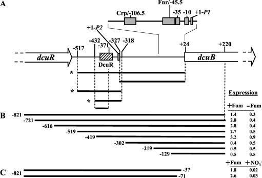
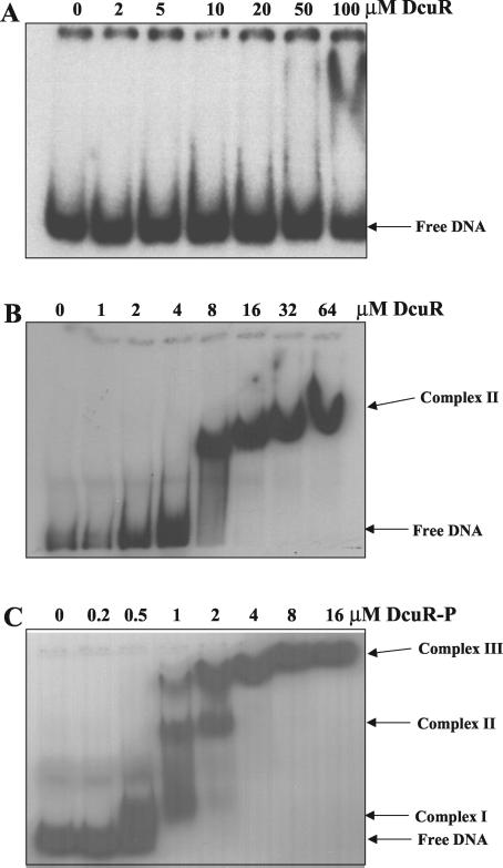
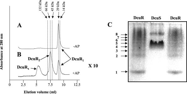
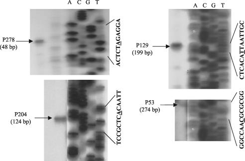
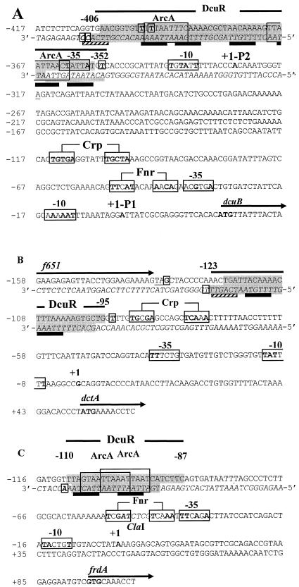
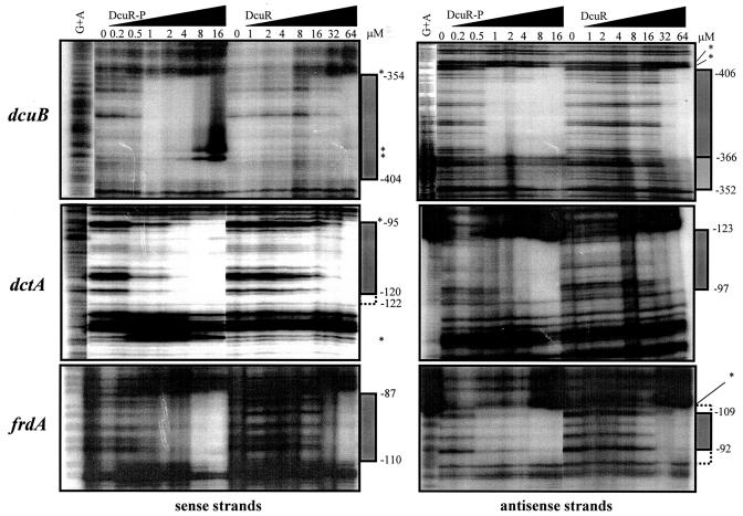
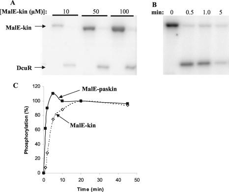
The DcuS-DcuR system of Escherichia coli is a two-component sensor-regulator that controls gene expression in response to external C(4)-dicarboxylates and citrate. The DcuS protein is particularly interesting since it contains two PAS domains, namely a periplasmic C(4)-dicarboxylate-sensing PAS domain (PASp) and a cytosolic PAS domain (PASc) of uncertain function. For a study of the role of the PASc domain, three different fragments of DcuS were overproduced and examined: they were PASc-kinase, PASc, and kinase. The two kinase-domain-containing fragments were autophosphorylated by [gamma-(32)P]ATP. The rate was not affected by fumarate or succinate, supporting the role of the PASp domain in C(4)-dicarboxylate sensing. Both of the phosphorylated DcuS constructs were able to rapidly pass their phosphoryl groups to DcuR, and after phosphorylation, DcuR dephosphorylated rapidly. No prosthetic group or significant quantity of metal was found associated with either of the PASc-containing proteins. The DNA-binding specificity of DcuR was studied by use of the pure protein. It was found to be converted from a monomer to a dimer upon acetylphosphate treatment, and native polyacrylamide gel electrophoresis suggested that it can oligomerize. DcuR specifically bound to the promoters of the three known DcuSR-regulated genes (dctA, dcuB, and frdA), with apparent K(D)s of 6 to 32 micro M for untreated DcuR and < or =1 to 2 microM for the acetylphosphate-treated form. The binding sites were located by DNase I footprinting, allowing a putative DcuR-binding motif [tandemly repeated (T/A)(A/T)(T/C)(A/T)AA sequences] to be identified. The DcuR-binding sites of the dcuB, dctA, and frdA genes were located 27, 94, and 86 bp, respectively, upstream of the corresponding +1 sites, and a new promoter was identified for dcuB that responds to DcuR.


Figures

FIG. 1.
FIG. 1.
DcuR-dependent regulation of the dcuB operator-promoter region. (A) The dcuB operator-promoter region is shown together with a summary of gel retardation experiments performed with DcuR and the indicated DNA fragments. Asterisks indicate those fragments that were specifically retarded by DcuR. The coordinates for each fragment are given with respect to the previously defined +1 site (11). The promoter and predicted Fnr and Crp sites are indicated by gray boxes. The experimentally determined location of the DcuR-binding site is indicated by the hatched box. (B and C) The indicated segments of the dcuB operator-promoter region were used to construct multicopy dcuB-lacZ transcriptional fusions (Table 1; also see Materials and Methods for details). Expression (in micromoles of o-nitrophenyl-β-d-galactopyranoside per minute per gram of protein) was tested anaerobically in M9 medium containing glycerol, Casamino Acids, and TMAO, with (+) or without (−) fumarate (for panel C, the fumarate-free medium included nitrate).
FIG. 2.
FIG. 2.
Gel retardation of the operator-promoter region of dcuB by DcuR. The results of gel retardation analysis of DcuR (A and B) and acetylphosphate-treated DcuR (C) with the −327 to +24 (A) and −517 to −318 (B and C) fragments of the dcuB promoter-operator region are shown. Similar gel retardation studies with −517 to +24, −432 to −318, and −432 to −371 dcuB operator-promoter fragments were performed (data not shown), and the results are summarized in Fig. 1A. The mobility positions of the free DNA and various DcuR-DNA complexes are indicated. For DcuR-P, the concentrations given are for the protein at the beginning of the phosphorylation reaction. The final concentrations of DcuR-P are likely to be lower due to precipitation induced by the acetylphosphate treatment.
FIG. 3.
FIG. 3.
Assembly status of DcuR. Elution profiles of acetylphosphate-treated (B) and untreated (A) DcuR during analytical gel permeation chromatography through a BioSelect-125 column (Bio-Rad). The eluant was phosphorylation buffer (see Materials and Methods), and the flow rate was 0.5 ml/min. The arrows at the top indicate the elution volumes of the native molecular mass markers (range, 14,000 to 5,000,000 Da; Sigma) that were used to standardize the column. Monomeric, dimeric, and high-molecular-weight (DcuRx) forms of DcuR are indicated. Note that acetylphosphate treatment resulted in the precipitation of ∼90% of the DcuR, so the scale of the corresponding elution profile has been increased 10-fold for panel B with respect to panel A (×10). (C) Native PAGE (10%) of DcuR (and DcuS). The gel was stained with Coomassie blue. Arrows indicate different oligomeric forms of DcuR (1 to 8). The high-mobility form (1) is presumed to be a monomer.
FIG. 4.
FIG. 4.
Determination of the distal transcriptional start site (P2dcuB) of dcuB by reverse-transcriptase-mediated primer extension. The reverse-transcriptase-mediated primer extension products were generated by using primers (P53, P129, P204, and P278) specific for the dcuB transcript (see Materials and Methods for details). Sequencing ladders (lanes A, C, G, and T) were generated by using the M13 universal primer and M13mp18 as the template. The relevant M13 nucleotide sequences (the underlined residues are those whose corresponding reaction products matched the size of the reverse transcription product) and the sizes of the primer extension products are shown.
FIG. 5.
FIG. 5.
Nucleotide sequences of the dcuB (A), dctA (B), and frdA (C) promoter-operator regions. The DcuR-protected regions are highlighted in dark gray (sequences for both strands are presented, with the noncoding strand in italics), with a weakly protected region underscored with a gray bar and adjacent areas that are potentially protected but were not subject to DNase I cleavage highlighted in light gray. Hypersensitive sites are shown in bold and enclosed in small boxes. Tandemly repeated (T/A)(A/T)(T/C)(A/T)AA sequences possibly corresponding to a DcuR-binding motif are indicated with black bars or, for weak matches, striped bars. Transcription and translation start sites are in bold, and promoters and potential binding sites for Crp and Fnr are in large boxes with residues matching the Crp and Fnr consensus sequences shown in bold. The f651 stop codon is underlined. Sequences within the DcuR-binding regions closely resembling the ArcA-binding consensus [(A/T)TAATTAAC(A/T)] are boxed. Numbering corresponds to distances from the transcriptional start site. A relevant restriction site is indicated in italics.
FIG. 6.
FIG. 6.
DNase I footprint analysis of the dcuB, dctA, and frdA promoter-operator regions with phosphorylated and unphosphorylated DcuR. The corresponding DNA fragments were PCR amplified by using a labeled forward or reverse primer to enable radiolabeling of the sense and antisense strands, respectively. The concentrations of DcuR subunit employed are shown, as are the lanes containing the G+A ladder. The regions of protection are indicated by bars on the right side of the autoradiographs (the lightly shaded bar indicates a region that is partially protected), and hypersensitive bands are highlighted with asterisks. Broken bars indicate regions that may be protected but were not cleaved by DNase I. The corresponding coordinates are distances (in base pairs) from the proximal transcription start site (see Fig. 5). For an explanation of DcuR-P concentrations, see the legend for Fig. 2.
FIG. 7.
FIG. 7.
Autophosphorylation of the DcuS-kinase domain and phosphotransfer to DcuR. (A and B) Phosphorimager-generated autoradiographs of phosphorylated DcuS (MalE-Kin) and DcuR separated by SDS-PAGE (reactions performed at 25°C). DcuS was phosphorylated with [γ-32P]ATP prior to the addition of DcuR. (A) DcuR (5 μM) was added to phosphorylated DcuS at time zero, resulting in the complete dephosphorylation of DcuS within 1 min and concomitant phosphotransfer to DcuR. The level of DcuR phosphorylation increased with increasing concentrations of phosphorylated DcuS. (B) DcuR (5 μM) was added to 20 μM phosphorylated DcuS at time zero. After the initial rapid phosphotransfer from DcuS to DcuR, the phosphorylated form of DcuR dephosphorylated, resulting in the almost complete dephosphorylation of DcuR after a 5-min incubation. (C) Graph comparing the relative rates of autophosphorylation for MalE-Kin and MalE-PAScKin at 25°C. Reactions for the two DcuS proteins were performed in the same tube, and the degree of phosphorylation was determined (as a percentage of the level achieved at 20 min) after the separation of the two proteins by SDS-PAGE. Note that the two proteins incorporated closely similar amounts of 32P after 20 and 45 min of incubation.

References

    1. Aiba, H., S. Adkya, and B. de Crombrugghe. 1981. Evidence for a functional gal promoter in intact Escherichia coli cells. J. Biol. Chem. 256:11905-11910. - PubMed
    1. Bartolomé, B., Y. Jubete, E. Martinez, and F. Delacruz. 1991. Construction and properties of a family of pACYC184-derived cloning vectors compatible with pBR322 and its derivatives. Gene 102:75-78. - PubMed
    1. Bott, M., M. Meyer, and P. Dimroth. 1995. Regulation of anaerobic citrate metabolism in Klebsiella pneumoniae. Mol. Microbiol. 18:533-546. - PubMed
    1. Bourret, R. B., K. A. Borkovich, and M. I. Simon. 1991. Signal transduction pathways involving protein phosphorylation in prokaryotes. Annu. Rev. Biochem. 60:401-441. - PubMed
    1. Davies, S. J., P. Golby, D. Omrani, S. A Broad, V. L. Harrington, J. R. Guest, D. J. Kelly, and S. C. Andrews. 1999. Inactivation and regulation of the aerobic C4-dicarboxylate transport (dctA) gene of Escherichia coli. J. Bacteriol. 181:5624-5635. - PMC - PubMed

Publication types

MeSH terms