Review
. 2004 Mar 8;90(5):944-9.
doi: 10.1038/sj.bjc.6601627.

Targeting oestrogen to kill the cancer but not the patient

Affiliations
Review

Targeting oestrogen to kill the cancer but not the patient

J S Lewis et al. Br J Cancer. .

Abstract

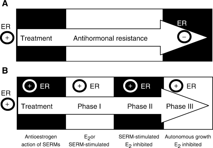
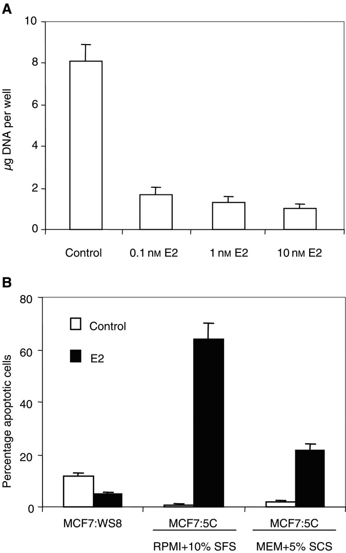
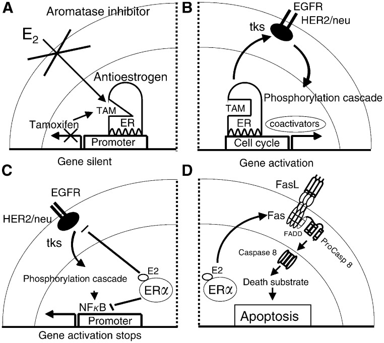
The link between sex steroids and the development and growth of breast cancer has proved to be an invaluable clue for advances in the prevention and treatment of breast cancer. The identification of the oestrogen receptor (ER) not only allowed advances in the molecular endocrinology of oestrogen action, but also provided a target for antioestrogenic therapeutic agents. However, the application of long-term or indefinite treatment regimens has consequences for the breast cancer. New forms of resistance, based upon enhanced cellular survival networks independent of ER and the suppression of apoptotic mechanisms, develop and then evolve. Remarkably, low concentrations of oestrogen collapse survival pathways and induce apoptosis in completely antihormonally refractory breast cancer. However, recurrent oestrogen-stimulated disease is again sensitive to antihormonal therapy. The novel reapplication of the ER as a therapeutic target for apoptosis is emerging as a new strategy for the long-term targeted maintenance treatment of breast cancer, and in formulating a targeted strategy for endocrine independent cancer.


Figures

Figure 1
Figure 1
The evolution of antihormonal resistance. (A) About 20 years ago, it was believed that oestrogen receptor-positive (ER+) tumours would usually be expected to respond to oestrogen withdrawal or a selective oestrogen receptor modulator (SERM) such as tamoxifen, but eventually resistance would occur because ER− cells would overgrow the tumour. (B) Emerging laboratory and clinical evidence suggests that SERM resistance evolves from acquired resistance (Phase I) to Phase II, where any SERM will maintain growth, whereas, unliganded ER does not provoke growth. However, oestrogen at physiological levels causes rapid apoptosis. In Phase III, tumours are completely resistant to all antihormonal therapies and grow spontaneously. Nevertheless, physiological concentrations of oestrogen causes rapid apoptosis.
Figure 2
Figure 2
Oestradiol inhibits the growth of MCF-7:5C cells and induces apoptosis. MCF-7:5C cells were cloned from wild-type MCF-7:WS8 cells following long-term growth (∼1 year) in oestrogen-free RPMI medium containing 10% (v v−1) dextran charcoal-stripped (DCC) foetal bovine serum (FBS), 2 mM glutamine, 100 U ml−1 penicillin-streptomycin, 6 ng ml−1 bovine insulin, and 1 × nonessential amino acids. (A) For DNA assays, MCF-7:5C cells were seeded into 12-well plates at a density of ∼20 000 cells per well in RPMI medium. The cells were left for 24 h to acclimatise, and then treated with 0.1, 1, or 10 nM oestradiol (E2) for a total of 6 days, with the control cells receiving <0.1% ethanol vehicle. Cells were re-fed on days 3 and 5. Total DNA (μg) per well was used to measure cell growth. The data represent the average of five separate experiments. (B) Apoptotic cells were identified/quantified by double staining with recombinant FITC-conjugated annexin V and propidium iodide (PI), using the Annexin V-FITC kit (Immunotech, Beckman Coulter). For experiments, MCF-7:WS8 and MCF-7:5C cells were seeded in 100 mm plates at a density of 1 × 10⁶ per plate in either oestrogen-free RPMI medium containing 10% DCC stripped fetal bovine serum (SFS) or MEM containing 5% DCC stripped calf serum (SCS). The cells were left for 24 h to acclimatise and then treated with either 1 nM E2 or less than 0.1% ethanol vehicle (control) for 72 h. Data shown represent three separate experiments. It should be noted that oestradiol treatment of MCF-7:WS8 cells in oestrogen-free MEM media containing 5% SCS did not have any significant effect on apoptosis (data not shown).
Figure 3
Figure 3
The development of tamoxifen (TAM)-resistant breast cancer and the changing role of oestradiol (E2) in the life and death of ER-positive cancer cells. (A) E2-stimulated growth is inhibited by the use of an aromatase inhibitor to block oestrogen synthesis or TAM to block the ER and prevent oestrogen-stimulated gene transcription. Optimal antioestrogenic effects occur in the absence of pre-existing cell surface signalling mechanisms. (B) Prolonged use of TAM promotes an increase in HER2/neu cell surface signalling that creates a survival pathway phosphorylating the TAM ER complex and coactivator proteins. The transcription complex becomes activated to enhance gene activation and TAM-stimulated growth. If this is Phase I resistance, then oestrogen will also promote growth; so an aromatase inhibitor is an appropriate second-line therapy. If it is Phase II resistance, E2 causes apoptosis. (C) In Phase II tamoxifen resistance, the E2 ER complex collapses the survival mechanisms by dramatically reducing the level of cell surface signalling by preventing HER2/neu mRNA transcription and the nuclear level of NFκB, a transcription factor. (D) In Phase II tamoxifen resistance, the E2 ER complex also enhances the synthesis of Fas receptor mRNA and protein which, in the presence of Fas ligand (FasL) activates caspase 8 and a cascade of events resulting in apoptosis. These figures are summaries of the mechanisms described in Osipo et al (2003) and Liu et al (2003).

References

    1. ATAC Trialists' Group (2002) Anastrozole alone or in combination with tamoxifen versus tamoxifen alone for adjuvant treatment of postmenopausal women with early breast cancer: first results of the ATAC randomized trial. Lancet 359: 2131–2139 - PubMed
    1. Bentrem D, Fox JE, Pearce ST, Liu H, Pappas S, Kupfer D, Zapf JW, Jordan VC (2003) Distinct moleular conformations of the estrogen receptor alpha complex exploited by environmental estrogens. Cancer Res 63: 7490–7496 - PubMed
    1. Cummings SR, Eckert S, Krueger KA, Grady D, Powles TJ, Cauley JA, Norton L, Nickelsen T, Bjarnason NH, Morrow M, Lippman ME, Black D, Glusman JE, Costa A, Jordan VC (1999) The effect of raloxifene on risk of breast cancer in postmenopausal women: results from the MORE randomized trial. Multiple Outcomes of Raloxifene Evaluation. JAMA 281: 2189–2197 - PubMed
    1. Cuzick J, Powles T, Veronesi U, Forbes J, Edwards R, Ashley S, Boyle P (2003) Overview of the main outcomes in breast-cancer prevention trials. Lancet 361: 296–300 - PubMed
    1. EBCTCG (1998) Tamoxifen for early breast cancer: an overview of the randomised trials. Lancet 351: 1451–1467 - PubMed

Publication types