. 2004 Mar 3:3:5.
doi: 10.1186/1475-925X-3-5.

Transverse propagation of action potentials between parallel chains of cardiac muscle and smooth muscle cells in PSpice simulations

Affiliations

Transverse propagation of action potentials between parallel chains of cardiac muscle and smooth muscle cells in PSpice simulations

Nicholas Sperelakis et al. Biomed Eng Online. .

Abstract

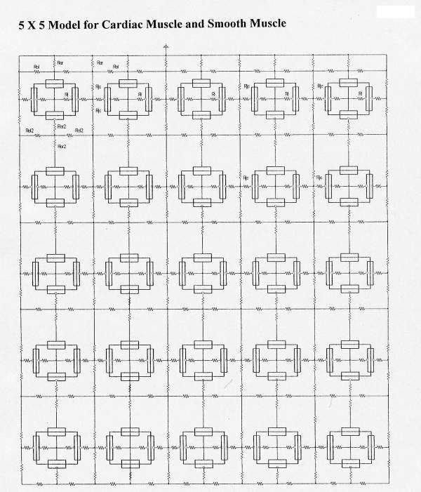
Background: We previously examined transverse propagation of action potentials between 2 and 3 parallel chain of cardiac muscle cells (CMC) simulated using the PSpice program. The present study was done to examine transverse propagation between 5 parallel chains in an expanded model of CMC and smooth muscle cells (SMC).

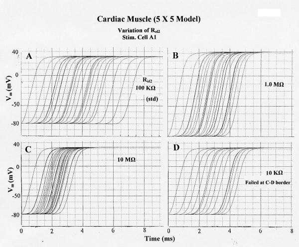
Methods: Excitation was transmitted from cell to cell along a strand of 5 cells not connected by low-resistance tunnels (gap-junction connexons). The entire surface membrane of each cell fired nearly simultaneously, and nearly all the propagation time was spent at the cell junctions, the junctional delay time being about 0.3-0.5 ms (CMC) or 0.8-1.6 ms (SMC). A negative cleft potential (Vjc) develops in the narrow junctional clefts, whose magnitude depends on the radial cleft resistance (Rjc), which depolarizes the postjunctional membrane (post-JM) to threshold. Propagation velocity (theta) increased with amplitude of Vjc. Therefore, one mechanism for the transfer of excitation from one cell to the next is by the electric field (EF) that is generated in the junctional cleft when the pre-JM fires. In the present study, 5 parallel stands of 5 cells each (5 x 5 model) were used.

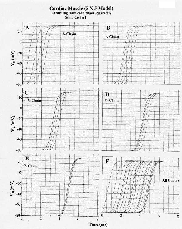
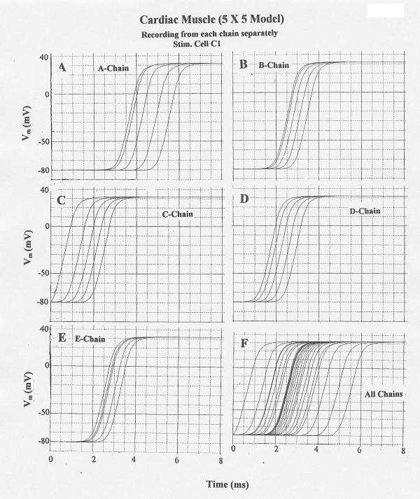
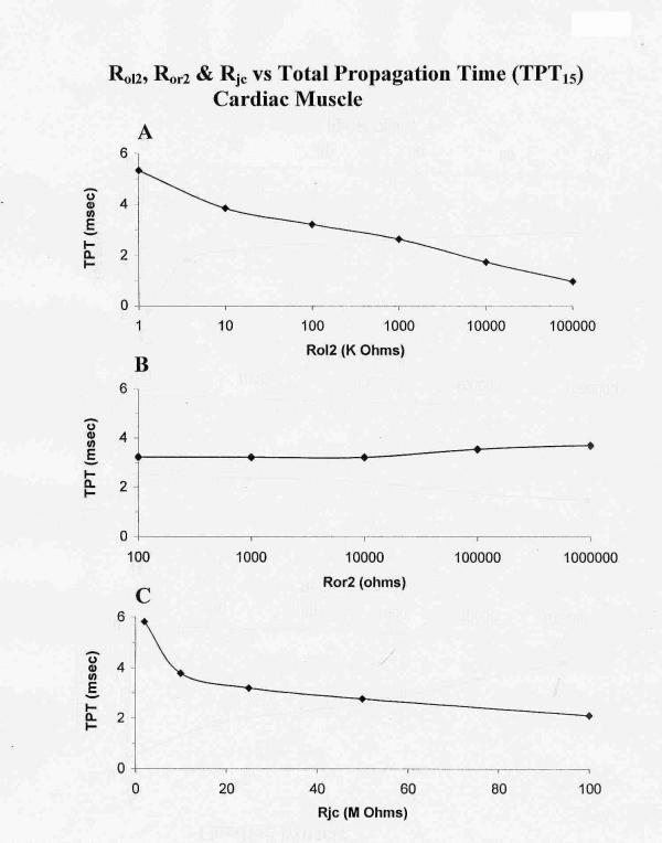
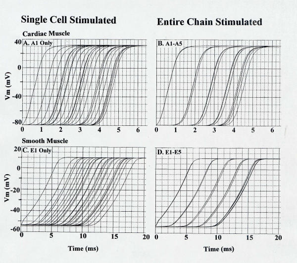
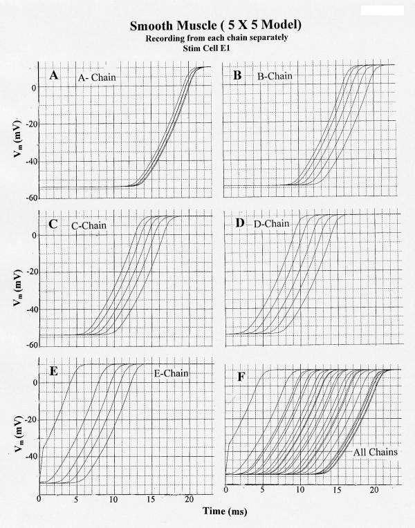
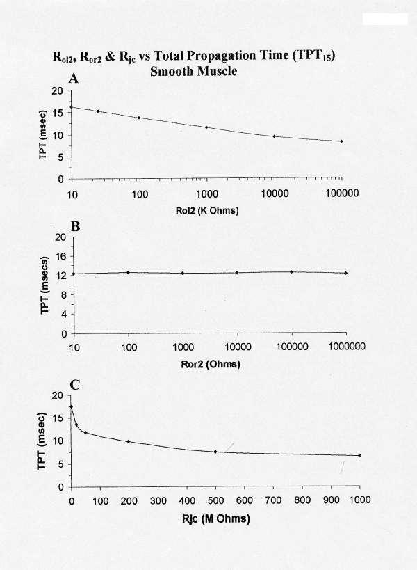
Results: With electrical stimulation of the first cell of the first strand (cell A1), propagation rapidly spread down that chain and then jumped to the second strand (B chain), followed by jumping to the third, fourth, and fifth strands (C, D, E chains). The rapidity by which the parallel chains became activated depended on the longitudinal resistance of the narrow extracellular cleft between the parallel strands (Rol2); the higher the Rol2 resistance, the faster the theta. The transverse resistance of the cleft (Ror2) had almost no effect. Increasing Rjc decreases the total propagation time (TPT) over the 25-cell network. When the first cell of the third strand (cell C1) was stimulated, propagation spread down the C chain and jumped to the other two strands (B and D) nearly simultaneously.

Conclusions: Transverse propagation of excitation occurred at multiple points along the chain as longitudinal propagation was occurring, causing the APs in the contiguous chains to become bunched up. Transverse propagation was more erratic and labile in SMC compared to CMC. Transverse transmission of excitation did not require low-resistance connections between the chains, but instead depended on the value of Rol2. The tighter the packing of the chains facilitated transverse propagation.


Figures

Figure 1
Figure 1
Diagrams of the arrangement of the myocardial cells and smooth muscle cells, basic units, and key resistances for the 5 × 5 model: 5 parallel chains (A – E) of 5 cells each (1 – 5). There were no low-resistance connections between cells within a chain or between chains. The surface membrane of each cell was represented by 2 basic units (one upwards and one inverted), and each junctional membrane by one unit each. The radial junctional cleft resistance (Rjc) is depicted, as is the longitudinal resistance of the interstitial space between the chains. Propagation of simulated action potentials was examined when only one cell of one chain was electrically stimulated (usually cell #1 of the A-chain (cell A1)).
Figure 2
Figure 2
Printout of a portion of the circuits used for PSpice simulation of 2-dimensional propagation of action potentials in smooth muscle in the 5 × 5 model. The electrical circuits consisted of many repeat units, so only the upper left portion of the 5 × 5 model is illustrated. Doing this allowed the circuit elements and labels to be larger, and therefore more easily resolved by the reader. Ror2 is the transverse resistance of the interstitial space between the chains. Ror is the radial resistance of the Ringer solution bathing the upper chain (A-chain) of the bundle, Rol is the corresponding longitudinal resistance. Ri is the longitudinal intracellular resistance. Rjc is depicted as two parallel pathways, one directed upwards and one downwards. Stimulus (0.5 ms; 0.5 nA) was usually applied at zero time to inside of cell #1 of the upper chain (cell A1), but for some experiments, stimulus was applied to the first cell of the other four chains.
Figure 3
Figure 3
Propagation of cardiac action potentials (APs) in the 5 × 5 model (5 parallel chains of 5 cells each). First cell of the A-chain (cell A1) was electrically stimulated by a rectangular current pulse (0.5 nA, 0.5 ms) applied internally. PSpice simulation. A: V recording from the A-chain only. B: V recording from the B-chain only. C: V recording from C-chain only. D: V-recording from the D-chain only. E: V-recording from the E-chain only. F: V recording simultaneously from all 5 chains. (The ordinate scale in this panel is slightly different from panels A – E because the computer must list more data numbers at the bottom of the plot, hence compressing the ordinate scale.) All parameters were the standard values (Rjc = 25 MΩ; Rol2 = 100 KΩ). Note that excitation spreads from the A-chain to the B-chain after a delay (from when cell A1 responded) of about 1.4 ms. Also note that once excitation entered the B-chain, it spread more quickly over the 5 cells in that chain. The same was true of the other chains, e.g. in the E-chain the 5 APs were bunched closely together.
Figure 4
Figure 4
Propagation of cardiac APs in the 5 × 5 model when cell C1 was stimulated. Rol2 = 1.0 MΩ. (When Rol2 was at the standard value of 100 KΩ, failure occurred at the border between chains D and E.) All other parameters were at standard values. A–E: V recording from only one chain at a time: A-chain (A), B-chain (B), C-chain (C), D-Chain (D), and E-chain (E). F: V recording simultaneously from all 5 chains. Transverse propagation occurred nearly simultaneously from the C-chain to the B and D chains, followed by excitation of the E and A chains.
Figure 5
Figure 5
Effect of varying the longitudinal resistance of the interstitial space (Rol2) between the 5 parallel chains on propagation of APs simulated by PSpice in the 5 × 5 model. All other parameters held at their standard values (e.g., Rjc = 25 MΩ). Stimulation applied to cell A1. A: Rol2 = 100 KΩ (standard). B: Rol2 = 1.0 MΩ. C: Rol2 = 10 MΩ. D: Rol2 = 10 KΩ. Failure occurred at the border between C-chain and D-Chain. Propagation over the 25-cell network was fastest in C (TPT = 2.6 ms) and slowest in A (TPT = 6.5 ms). Propagation in the stimulated A-chain was unaffected by changing Rol2.
Figure 6
Figure 6
Graphic summary of the effects of varying Rol2 (A), Ror2 (B), and Rjc (C) on the total propagation time (TPT) over the first 15 cells (TPT15) of the 25-cell network (5 × 5 model) of cardiac muscle cells. As shown in A, TPT decreased as Rol2 (longitudinal resistance of the interstitial space between chains of the bundle) was increased (reflecting tighter packing of the chains). Hence, average velocity of propagation was increased. Elevation of Ror2 (transverse resistance of the interstitial space) had only a slight effect on TPT (B), indicating that transverse current flow was small and not important to transverse transmission. Elevation of Rjc (radial shunt resistance of the junctional cleft) decreased TPT. A high Rjc value reflects a narrow junctional cleft. TPT was measured over only 15 cells, because in a few cases, failure occurred between chains C and D or between D and E.
Figure 7
Figure 7
Plot of total propagation time (TPT) over the entire 25-cell network as a function of the capacitance of the junctional membrane (Cj). Data were collected at more Cj values (e.g., 0.75, 8.0, and 10 pF), but these data are not plotted because all cells did not fire. Lowering Cj greatly decreased TPT25 and therefore increased velocity.
Figure 8
Figure 8
Simultaneous stimulation of all five cells of the A-Chain in cardiac muscle (B) and E-Chain in smooth muscle (D). This was done to obtain a more accurate measurement of tranverse propagation velocity. These values were compared with stimulations of only one cell(A and C).
Figure 9
Figure 9
Propagation of smooth muscle action potentials (APs) in the 5 × 5 model (5 parallel chains of 5 cells each) when the first cell of the E chain (cell E1) is electrically stimulated (0.5 nA, 0.5 ms rectangular current pulse applied internally). PSpice simulation. A: V recording markers placed on the A-chain cell units only. B: V markers on the B-chain only. C: V recording from the C-chain only. D: V recording from D-chain. E: V recording from E-chain. F: V recording from all 5 chains simultaneously. All parameters were the standard values (Rjc = 10 MΩ; Rol2 = 100 KΩ). Note that excitation spreads from the E-chain to the D-chain after a delay (from first response of A-chain) of about 5.0 ms, and that excitation spreads from the D-chain to the C-chain after a delay (from first response of D-chain) of about 3.7 ms. Once excitation entered the D, C, B and A chains, it propagated very quickly over the 5 cells of each chain. The effect of bunching of the APs is greatest in the last chain (A-chain).
Figure 10
Figure 10
Graphic summary of the effects of varying Rol2 (A), Ror2 (B), Rjc (C) on TPT over the first 15 cells of the network (5 × 5 model) of smooth muscle cells. Increasing Rol2 (A) produced a decrease in TPT. Increasing Ror2 (B) had only slight effect on TPT15 (as in cardiac muscle). Increasing Rjc (C) decreased TPT15, much like in cardiac muscle.

Similar articles

Cited by

References

    1. Sperelakis N, Mann JE. Evaluation of electric field changes in the cleft between excitable cells. J Theor Biol. 1977;64:71–96. - PubMed
    1. Sperelakis N, McConnell K. An electric field mechanism for transmission of excitation from cell to cell in cardiac muscle and smooth muscles. In: Mohan RM, editor. Research Advances in Biomedical Engineering. Vol. 2. Global Research Network; 2001. pp. 39–66.
    1. Sperelakis N, McConnell K. Electric field interactions between closely-abutting excitable cells. IEEE-EMB. 2002;21:77–89. doi: 10.1109/51.993199. - DOI - PubMed
    1. Sperelakis N. Cell Physiology Source Book. 1. Academic Press Publishers; 1995. Cable Properties and propagation of action potentials, ch. 18; pp. 245–254.
    1. Cohen SA. Immunocytochemical localization of rH1 sodium channel in adult rat heart atria and ventricle. Presence in terminal intercalated disks. Circulation. 1996;94:3083–3086. - PubMed

MeSH terms

LinkOut - more resources