. 2004 Mar 3;24(9):2102-11.
doi: 10.1523/JNEUROSCI.3988-03.2004.

Dissociating self-generated from passively applied head motion: neural mechanisms in the vestibular nuclei

Affiliations

Dissociating self-generated from passively applied head motion: neural mechanisms in the vestibular nuclei

Jefferson E Roy et al. J Neurosci. .

Abstract

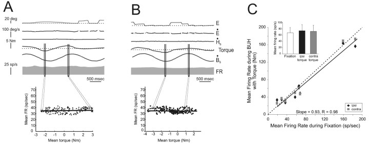
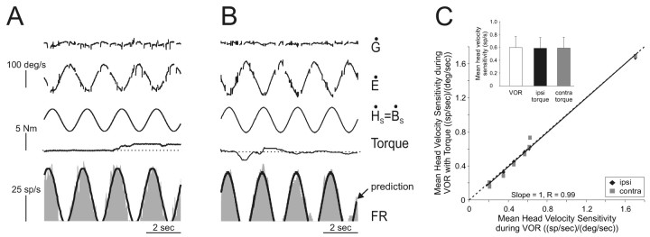
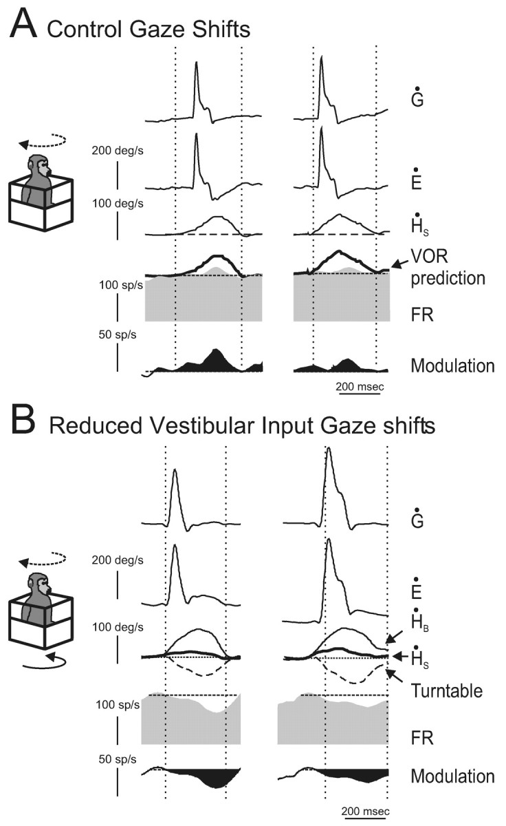
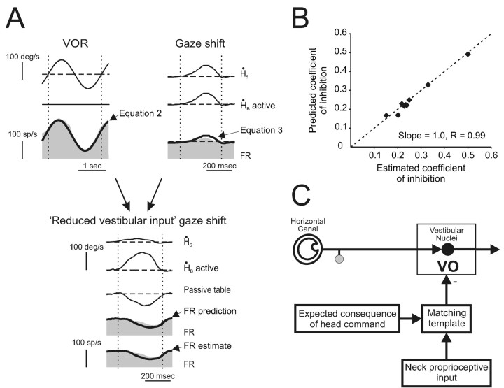
The ability to distinguish sensory inputs that are a consequence of our own actions from those that result from changes in the external world is essential for perceptual stability and accurate motor control. To accomplish this, it has been proposed that an internal prediction of the consequences of our actions is compared with the actual sensory input to cancel the resultant self-generated activation. Here, we provide evidence for this hypothesis at an early stage of processing in the vestibular system. Previous studies have shown that neurons in the vestibular nucleus, which receive direct inputs from vestibular afferent fibers, are responsive to passively applied head movements. However, these same neurons do not reliably encode head velocity resulting from self-generated movements of the head on the body. In this study, we examined the mechanism that underlies the selective elimination of vestibular sensitivity to active head-on-body rotations. Individual neurons were recorded in monkeys making active head movements. The correspondence between intended and actual head movement was experimentally controlled. We found that a cancellation signal was gated into the vestibular nuclei only in conditions in which the activation of neck proprioceptors matched that expected on the basis of the neck motor command. This finding suggests that vestibular signals that arise from self-generated head movements are inhibited by a mechanism that compares the internal prediction of the sensory consequences by the brain to the actual resultant sensory feedback. Because self-generated vestibular inputs are selectively cancelled early in processing, we propose that this gating is important for the computation of spatial orientation and control of posture by higher-order structures.


Figures

Figure 1.
Figure 1.
Activity of an example VO neuron (unit b84_3) during the head-restrained condition. A, B, Passive whole-body rotation was used to characterize the response of the neuron to head movements during VOR in the dark (A) and head movements while the monkey canceled its VOR by fixating a target that moved with the turntable (B). A model based on head-restrained head movement sensitivities during VOR in the dark (pWBR prediction, thick trace) is superimposed on the firing rate traces. C, The neuron was unresponsive to changes in eye position during ocular fixation (vertical arrows). Inset, Regression of mean eye position and mean firing rate. Traces directed upward are in the ipsilateral direction. E, Eye position; H, head position; Ė, eye-in-head velocity; Ḣ, head velocity; Ġ, gaze velocity (=Ė + ).
Figure 2.
Figure 2.
Four possible mechanisms that could be responsible for the observed attenuation of VO neurons during self-generated head-on-body motion. See Results for details.
Figure 3.
Figure 3.
Activity of example VO neuron cr37_3 during head-restrained saccades. A, B, The example neuron was unresponsive during ipsilaterally (left plots) and contralaterally (right plots) directed saccades when the monkey produced little torque (A, <0.3 N-m). Similarly, the response of the neuron was not changed when the monkey generated relatively higher levels of torque (B, >1 N-m). Traces are aligned on the start of the saccades. C, Saccades from B with the traces now aligned on torque onset. Individual action potentials, represented by tick marks, are shown below the eye traces (UA). The mean firing rate ± SD is plotted on the firing rate (bottom trace).
Figure 4.
Figure 4.
Summary of VO neuron responses during large saccades. A, The mean firing rate of individual VO neurons was comparable during ocular fixation and large ipsilaterally (Ipsi, solid diamond) and contralaterally (Contra, gray squares) directed saccades that were verified to be accompanied by neck torque. Top right inset, Sample mean firing rates were not significantly different during ocular fixation (open column) and during large ipsilaterally (filled column) and contralaterally (gray column) directed saccades. Bottom left inset, Torque generated when monkey's head was unexpectedly held stationary just before the onset of a gaze shift. B, A similar result was found for neurons during large saccades in which torque was not measured. Inset, Sample mean firing rates were not significantly different during fixation (open column) and during large ipsilaterally (filled column) and contralaterally (gray column) directed saccades. Dashed lines indicate unity (slope = 1).
Figure 5.
Figure 5.
Activity of two example VO neurons during passive body-under-head rotation. A, B, Example neurons cr37_3 (A) and cr42_2 (B) were typical in that they were not modulated when the monkey's body was passively rotated beneath its earth-stationary head even when the monkey generated neck torque. C, The mean firing rate of each neuron was comparable during ocular fixation and passive neck rotation accompanied by the generation of neck torque in either the ipsilateral (Ipsi, solid diamonds) or contralateral (Contra, gray squares) direction. Inset, Average mean firing rates were not significantly different during ocular fixation (open column) and when ipsilaterally (filled column) or contralaterally (gray column) directed torque was generated. Dashed line indicates unity (slope = 1).
Figure 6.
Figure 6.
Activity of two example VO neurons during intervals of passive whole-body rotation in which monkeys generated neck torque. A, B, Example neurons cr37_3 (A) and cr42_2 (B) were typical in that their responses to head velocity were comparable regardless of whether the monkey generated neck torque during passive whole-body rotation. A model based on neuronal responses to head velocity during VOR during periods when no torque was produced continued to describe the neuronal modulations during intervals in which torque was produced (prediction, thick trace). C, The mean head velocity sensitivity of each neuron was comparable during the VOR when no torque was produced and when torque was generated. Inset, Average head velocity sensitivities were not significantly different during VOR without torque (open column) and when either ipsilaterally (Ipsi, filled column) or contralaterally (Contra, gray column) directed torque was generated during passive whole-body rotation. Dashed line indicates unity (slope = 1).
Figure 7.
Figure 7.
Activity of an example neuron during active head-on-body movements. A, Responses of neuron cr122_3 during coordinated eye and head gaze shifts (active head movement is dashed line arrow in schema). The mean discharge rate during ocular fixation is superimposed on the firing rate (dashed line, second row from bottom). In addition, a prediction of the response of the neuron based on it response to passive whole-body rotation is superimposed (VOR prediction, thick trace). The black filled trace (bottom row) represents the difference between the mean firing rate during ocular fixation and gaze shifts. Note that the scale has changed. B, Response of the same neuron during the reduced vestibular input gaze shift paradigm. In this paradigm, the head motion actively generated by the monkey (dashed line arrow in schema) was fed to the vestibular turntable controller so that the whole monkey was simultaneously rotated in the opposite direction (solid arrow in schema). The resultant head-in-space motion (S = B + turntable; thick trace) was significantly reduced from what it would had been during a control gaze shift, but the movement of the head relative to the body (ḢB) was not affected. The mean discharge of the neuron during ocular fixation is superimposed on the firing rate (dashed line, second row from bottom). The black filled trace (bottom row) represents the difference between the mean firing rate during ocular fixation and reduced vestibular input gaze shifts. Note that the scale has changed.
Figure 8.
Figure 8.
The reduction in modulation during the reduced vestibular input gaze shifts could be predicted on the basis of the difference between responses to ḢS during passive whole-body rotation (VOR) and control gaze shifts. A, This neuron was typical in that the inhibition that occurred during reduced vestibular input gaze shifts (FR estimate) was identical to that predicted (FR prediction) on the basis of the same neurons response in two different experiments: VOR (Equation 2) and control gaze shifts (Equation 3). B, For each neuron in the sample tested, the estimated coefficient of inhibition during reduced vestibular input gaze shifts was comparable with the inhibition predicted by neural responses during VOR and control gaze shifts. C, Schematic to explain the present findings. A cancellation signal is gated into the vestibular nuclei only in conditions in which the activation of neck proprioceptors matched that expected on the basis of the neck motor command.

References

    1. Akbarian S, Grüsser O-J, Guldin WO (1993) Corticofugal projections to the vestibular nuclei in squirrel monkeys: further evidence of multiple cortical vestibular fields. J Comp Neurol 332: 89-104. - PubMed
    1. Akbarian S, Grüsser O-J, Guldin WO (1994) Corticofugal connections between the cerebral cortex and brainstem vestibular nuclei in the macaque monkey. J Comp Neurol 339: 421-437. - PubMed
    1. Alstermark B, Pinter MJ, Sasaki S (1992a) Descending pathways mediating disynaptic excitation of dorsal neck motoneurons in the cat: facilitatory interactions. Neurosci Res 15: 32-41. - PubMed
    1. Alstermark B, Pinter MJ, Sasaki S (1992b) Descending pathways mediating disynaptic excitation of dorsal neck motoneurons in the cat: brain stem relay. Neurosci Res 15: 42-57. - PubMed
    1. Anastasopoulos D, Mergner T (1982) Canal-neck interaction in vestibular nuclear neurons of the cats. Exp Brain Res 46: 269-280. - PubMed

Publication types