. 2004 Mar 3;24(9):2182-90.
doi: 10.1523/JNEUROSCI.5275-03.2004.

Minocycline treatment reduces delayed oligodendrocyte death, attenuates axonal dieback, and improves functional outcome after spinal cord injury

Affiliations

Minocycline treatment reduces delayed oligodendrocyte death, attenuates axonal dieback, and improves functional outcome after spinal cord injury

David P Stirling et al. J Neurosci. .

Abstract

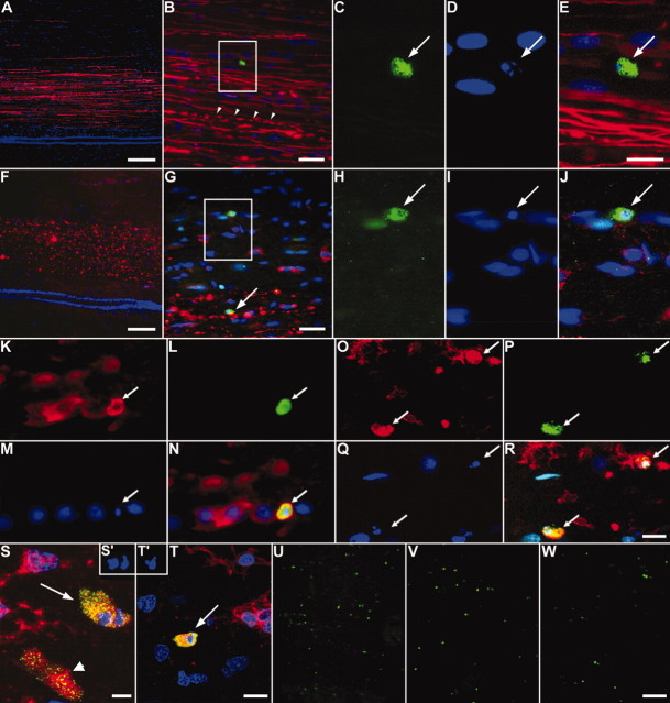
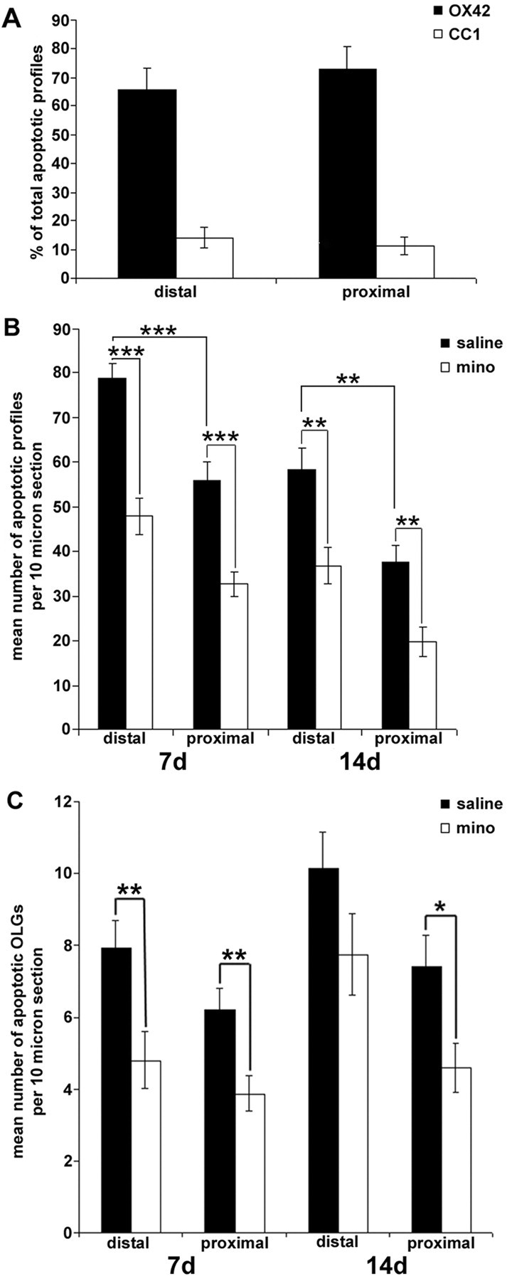
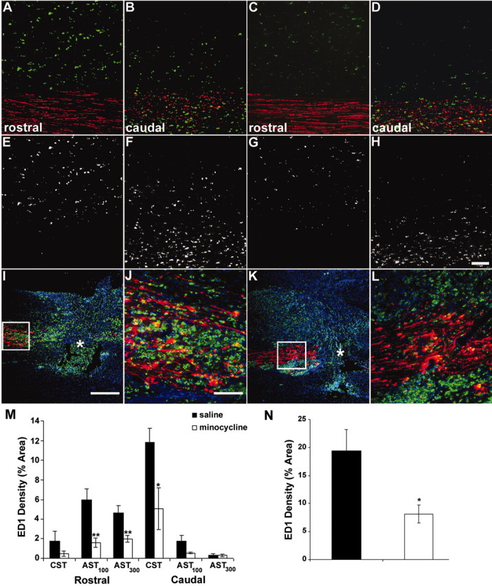
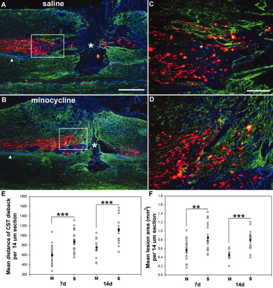
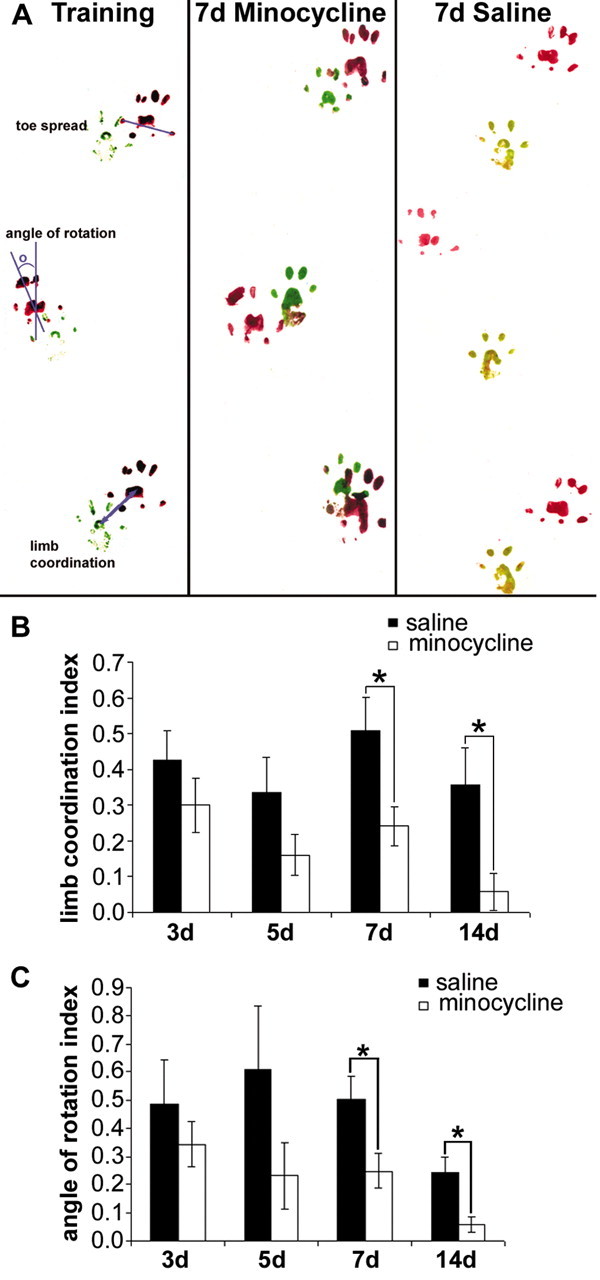
Minocycline has been demonstrated to be neuroprotective after spinal cord injury (SCI). However, the cellular consequences of minocycline treatment on the secondary injury response are poorly understood. We examined the ability of minocycline to reduce oligodendrocyte apoptosis, microglial/macrophage activation, corticospinal tract (CST) dieback, and lesion size and to improve functional outcome after SCI. Adult rats were subjected to a C7-C8 dorsal column transection, and the presence of apoptotic oligodendrocytes was assessed within the ascending sensory tract (AST) and descending CST in segments (3-7 mm) both proximal and distal to the injury site. Surprisingly, the numbers of dying oligodendrocytes in the proximal and distal segments were comparable, suggesting more than the lack of axon-cell body contiguity played a role in their demise. Minocycline or vehicle control was injected into the intraperitoneal cavity 30 min and 8 hr after SCI and thereafter twice daily for 2 d. We report a reduction of apoptotic oligodendrocytes and microglia within both proximal and distal segments of the AST after minocycline treatment, using immunostaining for active caspase-3 and Hoechst 33258 staining in combination with cell-specific markers. Activated microglial/macrophage density was reduced remote to the lesion as well as at the lesion site. Both CST dieback and lesion size were diminished after minocycline treatment. Footprint analysis revealed improved functional outcome after minocycline treatment. Thus, minocycline ameliorates multiple secondary events after SCI, rendering this clinically used drug an attractive candidate for SCI treatment trials.


Figures

Figure 1.
Figure 1.
Cell death of oligodendrocytes and microglia/macrophages within both proximal and distal segments of the CST and AST after SCI. The dorsal funiculus 5 mm rostral (A-E) or caudal (F-J) to the injury site, 14 d after the lesion, is shown. The BDA-labeled CST (red) is easily distinguished from the adjacent ascending fibers, allowing for analysis of apoptotic profiles within the CST or AST. B, Most CST fibers are intact, but some degenerate (arrowheads). C-E, Boxed area in B, an active caspase-3-positive profile (C, arrow) with a condensed nucleus (D, arrow). E, Merged image of C, D, and BDA (red). Caudal to the injury site, the CST completely degenerates (F). Apoptotic profiles are found within both the degenerating CST (arrow) and within the proximal segment of the ascending fibers (boxed area in G). H-J, Boxed area in G showing an active caspase-3-positive profile (H) with a condensed nucleus (I, arrow). J, Merged image of H, I, and BDA (red). K-N, Colocalization of CC1 (K), active caspase-3 (L), and a condensed nucleus (M) revealing an apoptotic oligodendrocyte within the proximal AST 7 d after injury (N, merged image of K-M). O-R, OX42 immunoreactivity (O) revealing that apoptotic microglia/macrophages were also present within the proximal AST (P, active caspase-3; Q, Hoechst 33528; R, merged image of O-Q). S, T, Representative merged confocal images of CC1-positive (S, arrow) and OX42-positive (T, arrow), active caspase-3-positive profiles with (S′, T′) or without a condensed nucleus (S, arrowhead). U-W, Numerous active caspase-3-positive profiles (green) are present within the proximal (U) and distal (V) AST at 7 d after the lesion. Minocycline treatment greatly reduces the number of active caspase-3-positive profiles within both the proximal and distal AST (W, distal AST). Scale bars: A, F, U-W, 100 μm; B, G, 25 μm; C-E, H-T, 10 μm.
Figure 2.
Figure 2.
Minocycline treatment inhibited dorsal column transection-induced glial cell death within the distal and proximal AST. A, Percentage of apoptotic microglia/macrophages (OX42-positive) or oligodendrocytes (CC1-positive) within the proximal and distal AST 7 d after the lesion (n = 6 per group). Microglia/macrophages are the main cell type to undergo apoptosis in this model. B, Quantification of the mean number of apoptotic profiles within the distal and proximal segments of the AST. Significantly fewer apoptotic profiles are located within the proximal versus distal segments of the AST. Minocycline (mino) treatment significantly reduced the mean number of apoptotic profiles per 10 μm section ± SEM within distal and proximal segments of the AST at both 7 and 14 d after injury compared with saline-treated controls. (*p < 0.05; **p < 0.01; ***p < 0.001; 7 d, n = 6 per group; 14 d, n = 4 or 5 per group). C, Apoptotic oligodendrocytes (CC1-positive), active caspase-3-positive profiles with a condensed nucleus, were significantly decreased in the minocycline-treated group compared with saline controls within the proximal AST at both 7 and 14 d after injury. However, only the proximal segment of the AST from minocycline-treated animals contained significantly fewer apoptotic oligodendrocytes at 14 d after injury. There was no difference between the mean number of apoptotic oligodendrocytes within the proximal versus distal segments of the AST at both 7 and 14 d after injury (data represent mean ± SEM; *p < 0.05; 7 d, n = 5 per group; 14 d, n = 4 or 5 per group).
Figure 3.
Figure 3.
Minocycline treatment reduced ED1-positive (microglial/macrophage) density 7 d after injury. A-H, ED1-positive profiles (green) 3 mm rostral (A, C, E, G) and caudal (B, D, F, H) to injury. Minocycline significantly reduced ED1 density within both rostral AST (100 and 300 μm dorsal to CST; C, G, M) and within the caudal CST (D, H, M) compared with saline-treated animals (A, B, E, F, M). Triple immunofluorescence images of the lesion site from saline-treated (I, J) and minocycline-treated (K, L) animals are shown. The proximal CST is red; ED1-positive microglia/macrophages are green; and Hoechst 33258 is blue. Note the reduced ED1-positive signal in K compared with I. J, L, Higher magnification of the boxed area in I and K. Less ED1-positive signal is evident within the proximal CST of minocycline-treated animals compared with saline-treated animals. N, Quantification of the density of the ED1-positive signal within the proximal CST. Data represent mean percentage ± SEM (n = 4 or 5 per group). *p < 0.05; **p < 0.01. Scale bars: A-H, J, L, 100 μm; I, K, 500 μm.
Figure 4.
Figure 4.
Minocycline treatment reduced CST dieback and lesion size both 7 and 14 d after the lesion. A, B, Images of the dorsal columns revealing the lesion site at 7 d after injury from saline-treated (A) versus minocycline-treated (B) rats. Note that the lesion area (delineated by GFAP staining, green) and CST dieback (red) is clearly reduced in minocycline- versus saline-treated animals. C, Boxed area in A; D, boxed area in B showing a higher magnification of the proximal BDA-traced CST axons (red). E, Quantification of CST dieback. Overall minocycline (M) treatment significantly reduced CST axonal dieback by ∼32 and ∼33% at 7 and 14 d, respectively. S, Saline. F, Quantification of lesion area. Minocycline treatment significantly reduced the lesion area by ∼34 and ∼43% at 7 and 14 d. Data represent mean ± SEM (n = 4-7 per group). **p < 0.01; **p < 0.001. Scale bars: A, B, 500 μm; C, D, 100 μm.
Figure 5.
Figure 5.
Minocycline treatment improved interlimb coordination and reduced hindlimb angle of rotation after SCI. A, Representative footprints collected from saline- and minocycline-treated animals. Minocycline treatment improved limb coordination (B) and reduced the hindlimb angle of rotation (C) at both 7 and 14 d after injury. Indexes were calculated as experimental value - baseline value/baseline value. Data represent mean ± SEM (7 d, n = 8 per group; 14 d, n = 4 per group). *p < 0.05.

References

    1. Abe Y, Yamamoto T, Sugiyama Y, Watanabe T, Saito N, Kayama H, Kumagai T (1999) Apoptotic cells associated with Wallerian degeneration after experimental spinal cord injury: a possible mechanism of oligodendroglial death. J Neurotrauma 16: 945-952. - PubMed
    1. Agrawal SK, Fehlings MG (1997) Role of NMDA and non-NMDA ionotropic glutamate receptors in traumatic spinal cord axonal injury. J Neurosci 17: 1055-1063. - PMC - PubMed
    1. Amin AR, Patel RN, Thakker GD, Lowenstein CJ, Attur MG, Abramson SB (1997) Post-transcriptional regulation of inducible nitric oxide synthase mRNA in murine macrophages by doxycycline and chemically modified tetracyclines. FEBS Lett 410: 259-264. - PubMed
    1. Balentine JD, Spector M (1977) Calcification of axons in experimental spinal cord trauma. Ann Neurol 2: 520-523. - PubMed
    1. Beattie MS, Harrington AW, Lee R, Kim JY, Boyce SL, Longo FM, Bresnahan JC, Hempstead BL, Yoon SO (2002) ProNGF induces p75-mediated death of oligodendrocytes following spinal cord injury. Neuron 36: 375-386. - PMC - PubMed

Publication types

MeSH terms