. 2004 Mar 3;24(9):2259-69.
doi: 10.1523/JNEUROSCI.5191-03.2004.

Rapid deletion of mossy cells does not result in a hyperexcitable dentate gyrus: implications for epileptogenesis

Affiliations

Rapid deletion of mossy cells does not result in a hyperexcitable dentate gyrus: implications for epileptogenesis

Anna d H Ratzliff et al. J Neurosci. .

Abstract

Loss of cells from the hilus of the dentate gyrus is a major histological hallmark of human temporal lobe epilepsy. Hilar mossy cells, in particular, are thought to show dramatic numerical reductions in pathological conditions, and one prominent theory of epileptogenesis is based on the assumption that mossy cell loss directly results in granule cell hyperexcitability. However, whether it is the disappearance of hilar mossy cells from the dentate gyrus circuitry after various insults or the subsequent synaptic-cellular alterations (e.g., reactive axonal sprouting) that lead to dentate hyperexcitability has not been rigorously tested, because of the lack of available techniques to rapidly remove specific classes of nonprincipal cells from neuronal networks. We developed a fast, cell-specific ablation technique that allowed the targeted lesioning of either mossy cells or GABAergic interneurons in horizontal as well as axial (longitudinal) slices of the hippocampus. The results demonstrate that mossy cell deletion consistently decreased the excitability of granule cells to perforant path stimulation both within and outside of the lamella where the mossy cell ablation took place. In contrast, ablation of interneurons caused the expected increase in excitability, and control aspirations of the hilar neuropil or of interneurons in the presence of GABA receptor blockers caused no alteration in granule cell excitability. These data do not support the hypothesis that loss of mossy cells from the dentate hilus after seizures or traumatic brain injury directly results in hyperexcitability.


Figures

Figure 1.
Figure 1.
Specific fluorescent labeling of mossy cells. A, Low-magnification image of the left hilus showing cells labeled with DiO (green) labeled by a contralateral injection of retrograde tracer. Note that the axonal projections of the mossy cells to the inner molecular layer are clearly visible (arrowheads). Inset, Schematic of the hippocampus in situ shows the injection sites (green circles on the right) and the approximate level used for horizontal slices (green rectangle on the left). Scale bar, 60 μm. B, C, Histology from injected animals demonstrated that the DiO-labeled cells (B) were immunoreactive for GluR2/3 (C), which strongly stains only the mossy cells in the hilus, as indicated by the arrows. Scale bar, 30 μm. D, Top, Cell counts revealed that most of the DiO-labeled cells were GluR2/3+; therefore, the retrograde labeling was specific to mossy cells. Bottom, The two contralateral injections labeled the majority of the GluR2/3+ cells on the ipsilateral side, because most of these cells were DiO labeled. E, Camera lucida drawings of biocytin-filled processes from both a DiO-labeled mossy cell (top) and a mossy cell post hoc identified by GluR2/3 immunoreactivity (bottom) showed that both cells exhibit classic anatomical features of mossy cells, including large multipolar somata, dendrites mostly confined to the hilus, and axonal projections to the inner molecular layer (green). Scale bars, 30 μm. F, Representative whole-cell current-clamp traces from DiO-labeled (top; black) and GluR2/3+ (middle; blue) mossy cells showed similar responses to positive and negative current injections. Bottom, There were no significant differences between the current-voltage curves for the DiO-labeled (black circles) and GluR2/3+ (blue triangles) mossy cells. OML, Outer molecular layer; IML, inner molecular layer; GCL, granule cell layer; HIL, hilus.
Figure 3.
Figure 3.
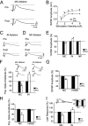
Mossy cell ablation decreases dentate granule cell excitability. A, Representative traces of perforant path evoked fEPSP recordings from the granule cell layer before (Pre; top panel) and after (Post; bottom panel) ablation of mossy cells in a horizontal slice showed decreased fEPSP peak amplitude in response to perforant path stimulation (3 mA). The dashed line indicates the pre-ablation peak amplitude. B, There was a significant decrease in the peak amplitude of the fEPSP after mossy cell ablation at all stimulation intensities. Note that because the minimal stimulation intensity to cause a maximal response was 3 mA, the 3 mA stimulation strength was used in all subsequent experiments illustrated in this figure. C, Representative traces demonstrating an increase in fEPSP amplitude after the deletion of granule cell layer-hilar border interneurons. D, Hilar neuropil ablation caused no change in the fEPSP amplitude. Scale bar bottom panel applies to C and D. E, Summary plot showing the effects of different ablations on fEPSP amplitude. F, The population spike amplitude after mossy cell ablation decreased significantly, whereas it increased after interneuron ablation. The insets show representative traces of the effects of mossy cell (left inset) and interneuron (right inset) ablations on population spike amplitude. G, H, Mossy cell ablation experiments performed blind resulted in a decrease in both the fEPSPs (G) and the population spikes (H). The sham ablations performed in these blind experiments constituted a control for transferring slices between the recording and ablation chambers (handling control); note the lack of a change in either fEPSPs (G) or population spikes (H) after sham ablations. I, The late component of the field response (between 7 and 10 msec after perforant path stimulation as indicated in the left inset) decreased after mossy cell ablation and increased significantly after interneuron ablation. When traces were normalized to the fEPSP peak amplitude, the late component still decreased after mossy cell ablation and increased after interneuron ablation (right inset). MC, Mossy cell; IN, interneuron; NP, neuropil; HAND, handling control.
Figure 4.
Figure 4.
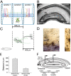
The axial slice preserves the extra-lamellar projections of mossy cells. A, Schematic illustration of the more recent version of the dormant basket cell hypothesis (Sloviter, 1994) that emphasizes the extra-lamellar effects of mossy cell loss. “Lamella 0” is where the cell body of the mossy cell resides (the width of a hippocampal lamella is 600 μm, i.e., 300 μm on either side of a mossy cell). Note that the dormant basket cell hypothesis predicts the loss of surround inhibition after the deletion of mossy cells in one lamella (indicated by the red X over the mossy cells). MC, Mossy cell; BC, basket cell; GC, granule cell. B, Nissl stain of a typical axial slice used in experiments to determine the effect of mossy cell ablation on remote granule cell responses. Scale bar, 0.5 mm. C, A representative biocytin-filled mossy cell is illustrated from an axial slice, with an axonal projection to the inner molecular layer located in the adjacent lamella (extent of axon from the soma: 823 μm). The inset in the bottom left indicates the position of the cell body in the axial slice. Scale bar: inset, 250 μm. GCL, Granule cell layer. D, The mossy cell axon in the inner molecular layer from the shaded area in C is shown in situ. The part of the axon that is located between the arrows is shown enlarged in the inset on the right. The en passant axon terminals (sites of presumed synaptic contacts) in the inner molecular layer are indicated by arrowheads. Scale bars, 50 μm. E, Morphometric analysis of the biocytin-filled mossy cell axons in axial slices. Left column indicates the spatial extent of the axons, and the right column shows that the filled cells were from the area where the mossy cell ablations took place in subsequent experiments (within 300 μm from the bottom of the crest of the granule cell layer at the temporal end of the axial slice). F, Schematic of the axial slice indicates the site of perforant path stimulation at the temporal end, and the intra- and extra-lamellar positions of the field potential recordings from the granule cell layer along the longitudinal axis of the hippocampus.
Figure 2.
Figure 2.
Mossy cell ablation techniques and viability. A, Images depicting a typical mossy cell ablation. A1, Two neurons were imaged under infrared visualization techniques in a living slice. A2, DiO-labeled mossy cell identified under fluorescent visualization (arrow). A3, Mossy cell ablated with enlarged patch pipette. A4, Neighboring cell (white asterisk) was still visible after ablation of the mossy cell. Scale bar, 10 μm. B, Some DiO-labeled mossy cells (indicated by the asterisk in B1, in green) were labeled with the ethidium homodimer stain (B2, in red), indicating damaged membranes, whereas other DiO-labeled cells (indicated by the arrow) were not stained by the ethidium homodimer. Scale bar, 10 μm. The proportion of mossy cells stained with the ethidium homodimer permitted the calculation of the percentage of the live mossy cells that were ablated in the subsequent experiments (see Results). C, Most DiO-labeled mossy cells that were judged to be viable under the IR-DIC were not labeled by the ethidium homodimer (EthD) (note that only those mossy cells that appeared viable under IR-DIC were targeted for ablation in the subsequent experiments).
Figure 5.
Figure 5.
Mossy cell ablation in one hippocampal lamella decreases granule cell responses in the neighboring lamella. A, The field responses to perforant path stimulation decreased at both the intra- and extra-lamellar recording positions (see Fig. 4) after mossy cell ablations (traces are from the same slice; stimulation intensity: 8 mA). B, Control ablation of interneurons caused an increase in the perforant path evoked field response at both the intra- and extra-lamellar recording positions. C, D, Summary plots showing that after mossy cell ablation there was a decrease in the amplitude of the fEPSP (C) and the population spike (D). E, F, Interneuron ablation increased the amplitude of the fEPSP (E) and the population spike (F). MC, Mossy cell; IN, interneuron.
Figure 6.
Figure 6.
Effects of mossy cell and interneuron deletions on granule cell responses in the presence of GABA receptor antagonists. A, The perforant path-evoked field responses were decreased after mossy cell ablation in the presence of GABAA and GABAB blockers (experiments done in axial slices; traces are from the intra-lamellar position). Note that the absence of GABAergic input to the granule cells caused a hyperexcitable response to perforant path stimulation. In these experiments (with the exception of D and H), the number of cells ablated was approximately doubled compared with the experiments shown in Figures 3 and 5. B, Mossy cell ablation (∼29 cells; see Results) decreased the amplitude of the fEPSP at both the intra- and extra-lamellar positions. C, D, Ablation of either a higher (n = 29; C) or lower (n = 15; D) number of mossy cells resulted in a decrease in the first population spike amplitude in GABA receptor antagonists. Note that population spikes were evoked only from the intra-lamellar position. E, Representative traces show no change in fEPSPs after interneuron ablation (n = 29 ablated cells) in the presence of GABAA and GABAB receptor antagonists (traces are from the intra-lamellar position). F, Summary data show the lack of an effect of interneuron ablation (n = 29 ablated cells) on granule cell fEPSPs at both positions in the presence of GABA receptor antagonists. G, H, Ablation of either a higher (n = 29; G) or lower (n = 15; H) number of interneurons resulted in no change in the population spike amplitude from the intra-lamellar position in the presence of GABAA and GABAB blockers. MC, Mossy cell; IN, interneuron.

References

    1. Amaral DG (1978) A Golgi study of cell types in the hilar region of the hippocampus in the rat. J Comp Neurol 182: 851-914. - PubMed
    1. Amaral DG, Witter MP (1989) The three-dimensional organization of the hippocampal formation: a review of anatomical data. Neuroscience 31: 571-591. - PubMed
    1. Andersen P, Soleng AF, Raastad M (2000) The hippocampal lamella hypothesis revisited. Brain Res 886: 165-171. - PubMed
    1. Arabadzisz D, Freund TF (1999) Changes in excitatory and inhibitory circuits of the rat hippocampus 12-14 months after complete forebrain ischemia. Neuroscience 92: 27-45. - PubMed
    1. Bernard C, Esclapez M, Hirsch JC, Ben Ari Y (1998) Interneurones are not so dormant in temporal lobe epilepsy: a critical reappraisal of the dormant basket cell hypothesis. Epilepsy Res 32: 93-103. - PubMed

Publication types

MeSH terms

LinkOut - more resources