. 2004 Mar 16;101(11):3886-91.
doi: 10.1073/pnas.0400347101. Epub 2004 Mar 3.

Identification of a specific self-reactive IgM antibody that initiates intestinal ischemia/reperfusion injury

Affiliations

Identification of a specific self-reactive IgM antibody that initiates intestinal ischemia/reperfusion injury

Ming Zhang et al. Proc Natl Acad Sci U S A. .

Abstract

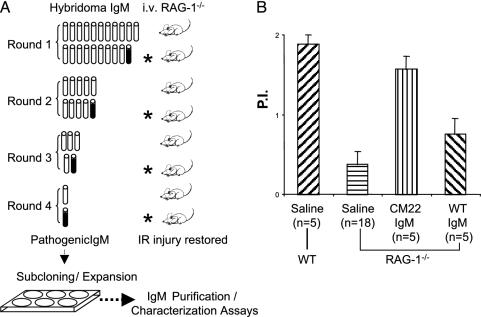
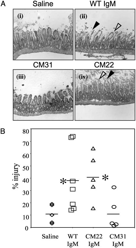
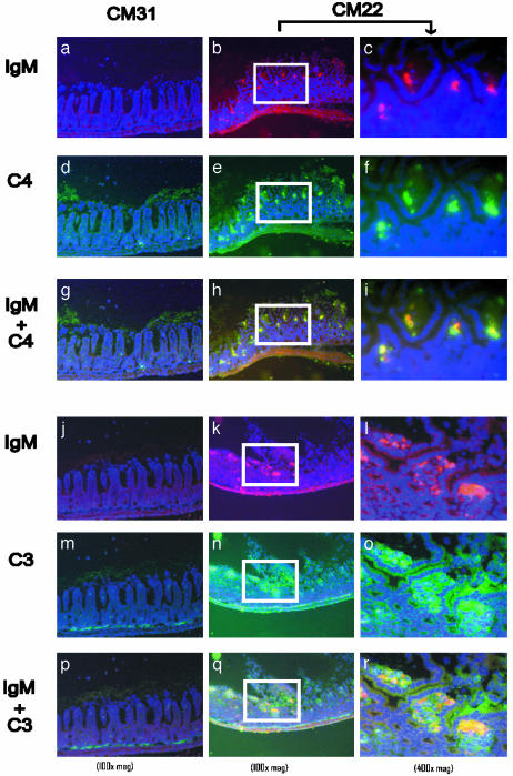
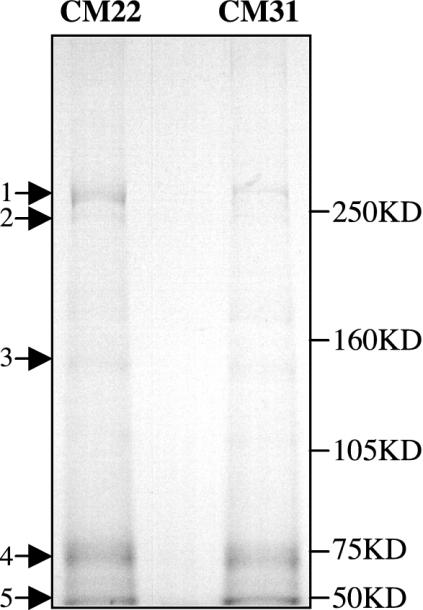
Reperfusion injury of ischemic tissue represents an acute inflammatory response that can cause significant morbidity and mortality. The mechanism of injury is not fully elucidated, but recent studies indicate an important role for natural antibody and the classical pathway of complement. To test the hypothesis that injury is initiated by specific IgM, we have screened a panel of IgM-producing hybridomas prepared from peritoneal cells enriched in B-1 cells. One clone, CM22, was identified that could restore pathogenic injury in RAG-1(-/-) mice in an intestinal model of ischemia/reperfusion (I/R). In situ activation of the classical pathway of complement was evident by deposition of IgM, complement C4, and C3 in damaged tissue after passive transfer of CM22 IgM. Sequence analysis of CM22 Ig heavy and light chains showed germ-line configurations with high homology to a V(H) sequence from the B-1 repertoire and a V(K) of a known polyreactive natural IgM. These data provide definitive evidence that I/R injury can be initiated by clonally specific natural IgM that activates the classical pathway of complement. This finding opens an avenue for identification of I/R-specific self-antigen(s) and early prevention of injury.


Figures

Fig. 1.
Fig. 1.
Identification of pathogenic hybridoma IgM from peritoneal B cells. (A) The strategy of assaying 21 hybridomas in groups in vivo. Intestinal vascular permeability is used as an index for reperfusion injury. The hybridoma group that tested positive (filled symbol) for induction of injury is further divided into two subgroups for next round test in animals. The positive hybridoma (CM22) is then subcloned by limiting dilution followed by expansion of the clone and purification of IgM. (B) Permeability indices of intestinal I/R comparing RAG-1-/- mice that received saline (negative control) or reconstituted with IgM from clone CM22 or WT control. Permeability index (P.I.) = (cpm-1·g-1 of jejunal loop)/(cpm-1·g-1 of blood).
Fig. 2.
Fig. 2.
IgM from a single B cell hybridoma (clone CM22) induces reperfusion injury in RAG-1-/- mice. (A) Representative cryosections were prepared from intestinal tissue of RAG-1-/- mice injected i.v. with saline (i), 400 μg of WT serum IgM (ii), 200 μg of IgM from clone CM31 (iii), or 200 μg of IgM from clone CM22 (iv) before treatment in the intestinal I/R model (see Materials and Methods). After surgery, intestinal tissues were harvested and stained with hematoxylin and eosin. Pathologic features of injury were indicated by arrowheads: filled arrowhead, epithelial disruption (disintegrated epithelium layer of villi); open arrowhead, subepithelial spaces (lack of cellular content beneath continuous epithelial layer). (Magnification, ×100). (B) Pathological scores of intestinal injury were calculated as described in Materials and Methods. *, Statistical significance determined by Student's t test of the Ig-treated versus saline-only groups.
Fig. 3.
Fig. 3.
Deposition of CM22 IgM, C4,and C3 on injured intestinal tissues. Representative cryosections of intestinal tissues were harvested after intestinal I/R from RAG-1-/- mice pretreated with either IgM from CM31 (a, d, g, j, m, and p) or CM22 (b, c, e, f, h, i, k, l, n, o, q, and r). All sections were stained with anti-IgM-biotin followed by streptavidin-Alexa 568 (red) and counterstained with 4′,6-diamidino-2-phenylindole (violet). Cryosections were costained with anti-C4-FITC (green in a-i) and anti-C3-FITC (green in j-r). The colocalization of IgM and C4 (red + green = yellow) is represented in g-i. Colocalization of IgM and C3 is represented in p-r (red + green = yellow). High-magnification (×400) images of CM22 treatment (c, f, i, l, o, and r) were taken from the same region as ×100 magnification marked by white boxes.
Fig. 4.
Fig. 4.
Immunoprecipitation of I/R-specific antigen(s). Intestinal lysates were obtained from CM22- or CM31-reconstituted RAG-1-/- mice after 15 min of reperfusion. Immunoprecipitation was performed as described in Materials and Methods, and IgM complexes were analyzed by SDS/6% PAGE under reducing conditions. Molecular mass markers are indicated on the right.

References

    1. Cotran, R. S. (1999) in Robbins Pathologic Basis of Disease (Saunders, St. Louis), pp. 7-12.
    1. Lawrence, J. B. (2003) in Current Diagnosis & Treatment in Gastroenterology, ed. Friedman, S. L. (McGraw-Hill, New York), Section I.9.
    1. Li, C. & Jackson, R. M. (2002) Am. J. Physiol. 282, C227-C241. - PubMed
    1. Koike, K., Moore, E. E., Moore, F. A., Franciose, R. J., Fontes, B. & Kim, F. J. (1995) J. Trauma 39, 23-27; discussion, 27-28. - PubMed
    1. Simpson, R., Alon, R., Kobzik, L., Valeri, C. R., Shepro, D. & Hechtman, H. B. (1993) Ann. Surg. 218, 444-453; discussion, 453-454. - PMC - PubMed

Publication types

Associated data