. 2004 Mar 16;101(11):3821-6.
doi: 10.1073/pnas.0400512101. Epub 2004 Mar 4.

Structural basis for the function of a minimembrane protein subunit of yeast oligosaccharyltransferase

Affiliations

Structural basis for the function of a minimembrane protein subunit of yeast oligosaccharyltransferase

Sergey Zubkov et al. Proc Natl Acad Sci U S A. .

Abstract

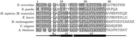
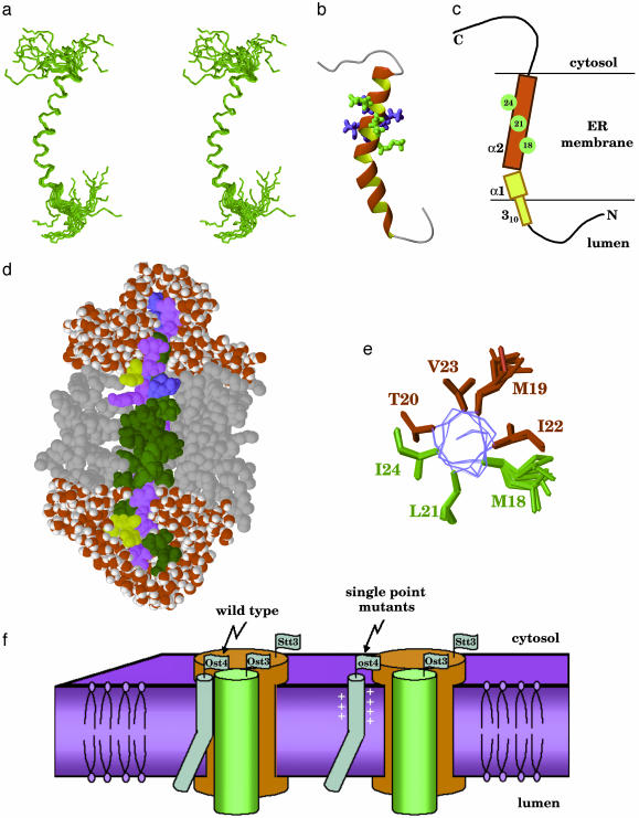
N-glycosylation of proteins is an essential, highly conserved modification reaction that occurs in all eukaryotes and some prokaryotes. This process is catalyzed by oligosaccharyltransferase (OT), a multisubunit enzyme localized in the endoplasmic reticulum. Complete loss of N-glycosylation is lethal in all organisms. In Saccharomyces cerevisiae, OT is composed of nine nonidentical membrane proteins. Here, we report the atomic structure of an OT subunit from S. cerevisiae, Ost4p. This unusually small membrane protein containing only 36 residues folds into a well formed, kinked helix in the model-membrane solvent system used in this study. The residues critical for the OT activity and the stability of Stt3p-Ost4p-Ost3p subcomplex are located in helix alpha2, the larger cytosolic half of this kinked helix. The residues known to disrupt Ost4p-Stt3p complex form a well defined ridge in the 3D structure. Taking together prior mutational studies and the NMR structure of Ost4p, we propose that in the OT complex Stt3p is packed against the alpha 2-helix of Ost4p by using a "ridges-into-grooves" model, with Met-18, Leu-21, and Ile-24 as the packing interface on one face, whereas Ost3p is involved in interactions with Met-19, Thr-20, Ile-22, and Val-23 on the other face.


Figures

Fig. 1.
Fig. 1.
Sequence alignment of Ost4p from the yeast S. cerevisiae and Ost4p analogs from other species: Schizosaccharomyces pombe (fission yeast), Homo sapiens (human), Mus musculus (house mouse), Xenopus laevis (clawed frog), Drosophila melanogaster (fruit fly), C. elegans (nematode), and Arabidopsis thaliana (thale cress). The secondary structure elements of Ost4p are shown below the sequence alignment.
Fig. 2.
Fig. 2.
(a) Fingerprint region of the 2D NOESY spectrum of Ost4p showing sequential connectivities dαN(i,i + 1), dβN(i,i + 1), and dαN(i,i + 3). (b) Amide proton region of the 2D NOESY spectrum of Ost4p showing sequential connectivities dNN(i,i + 1) and dNN(i,i + 2).
Fig. 3.
Fig. 3.
Ost4p secondary structure as determined by solution NMR spectroscopy. (a) Temperature coefficient for amide protons. Filled circles indicate coefficients more positive than -4.5 ppb/K (hydrogen bond present), and open circles indicate coefficients more negative than -4.5 ppb/K. (b) Summary of sequential and medium-range NOE connectivities. (c) Deviations of Hα chemical shifts from random coil values. (d) PsiCSI prediction of secondary structure. Negative bars correspond to helical conformation. (e) Secondary structure of Ost4p as observed in the 3D structure.
Fig. 4.
Fig. 4.
(a) Stereoview of the ensemble of the 20 lowest-energy NMR structures of Ost4p after solvent refinement. (b) Ribbon view of the structure of Ost4p. Residues 18, 21, and 24, expected to interact with Stt3p, are shown in green. Other residues found to be important for optimal protein activity are in blue. These residues may be interacting with Ost3p. (c) Schematic representation of Ost4p structure in membrane. Residues expected to interact with Stt3p are shown in green. (d) Structure of Ost4p from the restricted molecular dynamics calculation in a water/octane/water simulation cell. Octane molecules are shown in gray, and water molecules are shown in red and white. The hydrophobic residues V, I, L, A, F, M, and G are shown in green, hydrophilic S, T, N, Q, and Y are shown in purple, negatively charged D and E are shown in yellow, and positively charged K and H are shown in blue. (e) Location of the mutation-sensitive residues on the helix α2, viewed along the helix axis. (f) Model of the putative structure of Stt3p-Ost4p-Ost4p complex.

References

    1. Wacker, M., Linton, D., Hitchen, P. G., Nita-Lazar, M., Haslam, S. M., North, S. J., Panico, M., Morris, H. R., Dell, A., Wren, B. W., et al. (2002) Science 298, 1790-1793. - PubMed
    1. Kornfeld, R. & Kornfeld, S. (1985) Annu. Rev. Biochem. 54, 631-664. - PubMed
    1. Knauer, R. & Lehle, L. (1999) Biochim. Biophys. Acta 1426, 256-273. - PubMed
    1. Marshall, R. D. (1972) Annu. Rev. Biochem. 41, 673-702. - PubMed
    1. Kelleher, D. J. & Gilmore, R. (1997) Proc. Natl. Acad. Sci. USA 94, 4994-4999. - PMC - PubMed

Publication types

Substances

Associated data