

The caveolin proteins
- PMID: 15003112
- PMCID: PMC395759
- DOI: 10.1186/gb-2004-5-3-214
The caveolin proteins
Abstract
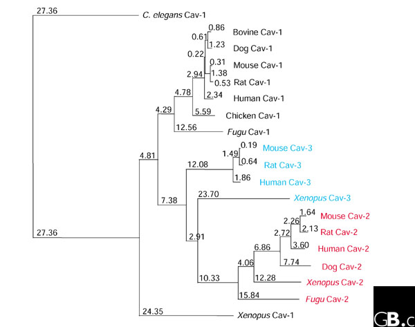
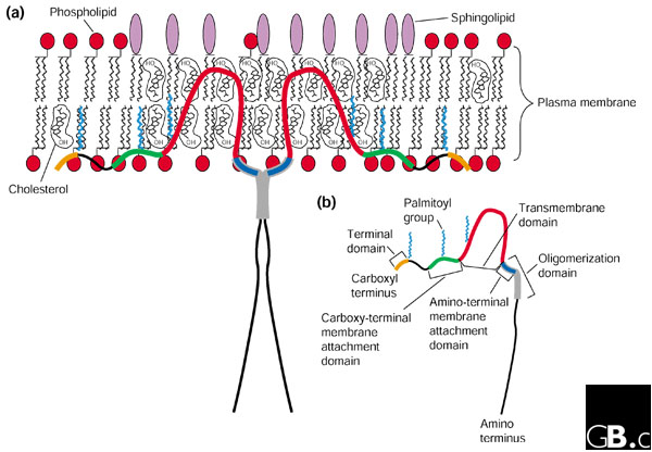
The caveolin gene family has three members in vertebrates: caveolin-1, caveolin-2, and caveolin-3. So far, most caveolin-related research has been conducted in mammals, but the proteins have also been found in other animals, including Xenopus laevis, Fugu rubripes, and Caenorhabditis elegans. Caveolins can serve as protein markers of caveolae ('little caves'), invaginations in the plasma membrane 50-100 nanometers in diameter. Caveolins are found predominantly at the plasma membrane but also in the Golgi, the endoplasmic reticulum, in vesicles, and at cytosolic locations. They are expressed ubiquitously in mammals, but their expression levels vary considerably between tissues. The highest levels of caveolin-1 (also called caveolin, Cav-1 and VIP2I) are found in terminally-differentiated cell types, such as adipocytes, endothelia, smooth muscle cells, and type I pneumocytes. Caveolin-2 (Cav-2) is colocalized and coexpressed with Cav-1 and requires Cav-1 for proper membrane targeting; the Cav-2 gene also maps to the same chromosomal region as Cav-1 (7q31.1 in humans). Caveolin-3 (Cav-3) has greater protein-sequence similarity to Cav-1 than to Cav-2, but it is expressed mainly in muscle cells, including smooth, skeletal, and cardiac myocytes. Caveolins participate in many important cellular processes, including vesicular transport, cholesterol homeostasis, signal transduction, and tumor suppression.
Figures
References
-
- Palade GE. Fine structure of blood capillaries. J Appl Phys. 1953;24:1424–1436. The first morphological description of caveolae.
-
- Rothberg KG, Heuser JE, Donzell WC, Ying YS, Glenney JR, Anderson RG. Caveolin, a protein component of caveolae membrane coats. Cell. 1992;68:673–682. Identification of caveolin. - PubMed
-
- Glenney JR, Jr, Soppet D. Sequence and expression of caveolin, a protein component of caveolae plasma membrane domains phosphorylated on tyrosine in Rous sarcoma virus-transformed fibroblasts. Proc Natl Acad Sci USA. 1992;89:10517–10521. The initial cloning and expression of caveolin-1/VIP21 (from chicken). - PMC - PubMed
Publication types
MeSH terms
Substances
Grants and funding
LinkOut - more resources
Full Text Sources
Other Literature Sources
