

doi: 10.1101/gad.291204.
WUSCHEL induces shoot stem cell activity and developmental plasticity in the root meristem
Affiliations
- PMID: 15004006
- PMCID: PMC359391
- DOI: 10.1101/gad.291204
Item in Clipboard
WUSCHEL induces shoot stem cell activity and developmental plasticity in the root meristem
Genes Dev.
.
Abstract
Most of the plant shoot originates f om a small group of stem cells, which in A abidopsis are specified by WUSCHEL (WUS). It is unknown whether these cells have an inrinsic potential to generate shoot tissues, or whether differentiation is guided by signals from more mature tissues. He e we show that WUS expression in the root induced shoot stem cell identity and leaf development (without additional cues), floral development (together with LEAFY), or embryogenesis (in response to increased auxin). Thus, WUS establishes stem cells with intrinsic shoot identity and responsive to developmental inputs that normally do not change root identity.
Figures

Ectopic WUS activated a shoot stem cell marker in roots. (A) RT–PCR detection of WUS, CLV3, and APT (constitutive control) mRNAs in roots from WUSMOS seedlings at different times after heat shock (+) or in non-heat-shocked controls (–). (B,C) RNA in situ hybridization on longitudinal sections of WUSMOS root tips, 3 d after heat shock (C) or non-heat-shocked control (B; same genotype as in C); the dark signal in C reveals WUS-expressing cells. (D) Longitudinal section of WUSMOS root tips, stained for GUS 3 d after heat shock; arrows indicate GUS-negative cells. (E,F) As in B,C, but hybridized with CLV3 antisense probe. (G–I) Double-labeling RNA in situ hybridization, with WUS signal in red (arrows) and CLV3 signal in blue (arrowheads). (G) Non-heat-shocked control. (H,I) Fixed 3 d after heat shock. Bar, 40 μm.
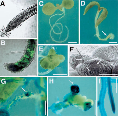
WUS induced the development of shoot tissues from roots. (A,B) Optical sections (combined bright field and GFP channels) of WUSMOS, ANT:GFP root tips. (A) Non-heat-shocked control. (B) Four days after heat shock. (C,D) Eight-day-old WUSMOS plants. (C) Non-heat-shocked control. (D) Six days after heat shock, with green tissue near the root tip (arrow). (E) Ectopic leaves on root, 18 d after heat shock. (F) Electromicrograph of ectopic leaves on WUSMOS root, 21 d after heat shock; arrows indicate leaf cell types such as interdigitated epidermal cells, guard cells, and a trichome. (G–I) GUS staining of WUSMOS roots of 20-day-old plants, 18 d after heat shock (G,H) or in non-heat-shocked control (I). (G) Mosaic GUS expression on the primary root and ectopic shoot tissue developing on a lateral root tip (arrow). (H) Ectopic shoot tissue on the primary root tip, 18 d after heat shock, with a mixture of GUS-positive and GUS-negative tissues; the arrow points at a GUS-positive ectopic leaf. Bars: A,B, 200 μm; C–E, 1 mm; F, 100 μm; G–I, 500 μm.
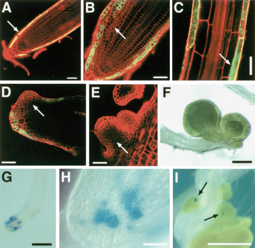
Expression of WUS in the lateral root cap induced ectopic leaf development. (A–C) Optical sections of J2301 roots (GFP and propidium iodide channels combined). (A) J2301, UAS:GFP root tip, 4 d after germination; the arrow indicates GFP expression in the lateral root cap. (B) J2301, UAS:GFP, UAS::WUS root tip, 7 d after germination; the arrow indicates abnormal cell proliferation. (C) Mature section of J2301, UAS:GFP, UAS::WUS root, 7 d after germination; the arrow indicates GFP expression in atrichoblasts. (D–E) Ectopic leaf primordia (arrows) on secondary root tips of J2301, UAS:GFP, UAS::WUS plants, 21 d after germination. (F) Ectopic leaves on the primary root tip of J2301, UAS:GFP, UAS:WUS plant, 21 d after germination. (G–I) GUS staining of J2301, UAS:GFP, UAS:WUS, CLV3:GUS root tips. (G,H) Primary root tip, 7 d after germination. (I) Secondary root tip, 21 d after germination; arrows indicate CLV3:GUS expression (blue signal) associated with ectopic leaf primordia. Bars: A–E,H, 40 μm; F,G,I, 200 μm.
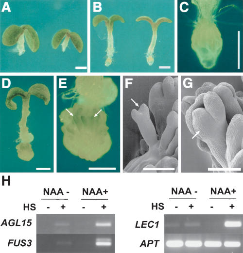
WUS expression combined with auxin induced somatic embryogenesis in roots. (A–C) Eight-day-old WUSMOS seedlings grown on medium with 5 μM NAA, without heat shock (A) or 6 d after heat shock (B); C is a higher-magnification view of the root tip of a seedling equivalent to B. (D,E) Twenty-day-old WUSMOS seedling grown with 5 μM NAA, 18 d after heat shock; the root tip of D is shown at higher magnification in E, with arrows indicating somatic embryos. (F,G) Cryo-scanning electron micrographs of 2-week-old heat shocked WUSMOS plants grown on medium containing 5 μM NAA. Arrows indicate somatic embryos. (H) RT–PCR detection of embryo marker mRNAs and constitutive control (APT) in roots dissected from WUSMOS seedlings, not heat shocked (HS–) or 2 wk after heat shock (HS+), grown in GM medium (NAA–) or medium supplemented with NAA 5 μM (NAA+). Bars: A–E, 1 mm; F,G, 100 μm.

Floral tissues in the roots of 21-day-old WUSMOS, 35S:LFY plants, 19 d after heat shock. (A) Sepals on lateral root tip. (B) Sepal and carpel-like organs on lateral root tip. (C) Arrow indicates anther originated from lateral root tip. (D–F) Cryo-scanning electron micrographs showing carpelloid tissue on primary root tip (D), sepaloid (se) and carpelloid (ca) tissues on lateral root tip, with arrow indicating stigmatic papillae (E), and an epidermal cell with the epicuticular ridges typically seen in floral organs (F). Bars: A–C, 1 mm; D,E, 200 μm; F, 10 μm.
References
-
- Brand U., Fletcher, J.C., Hobe, M., Meyerowitz, E.M., and Simon, R. 2000. Dependence of stem cell fate in Arabidopsis on a feedback loop regulated by CLV3 activity. Science 289: 617–619. - PubMed
-
- Clough S.J. and Bent, A.F. 1998. Floral dip: A simplified method for Agrobacterium-mediated transformation of Arabidopsis thaliana. Plant J. 16: 735–743. - PubMed
-
- Feher A., Pasternak, T.P., and Dudits, D. 2003. Transition of somatic plant cells to an embryogenic state. Plant Cell Tissue Organ Cult. 74: 201–228.
Publication types
MeSH terms
Substances
Grants and funding
LinkOut - more resources
Full Text Sources
Other Literature Sources
Molecular Biology Databases