. 2004 May;15(5):2253-63.
doi: 10.1091/mbc.e04-01-0002. Epub 2004 Mar 5.

Uncoated endocytic vesicles require the unconventional myosin, Myo6, for rapid transport through actin barriers

Affiliations

Uncoated endocytic vesicles require the unconventional myosin, Myo6, for rapid transport through actin barriers

Laura Aschenbrenner et al. Mol Biol Cell. 2004 May.

Abstract

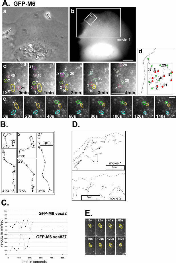
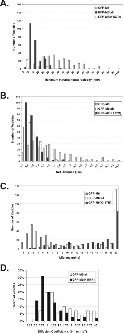
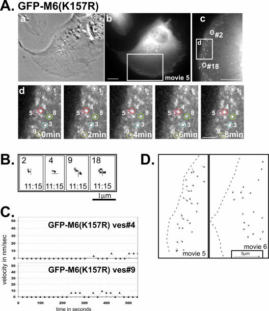
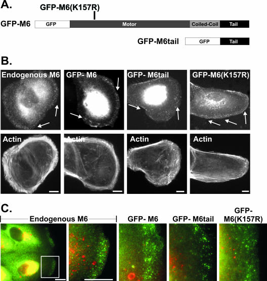
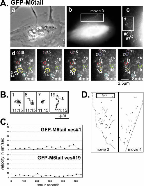
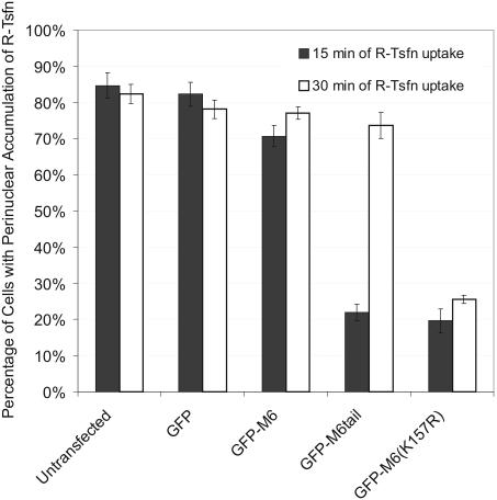
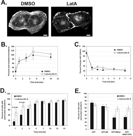
After clathrin-mediated endocytosis, clathrin removal yields an uncoated vesicle population primed for fusion with the early endosome. Here we present the first characterization of uncoated vesicles and show that myo6, an unconventional myosin, functions to move these vesicles out of actin-rich regions found in epithelial cells. Time-lapse microscopy revealed that myo6-associated uncoated vesicles were motile and exhibited fusion and stretching events before endosome delivery, processes that were dependent on myo6 motor activity. In the absence of myo6 motor activity, uncoated vesicles remained trapped in the actin mesh, where they exhibited Brownian-like motion. Exit from the actin mesh occurred by a slow diffusion-based mechanism, delaying transferrin trafficking to the early endosome. Expression of a myo6 mutant that bound tightly to F-actin produced immobilized vesicles and blocked trafficking. Depolymerization of the actin cytoskeleton rescued this block and specifically accelerated transferrin delivery to the early endosome without affecting earlier steps in endocytosis. Therefore actin is a physical barrier impeding uncoated vesicle trafficking, and myo6 is recruited to move the vesicles through this barrier for fusion with the early endosome.


Figures

Figure 2.
Figure 2.
GFP-M6–associated vesicles are motile and exhibit fusion and stretching. (A) A GFP-M6–expressing ARPE-19 cell analyzed by time-lapse videomicroscopy presented as a phase contrast (a) and a fluorescence image (b). The larger box corresponds to the second frame of Supplementary Movie 1. Scale bar for a and b, 10 μm. (c) Isolated time-lapse images of the small region boxed in panel b. The cell edge is at the left. The relative timing of each image is presented on each panel. Individual vesicles are numbered to allow tracking between frames. Vesicle fusion events are demarcated with arrows. (d) Schematic showing the tracks of numbered vesicles monitored in panel c (boxed area), with the starting point (green dot) and end point (red dot) emphasized. A dotted gray line depicts the cell border. Scale bar for c and d, 2.5 μm. (e) Isolated time-lapse images showing three examples of vesicle fusion. Scale bar, 2.5 μm. (B) Representative tracks of GFP-M6–decorated vesicles, numbered in A, panel c. Each arrow is a 20-s interval. The start point is demarcated with an “o,” the end point with a line perpendicular to the direction of the arrow. The total lifetime of the vesicle tracked is in min:sec. (C) Plots showing the change in the instantaneous velocity of individual vesicles tracked over time. The vesicle number corresponds to those shown in A and B. (D) Line-drawing showing a representative subset of distinct GFP-M6–associated vesicles evident during a 13-min (Movie 1) or a 16-min window (Movie 2). Each displacement vector shows the position of the vesicle, the direction traveled, and the distance covered over the vesicle's lifetime. The outline of the cell border is a dotted gray line. (E) Isolated time-lapse images showing vesicle stretching. The relative timing is presented on each panel. Scale bar, 1 μm.
Figure 3.
Figure 3.
Uncoated vesicles exhibit significant movement and a short lifetime only when associated with functional myo6. Two hundred fifty vesicles were tracked in cells expressing either GFP-M6 (gray bars), GFP-M6tail (black bars), or GFP-M6(K157R) (white bars). (A) Histogram showing the maximum instantaneous velocity in nm/s for each vesicle tracked. (B) Histogram showing net distance in micrometers traveled by each vesicle. (C) Histogram of the total lifetime in minutes exhibited by each vesicle. (D) Histogram of the diffusion coefficients obtained from tracking vesicles.
Figure 5.
Figure 5.
Analysis of GFP-M6(K157R)–associated vesicles reveals no movement, suggesting this mutation causes tight binding to the actin cytoskeleton. (A) Phase contrast (a) and fluorescence images (b) of a GFP-M6(K157R)–expressing ARPE-19 cell monitored by time-lapse video. Boxed region corresponds to Supplementary Movie 5 and is enlarged and rotated clockwise in panel (c). (d) Isolated images from the time-lapse movie of the cell portion boxed in panel (c). The cell edge is at the left of the panel and the relative timing of each image is presented on each panel. Individual vesicles are numbered. The scale bars are as in Figure 4. (B) Representative tracks showing the movements of GFP-M6(K157R)–decorated vesicles. The numbers correspond to the vesicles circled in A, panels (c) and (d), and tracks are as described for Figure 2. (C) Plots showing the change in the instantaneous velocity of individual vesicles tracked over time. (D) Line-drawing depicting the direction and distance traveled for a series of distinct GFP-M6(K157R)–associated vesicles present simultaneously during a 20-min period in two cells (Supplementary Movies 5 and 6; see legend for Figure 2D).
Figure 1.
Figure 1.
Myo6 and GFP-tagged myo6 constructs localize to peripheral uncoated vesicles. (A) Schematic of GFP-myo6 constructs. The motor domain point mutation, K157R, is indicated. (B) Staining of ARPE-19 cells using rabbit anti-myo6 antibodies (endogenous M6) or cells transfected with the GFP fusion constructs, counterstained for F-actin with rhodamine-conjugated phalloidin. Arrows point out myo6-associated peripheral vesicles. (C) Double-labeled images visualizing endogenous myo6 or GFP-myo6 fusion constructs (green) and the early endosome marker EEA1 (red). Scale bars, 10 μm.
Figure 4.
Figure 4.
Analysis of GFP-M6tail–associated vesicles reveals a requirement for the myo6 motor domain for vesicle movement. (A) Images of a GFP-M6tail–expressing ARPE-19 cell monitored by time lapse, presented as a phase contrast (a) and a fluorescence image (b). The boxed region corresponds to Supplementary Movie 3 and is enlarged and rotated counterclockwise in panel (c). (d) Isolated images from the time-lapse movie of the cell portion boxed in panel (c). The cell edge is at the left of the panel and the relative timing of each image is indicated. Representative individual vesicles are numbered in panels (c) and (d). Vesicle fusion events evident in panel (d) are demarcated with arrows. Scale bar for a and b, 10 μm; c, 5 μm; d, 2.5 μm. (B) Representative tracks of GFP-M6tail–decorated vesicles. The numbers correspond to the vesicles circled in A, panels (c) and (d), and tracks are as described for Figure 2. (C) Plots showing the change in the instantaneous velocity of individual vesicles tracked over time. (D) Line drawing depicting the direction and distance traveled for a series of distinct GFP-M6tail–associated vesicles present simultaneously over a 20-min period in two cells (Supplementary Movies 3 and 4; see legend for Figure 2D).
Figure 6.
Figure 6.
Expression GFP-M6(K157R) blocks transferrin trafficking and it cannot be rescued by extending uptake time. GFP-, GFP-M6–, GFP-M6tail–, and GFP-M6(K157R)–transfected cultures were incubated with rhodamine-conjugated transferrin (R-Tsfn). The histogram shows the percentage of transfected and untransfected cells exhibiting labeling of the pericentriolar endosomes with the endocytosed R-Tsfn after 15 min (black bars) or 30 min (white bars). See Supplementary Figure 2 for primary data. More than 300 cells were counted per time point and the data are representative of three experiments.
Figure 7.
Figure 7.
Removal of the actin barrier in ARPE-19 cells accelerates trafficking to the early endosome and rescues the trafficking defects seen upon GFP-M6tail and GFP-M6(K157R) expression. (A) Fluorescence staining of F-actin in cells treated for 30 min with DMSO or 0.015 μM latrunculin A. Scale bars, 10 μm. (B and C) Kinetics of transferrin endocytosis and transferrin exit from clathrin-coated vesicles as judged by pulse-chase experiments in cells incubated in the presence of DMSO (•) or latrunculin A (□). (B) Quantification of the endocytosis of biotinylated transferrin. Shown is a representative example of four experiments. (C) Quantitation of the exit of transferrin from clathrin-coated vesicles, as assayed by the percent overlap between clathrin and endocytosed R-Tsfn during the course of a pulse-chase experiment. (D and E) Delivery of transferrin to the pericentriolar early endosome, as judged by steady state uptake experiments in the presence of DMSO (black bars) or latrunculin A (white bars). (D) Histogram showing the percent of cells exhibiting delivery of R-Tsfn to the pericentriolar early endosome at fixed uptake times. More than 600 cells were counted per time point. (E) Histogram showing the percent of transfected cells exhibiting delivery of endocytosed R-Tsfn to the pericentriolar early endosome after 15 min of uptake. More than 500 cells were counted per construct.

Similar articles

Cited by

References

    1. Abney, J.R., Meliza, C.D., Cutler, B., Kingma, M., Lochner, J.E., and Scalettar, B.A. (1999). Real-time imaging of the dynamics of secretory granules in growth cones. Biophys. J. 77, 2887–2895. - PMC - PubMed
    1. Aschenbrenner, L., Lee, T., and Hasson, T. (2003). Myo6 facilitates the translocation of endocytic vesicles from cell peripheries. Mol. Biol. Cell 14, 2728–2743. - PMC - PubMed
    1. Bose, A., Guilherme, A., Robida, S.I., Nicoloro, S.M.C., Zhou, Q.L., Jiang, Z.Y., Pomerleau, D.P., and Czech, M.P. (2002). Glucose transporter recycling in response to insulin is facilitated by mysoin Myo1c. Nature 420, 821–824. - PubMed
    1. Clague, M.J. (1998). Molecular aspects of the endocytic pathway. Biochem. J. 336, 271–282. - PMC - PubMed
    1. Cramer, L.P. (1997). Molecular mechanism of actin-dependent retrograde flow in lamellipodia of motile cells. Front. Biosci. 2, d260–d270. - PubMed

Publication types

MeSH terms