. 2004 May;15(5):2375-87.
doi: 10.1091/mbc.e03-12-0886. Epub 2004 Mar 5.

Dynamic regulation of a GPCR-tetraspanin-G protein complex on intact cells: central role of CD81 in facilitating GPR56-Galpha q/11 association

Affiliations

Dynamic regulation of a GPCR-tetraspanin-G protein complex on intact cells: central role of CD81 in facilitating GPR56-Galpha q/11 association

Kevin D Little et al. Mol Biol Cell. 2004 May.

Abstract

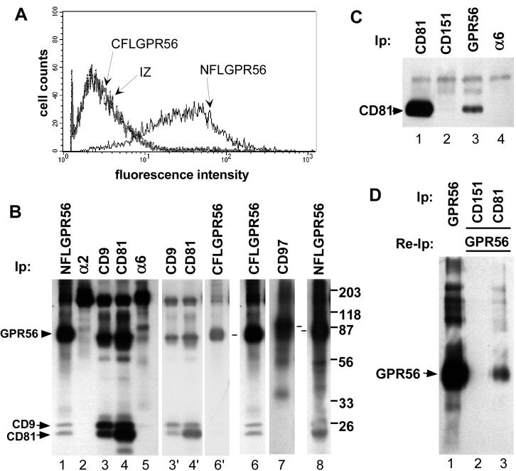
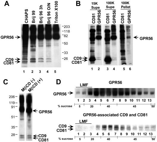
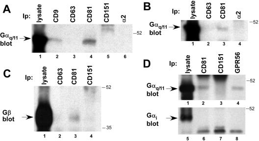
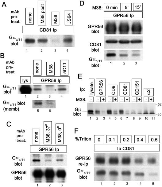
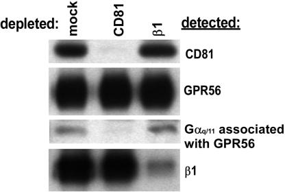
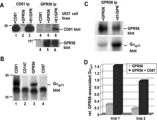
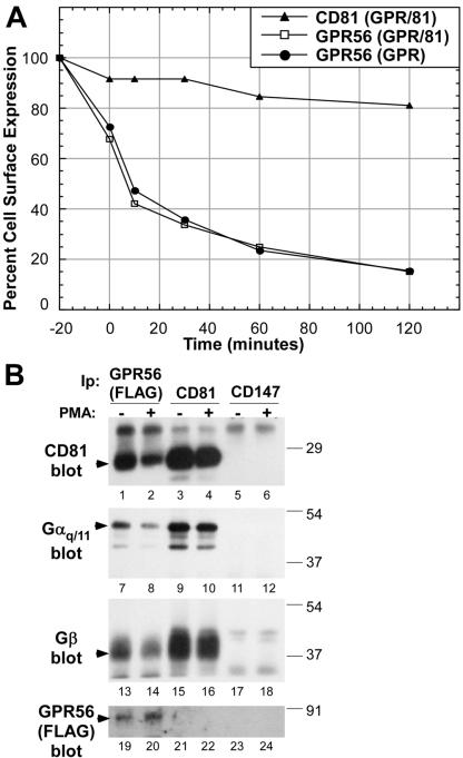
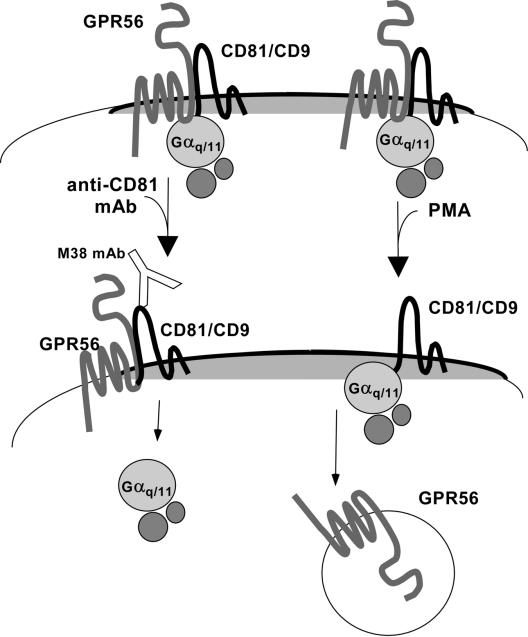
By means of a variety of intracellular scaffolding proteins, a vast number of heterotrimeric G protein-coupled receptors (GPCRs) may achieve specificity in signaling through a much smaller number of heterotrimeric G proteins. Members of the tetraspanin family organize extensive complexes of cell surface proteins and thus have the potential to act as GPCR scaffolds; however, tetraspanin-GPCR complexes had not previously been described. We now show that a GPCR, GPR56/TM7XN1, and heterotrimeric G protein subunits, Galpha(q), Galpha(11), and Gbeta, associate specifically with tetraspanins and CD81, but not with other tetraspanins. CD9 Complexes of GPR56 with CD9 and CD81 remained intact when fully solubilized and were resistant to cholesterol depletion. Hence they do not depend on detergent-insoluble, raft-like membrane microdomains for stability. A central role for CD81 in promoting or stabilizing a GPR56-CD81-Galpha(q/11) complex was revealed by CD81 immunodepletion and reexpression experiments. Finally, antibody engagement of cell surface CD81 or cell activation with phorbol ester revealed two distinct mechanisms by which GPR56-CD81-Galpha(q/11) complexes can be dynamically regulated. These data reveal a potential role for tetraspanins CD9 and CD81 as GPCR scaffolding proteins.


Figures

Figure 1.
Figure 1.
Specific association of CD9 and CD81 with GPR56. (A) Nonpermeabilized 293-NFLGPR56 cells (solid black line), 293-CFLGPR56 cells (solid gray line), or 293 IZ empty vector control cells (broken black line) were stained with M2 anti-FLAG antibody followed by FITC-goat anti-mouse 2o antibody and analyzed by flow cytometry. (B) The indicated proteins were immunoprecipitated from a Brij 96 lysate of biotinylated 293-NFLGPR56 cells (lanes 1–5 and 8), 293-CFLGPR56 cells (lane 6), or 293-CD97 cells (lane 7). Dashes between lanes 7 and 8 designate CD97 and GPR56, respectively. For lanes 3, 4, and 6, lighter exposures are shown (lanes 3′, 4′ and 6′) to allow better resolution of the 70–80-kDa region. (C) The indicated proteins were immunoprecipitated from a 1% Brij 96 detergent extract of 293-NFLGPR56 cells, followed by immunoblotting with the M38 anti-CD81 mAb. Cell equivalents, 4.5 × 10⁵, were used for the immunoprecipitation in lane 1, and 4.3 × 10⁶ cell equivalents were used in lanes 2–4. Analysis of the blot by densitometry indicated that ∼2% of total CD81 may be GPR56-associated. (D) Because of the limited sensitivity of the FLAG antibody in immunoblotting, coprecipitation of GPR56 with CD81 was analyzed by reimmunoprecipitation. About 1.8 × 10⁷ biotinylated 293-NFLGPR56 cells were lysed in 1% Brij 96 and GPR56 was immunoprecipitated directly from 5% of the lysate (lane 1). The remainder of the lysate was divided equally between CD151 and CD81 immunoprecipitations (lanes 2 and 3). Next, CD151- and CD81-associated proteins were eluted with Triton X-100 from protein G–bound CD151 and CD81 immune complexes and reimmunoprecipitated for FLAG-GPR56. Analysis by densitometry indicated that ∼2% of total GPR56 may be CD81-associated. In A through D, antibodies used were M2 anti-FLAG agarose (for GPR56), A2-IIE10 anti-α2 integrin, Alb-6 anti-CD9, M38 anti-CD81, A6-ELE anti-α6 integrin, Vim3B anti-CD97, and 5C11 anti-CD151. Material in GPR56 lanes that is >150 kDa varied between experiments and may be SDS-resistant oligomers of GPR56.
Figure 2.
Figure 2.
Fully soluble GPR56 complexes with CD9 and CD81. (A) Cell surface–biotinylated 293-NFLGPR56 cells were extracted with the indicated detergents, and GPR56 complexes were immunoprecipitated with anti-FLAG agarose (∼2.6 × 10⁶ cells per IP). (B) A 1% Brij 96 extract of biotinylated 293-NFLGPR56 cells was centrifuged at 15,000 × g for 15 min. The resulting supernatant was used for M38 anti-CD81 or M2 anti-FLAG (GPR56) immunoprecipitation (lanes 1 and 2) or was further centrifuged for 40 min at 100,000 × g. The 100K supernatant (lanes 3 and 4) and the resuspended pellet (lanes 5 and 6) were then immunoprecipitated for CD81 or GPR56. Supernatant from ∼5.75 × 10⁵ cell equivalents was analyzed in lanes 1–4, and pelleted material from ∼2.9 × 10⁶ cell equivalents was analyzed in lanes 5 and 6. (C) A 1% Brij 96 extract of biotinylated 293-NFLGPR56 cells was divided into two aliquots and left untreated or treated with methyl-β-cyclodextrin (13 mg/ml final concentration). After a 10-min incubation at room temperature, GPR56 complexes were immunoprecipitated overnight at 4°C(∼1 × 10⁶ cell equivalents per IP). (D) A 1% Brij 96 lysate of ∼ 9 × 10⁶ biotinylated 293-NFLGPR56 cells was loaded in 45% sucrose over a 50% sucrose cushion and overlaid with a detergent-free step gradient of 40, 20, and 5% sucrose. After overnight centrifugation, Brij 96 was added to a final concentration of 1% to each gradient fraction, and GPR56 complexes were immunoprecipitated. Top panel: the distribution of the 70–80-kDa GPR56 band in the gradient; bottom panel: the distribution of coprecipitating CD9 and CD81 bands, shown separately to allow for a darker exposure. LMF, “light membrane fraction,” the region of the gradient where cholesterol-dependent detergent-insoluble membrane microdomains are localized.
Figure 3.
Figure 3.
Specific association of Gαq/11 and Gβ with CD9, CD81, and GPR56. (A) A Brij 96 detergent lysate of 293 cells was immunoprecipitated for the indicated molecules, followed by blotting for Gαq/11. Lysate from ∼4 × 10⁶ cells was used for each immunoprecipitation in lanes 2–6 and was compared with ∼2 × 10⁵ cell equivalents analyzed directly in lane 1. (B) A Brij 96 detergent extract of HT1080 cells was analyzed by immunoprecipitation and Gαq/11 immunoblotting, as in A. About 1.7 × 10⁵ cell equivalents of lysate were used in lane 1, and 3.3 × 10⁶ in each of lanes 2–4. (C) A Brij 96 lysate of A431 cells was immunoprecipitated for the indicated molecules, followed by blotting for the Gβ subunit. About 4 × 10⁶ cell equivalents were used for the immunoprecipitations in lanes 2–4, compared with ∼ 2 × 10⁵ analyzed directly in lane 1. (D) A Brij 96 lysate from ∼1.8 × 10⁷ 293-NFLGPR56 cells was immunoprecipitated for the indicated proteins, followed by blotting for Gαq/11 (lanes 1–4) or Gαi (lanes 5–8). In each experiment 2% of the lysate was analyzed directly (lanes 1 and 5), and the remainder was divided evenly among lanes 2–4 and 6–8. Antibodies used were as in Figure directly (lanes 1 and 5), and the remainder was divided evenly among lanes was analyzed 1, along with polyclonal anti-Gαq/11, Gαi, and Gβ antisera.
Figure 6.
Figure 6.
An anti-CD81 antibody triggers dissociation of Gαq/11 from GPR56-CD81. (A) About 5 × 10⁶ HT1080 cells were treated for 20 min at 37°C with 5 μg/ml anti-CD81 mAbs M38 (lane 3) or JS64 (lane 4) or were left untreated (lanes 1 and 2). Cells were rinsed with ice-cold PBS and lysed on ice in 1% Brij 96. After centrifugation, lysates were precleared with mouse IgG directly conjugated to agarose. For the sample in lane 2, 5 μg/ml M38 was added during this preclearing step. After preclearing, JS64 and M38 concentrations in each lysate were adjusted to 5 μg/ml, protein G was added, and CD81 immune complexes were collected. The presence of Gαq/11 in each immunoprecipitate was measured by immunoblotting. (B) Top panel: ∼7 × 10⁶ 293-NFLGPR56 cells were treated for 15 min at 37°C with 5 μg/ml anti-CD81 mAb, M38 (lane 3), anti-CD151 mAb, 5C11 (lane 4), or no antibody (lane 2), before rinsing in cold PBS and lysing in 1% Brij 96. GPR56 was immunoprecipitated with anti-FLAG agarose, followed by blotting for Gαq/11. In lane 1, lysate from ∼ 1.5 × 10⁵ cell equivalents was loaded. Lanes 5 and 6: cell membrane fraction (see MATERIALS AND METHODS) was treated with M38 (lane 2) or left untreated (lane 1) and then extracted with Brij 96. GPR56 was immunoprecipitated from the extracts, and Gαq/11 was blotted, as in lanes 1–4. (C) 293-NFLGPR56 cells were left untreated (lane 1), treated with M38 at 37°C for 15 min (lane 2), or prechilled on ice and treated with M38 in ice-cold buffer for 15 min (lane 3). Cells were then lysed in Brij 96, and GPR56-Gαq/11 association was analyzed as in B. By densitometry, Gαq/11 association was reduced by 84% (lane 2) and 63% (lane 3) after correction for small variations in recovered GPR56. (D) About 7 × 10⁶ 293-NFL-GPR56 cells were treated with the M38 antibody for the indicated times (0 min was not treated) before lysis in Brij 96 and GPR56 immunoprecipitation as in B. The levels of GPR56, CD81, and Gαq/11 in each immunoprecipitate were assayed by immunoblotting. (E) The indicated proteins were immunoprecipitated from 1% Brij 99 lysates of 293-NFLGPR56 cells that had been treated with M38 anti-CD81 (15 min. at 37°C) or left untreated. Coprecipitating Gβ subunit was detected by immunoblotting. About 2 × 10⁶ cell equivalents were analyzed in lanes 2–11, compared with ∼ 2 × 10⁵ in lane 1. (F) A clarified Brij 99 extract of ∼5.4 × 10⁷ biotinylated 293-NFLGPR56 cells was divided into five equal pools, and Triton X-100 was added to the indicated concentrations. After CD81 immunoprecipitation and rinsing with the appropriate detergent mixture, 90% of the bead-bound CD81 immune complexes were resuspended in 1% Triton X-100 (no Brij 99) to elute CD81-associated proteins, and then GPR56 was reimmunoprecipitated and detected by blotting with HRP-ExtrAvidin (top panel). The remaining 10% of the CD81 immune complexes were blotted for Gαq/11 (bottom panel).
Figure 4.
Figure 4.
Evidence for GPR56-CD81-Gαq/11 complexes. 293-NFL-GPR56 cells, 1.8 × 10⁷, were biotinylated and extracted with Brij 96. Equal portions of the lysate were depleted with protein G-Sepharose (mock), M38 anti-CD81, or TS2/16 anti-β1 integrin agarose. After each depletion, CD81 (top panel), GPR56 (second panel), and β1 integrin (bottom panel) were detected by immunoprecipitation followed by ExtrAvidin-HRP chemiluminescence. GPR56-associated Gαq/11 (third panel) was detected by FLAG immunoprecipitation followed by Gαq/11 blotting. Lysate from 6 × 10⁵ cell equivalents was used for each immunoprecipitation in panels 1, 2, and 4, and lysate from 4.2 × 10⁶ cell equivalents was used for each immunoprecipitation in panel 3.
Figure 5.
Figure 5.
CD81 enhances GPR56-Gαq/11 association. U937 cells were transduced with CD81 (+CD81), GPR56 (+GPR56), or both proteins (+81/GPR). (A) U937 cells were lysed with 1% Brij 99 and immunoprecipitated for CD81 (lanes 1–3; 1 × 10⁷ cells/lane) or GPR56 (lanes 4–6; 2 × 10⁷ cells/lane), followed by blotting with the M38 anti-CD81 antibody (top panel) or with the M2 anti-FLAG antibody, to detect GPR56 (bottom panel). (B) The indicated proteins were immunoprecipitated from a Brij 99 extract of 4 × 10⁷ U937 + 81/GPR cells, followed by immunoblotting Gαq/11. (C) 2 × 10⁷ of the indicated U937 cell types were lysed in 1% Brij 99, and GPR56 was immunoprecipitated with anti-FLAG agarose. GPR56 and coprecipitating Gαq/11 were blotted with biotinylated M2 anti-FLAG antibody or with Gαq/11 polyclonal antibody respectively. (D) Semiquantitative densitometry of results in C above and of a second independent trial. The y-axis equals the Gαq/11:GPR56 ratio. GeneTools software (Syngene) was used to analyze digitized images captured from trans-illuminated films.
Figure 7.
Figure 7.
PMA stimulation causes dissociation of GPR56 from CD81-Gαq/11. (A) U937 cells expressing NFLGPR56 alone (GPR), or together with CD81 (81/GPR), were treated with 100 nM PMA for 20 min at 37°C and then rinsed and returned to 37°C. GPR56 and CD81 cell surface levels were monitored by staining nonpermeabilized cells with M2 anti-FLAG mAb or M38 anti-CD81 mAb, followed by FITC-goat anti-mouse 2o antibody and flow cytometry. The y-axis is the mean fluorescence intensity as a percentage of the values for untreated cells. The 0-min time point corresponds to the end of the PMA treatment. (▴) U937 CD81/GPR56 cells stained for CD81; (□), U937 CD81/GPR56 cells stained for GPR56; (•), U937 GPR56 cells stained for GPR56. (B) 6 × 10⁷ U937 cells expressing both CD81 and NFLGPR56 were divided into two equal pools and maintained at 37°C while one pool was treated for 20 min with 200 nM PMA. Next, cells were extracted with 1% Brij 99, and equal volumes of lysates were immunoprecipitated with M38 anti-CD81 mAb, M2 anti-FLAG agarose, or 8G6 anti-CD147 mAb. The immunoprecipitates were blotted with M38 anti-CD81, anti-Gαq/11 clonal antibody, anti-Gβ polyclonal antibody, or biotinylated M2 anti-FLAG antibody.
Figure 8.
Figure 8.
Dynamic regulation of GPR56-CD81-Gαq/11 complexes. Left: An anti-CD81 mAb triggers dissociation of Gαq/11 from CD81-GPR56. This might involve mimicking some aspects of events that occur downstream of GPCR ligand binding, such as a mobilization of G proteins or transit of GPCRs into or out of different cell surface microdomains. Right: By a distinct mechanism, which resembles heterologous GPCR desensitization, PMA treatment triggers the apparent loss of GPR56 from the cell surface, resulting in a loss of GPR56 from CD81-Gαq/11.

Similar articles

Cited by

References

    1. Berditchevski, F. (2001). Complexes of tetraspanins with integrins: more than meets the eye. J. Cell Sci. 114, 4143–4151. - PubMed
    1. Berditchevski, F., Bazzoni, G., and Hemler, M.E. (1995). Specific association of CD63 with the VLA-3 and VLA-6 integrins. J. Biol. Chem. 270, 17784–17790. - PubMed
    1. Berditchevski, F., Chang, S., Bodorova, J., and Hemler, M.E. (1997a). Generation of monoclonal antibodies to integrin-associated proteins. Evidence that alpha3beta1 complexes with EMMPRIN/basigin/OX47/M6. J. Biol. Chem. 272, 29174–29180. - PubMed
    1. Berditchevski, F., Tolias, K.F., Wong, K., Carpenter, C.L., and Hemler, M.E. (1997b). A novel link between integrins, transmembrane-4 superfamily proteins (CD63 and CD81), and phosphatidylinositol 4-kinase. J. Biol. Chem. 272, 2595–2598. - PubMed
    1. Berditchevski, F., Zutter, M.M., and Hemler, M.E. (1996). Characterization of novel complexes on the cell surface between integrins and proteins with 4 transmembrane domains (TM4 proteins). Mol. Biol. Cell 7, 193–207. - PMC - PubMed

Publication types

MeSH terms