. 2004 May;15(5):2287-301.
doi: 10.1091/mbc.e03-10-0728. Epub 2004 Mar 5.

Identification and characterization of two novel proteins affecting fission yeast gamma-tubulin complex function

Affiliations

Identification and characterization of two novel proteins affecting fission yeast gamma-tubulin complex function

Srinivas Venkatram et al. Mol Biol Cell. 2004 May.

Abstract

The gamma-tubulin complex, via its ability to organize microtubules, is critical for accurate chromosome segregation and cytokinesis in the fission yeast, Schizosaccharomyces pombe. To better understand its roles, we have purified the S. pombe gamma-tubulin complex. Mass spectrometric analyses of the purified complex revealed known components and identified two novel proteins (i.e., Mbo1p and Gfh1p) with homology to gamma-tubulin-associated proteins from other organisms. We show that both Mbo1p and Gfh1p localize to microtubule organizing centers. Although cells deleted for either mbo1(+) or gfh1(+) are viable, they exhibit a number of defects associated with altered microtubule function such as defects in cell polarity, nuclear positioning, spindle orientation, and cleavage site specification. In addition, mbo1Delta and gfh1Delta cells exhibit defects in astral microtubule formation and anchoring, suggesting that these proteins have specific roles in astral microtubule function. This study expands the known roles of gamma-tubulin complex components in organizing different types of microtubule structures in S. pombe.


Figures

Figure 1.
Figure 1.
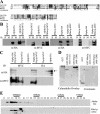
Gfh1p is homologous to hGCP4/Dgrip75 and is part of the S. pombe γ-TuC. (A) Alignment of Gfh1p with human GCP4 and Drosophila melanogaster Grip75. Identical residues are indicated by black boxes and conservative substitutions are shaded in gray. (B) Protein lysates were prepared from cells expressing tagged alleles of alp4+, alp6+, gfh1+, or both alp4+ and gfh1+ and alp6+ and gfh1+. These were subjected to immunoprecipitation using either 12CA5 (IP:HA) or 9E10 (IP:MYC) antibodies. The immunoprecipitates were resolved by SDS-PAGE and immunoblotted with either α-HA or α-Myc antibodies as indicated. (C) Protein lysates prepared from either control cells or cells expressing a MYC-tagged allele of gfh1+ were subjected to immunoprecipitation using 9E10 antibodies. The immunoprecipitates were resolved by SDS-PAGE and immunoblotted with either anti–γ-tubulin or α-Myc antibodies as indicated. The arrow in the bottom panel indicates the band corresponding to γ-tubulin and asterisk (*) indicates IgG heavy chain. (D) Protein lysates were prepared from cells expressing tagged alleles of alp6+ and gfh1+ and were subjected to gel filtration on a Superose-6 column. Thirty-four 0.5-ml fractions were collected, and fractions 11–34 were resolved by SDS-PAGE and immunoblotted with either 12CA5 (anti-HA) or 9E10 (anti-MYC) antibodies to detect the tagged proteins as indicated. The peak positions of calibration markers are also indicated on top of the fractions.
Figure 2.
Figure 2.
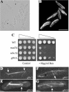
gfh1Δ cells are viable and exhibit astral microtubule defects. (A) DIC image of exponentially growing gfh1Δ cells. Asterisks indicate bent cells. (B) A field of gfh1Δ cells in interphase stained with TAT-1 antibodies to visualize microtubules. Scale bar, 10 μm. (C) Serial 10-fold dilutions of the cells of the indicated genotype were spotted on YE plate (control) or YE with 10 μg/ml benomyl. (D and E) Live imaging of cells expressing GFP-α-tubulin, illustrating an example of a detached astral microtubule in gfh1Δ cells compared with that gfh1+ cells. The arrows indicate the position of the SPB. (F and G) Live imaging of cells expressing GFP-α-tubulin, illustrating an example of a disorganized EMTOC in gfh1Δ cells compared with that of gfh1+ cells. Arrows indicate the position of the EMTOC ring.
Figure 3.
Figure 3.
Gfh1p localizes to the SPB and the EMTOC and its overexpression results in polarity and microtubule defects. (A) Cells of a microcolony overexpressing gfh1+. (B) Cells overexpressing gfh1+ were fixed and stained with either TAT-1 antibodies to visualize microtubules (tubulin) or DAPI to visualize DNA. (C and D) Cells expressing Gfh1p-GFP were subjected to live imaging using a GFP filter set. The localization of the fusion protein to SPBs is indicated by arrows (C) and to the medial EMTOC ring structure in a binucleate cell is indicated by an asterisk (D). Scale bar, 10 μm.
Figure 7.
Figure 7.
Localization of γ-TuC components, associated proteins, and their interdependence. (A) Cells expressing Alp4p-GFP were subjected to live imaging using a GFP filter set. The localization of the fusion protein was analyzed in wild-type and alp6-719 cells at 36°C and in mbo1Δ cells was analyzed at 27°C. (B) Cells expressing Alp6p-GFP were subjected to live imaging using a GFP filter set. The localization of the fusion protein was analyzed in wild-type and alp4-1891 cells at 36°C and in mbo1Δ cells was analyzed at 27°C. Scale bar, 10 μm. (C) Summary of the localization studies. The strains of the indicated genotype expressing different GFP-fusion proteins were subjected to live imaging using a GFP filter set. Localization studies in wild-type, alp4-1891, and alp6-719 strains were carried out at 36°C, whereas that in alp16Δ, mbo1Δ, and gfh1Δ cells were carried out at 27°C.
Figure 4.
Figure 4.
Mbo1p is related to ScSpc110p and SpPcp1p and both S. pombe proteins associate with the γ-TuC. (A) Alignment of the C-terminal region of Mbo1p, SpPcp1p, and ScSpc110p. Identical residues are shaded in black and conserved residues are shaded in gray. The regions used for this alignment correspond to aa1001–1115 of Mbo1p, aa1093–1208 of Pcp1p, and aa805–944 of Spc110p. (B) Protein lysates prepared from cells expressing tagged alleles of alp4+, alp6+, mbo1+, or both alp4+ and mbo1+ and alp6+ and mbo1+ were subjected to immunoprecipitation using either 12CA5 (IP:H) or 9E10 (IP:M) antibodies. The immunoprecipitates were resolved by SDS-PAGE and immunoblotted with either α-HA or α-Myc antibodies as indicated. (C) Protein lysates prepared from cells expressing tagged alleles of alp4+, pcp1+, or both alp4+ and pcp1+ were subjected to immunoprecipitation using either 12CA5 (IP:HA) or 9E10 (IP:MYC) antibodies. The immunoprecipitates were resolved by SDS-PAGE and immunoblotted with either α-HA or α-Myc antibodies as indicated. The arrow in the top panel indicates the band corresponding to the migration of Alp4p-HA. (D) The C-terminal regions (as indicated) of Mbo1p and Pcp1p were fused to GST and expressed in bacteria were purified along with GST alone. These proteins were resolved by SDS-PAGE and transferred to Immobilon, and the blot was subjected to a Calmodulin overlay assay as described in MATERIALS AND METHODS. A parallel gel was run and stained with Coomassie to visualize the recombinant proteins. The asterisks indicate the position of GST-containing proteins. (E) Protein lysates were prepared from cells expressing tagged alleles of alp6+ and mbo1+ and were subjected to gel filtration on a Superose 6 column. Thirty-four 0.5-ml fractions were collected, and fractions 11–34 were resolved by SDS-PAGE and immunoblotted with either 12CA5 (anti-HA) or 9E10 (anti-MYC) antibodies to detect the tagged proteins as indicated. The peak positions of calibration markers are also indicated on top of the fractions.
Figure 5.
Figure 5.
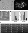
mbo1Δ cells are viable but exhibit polarity and microtubule and nuclear positioning defects. (A) DIC image of exponentially growing mbo1Δ cells. The numbers 1, 2, and 3 indicate three cells dividing at different cell lengths, and the asterisks indicate bent cells. The inset illustrates two cells with mispositioned septa indicated by arrows. Scale bar, 10 μm. (B) DAPI image of exponentially growing mbo1Δ cells. Asterisks indicate cells in which the nucleus is not positioned in the middle of the cell. (C) Live imaging of interphase mbo1Δ cells expressing GFP-α-tubulin. The arrow indicates a cell in which the microtubule bundle is curved around the tip of the cell. (D) Quantification of cell length at division for wild-type cells (dark bars) and mbo1Δ cells (light bars). (E) Quantification of the cytoplasmic microtubule bundles in wild-type cells (dark bars) and mbo1Δ cells (light bars). (F) Time-lapse confocal microscopy of live mbo1Δ cells expressing GFP-tubulin. The numbers on each panel correspond to the time (in min) elapsed since the capturing of the first frame.
Figure 6.
Figure 6.
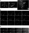
Localization of Mbo1p-GFP. (A) Cells expressing Mbo1p-GFP were subjected to live imaging using a GFP filter set. The localization of the fusion protein to SPBs juxtaposed to nuclei and to that of the medial EMTOC is indicated by arrowheads. Scale bar, 10 μm. (B) Time-lapse live imaging of cells expressing Mbo1p-GFP released from a cdc25-22 block to follow synchronous mitosis. Arrows indicate SPB localization, and asterisks indicate the medial ring structure. The numbers indicate the time (in min) elapsed from the capturing of the first frame. (C) 3D-deconvolution image of the medial ring structure that Mbo1p-GFP decorates. The arrowheads indicate the circumferential Mbo1p-GFP ring (D) Time-lapse live imaging of cells expressing Mbo1p-GFP released from a cdc25-22 block to follow synchronous mitosis. The arrow indicates the medial ring structure, and the numbers in each panel indicates the time (in min) elapsed since the capturing of the first frame.
Figure 8.
Figure 8.
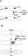
Models for Gfh1p and Mbo1p functions. (A) Gfh1p might play a specialized role as part of a γ-TuC cap structure at the cytoplasmic face of the SPB. This aids proper anchoring of astral microtubules to the SPB (top cell). In the absence of Gfh1p function (bottom cell), the cap would be altered resulting in the detachment of astral microtubules from the SPB. (B) During mitosis, Mbo1p localizes to a discontinuous ring at the medial region (top cell) and recruits other members of the γ-TuC (middle cell). The assembled complex might help facilitate the coalescence of the ring to an EMTOC and nucleate the PAA (bottom cell).

Similar articles

Cited by

References

    1. Bahler, J., Wu, J.Q., Longtine, M.S., Shah, N.G., McKenzie, A., 3rd, Steever, A.B., Wach, A., Philippsen, P., and Pringle, J.R. (1998). Heterologous modules for efficient and versatile PCR-based gene targeting in Schizosaccharomyces pombe. Yeast 14, 943–951. - PubMed
    1. Balasubramanian, M.K., McCollum, D., Chang, L., Wong, K.C.Y., Naqvi, N.I., He, X., Sazer, S., and Gould, K.L. (1998). Isolation and characterization of new fission yeast cytokinesis mutants. Genetics 149, 1265–1275. - PMC - PubMed
    1. Basi, G., Schmid, E., and Maundrell, K. (1993). TATA box mutations in the Schizosaccharomyces pombe nmt1 promoter affect transcription efficiency but not the transcription start point or thiamine repressibility. Gene 123, 131–136. - PubMed
    1. Chang, L., and Gould, K.L. (2000). Sid4p is required to localize components of the septation initiation pathway to the spindle pole body in fission yeast. Proc. Natl. Acad. Sci. USA 97, 5249–5254. - PMC - PubMed
    1. Fava, F., Raynaud-Messina, B., Leung-Tack, J., Mazzolini, L., Li, M., Guillemot, J.C., Cachot, D., Tollon, Y., Ferrara, P., and Wright, M. (1999). Human 76p: a new member of the gamma-tubulin-associated protein family. J. Cell Biol. 147, 857–868. - PMC - PubMed

Publication types

MeSH terms

LinkOut - more resources