. 2004 May;15(5):2176-88.
doi: 10.1091/mbc.e03-07-0503. Epub 2004 Mar 5.

Tyrosine phosphorylation of Sprouty proteins regulates their ability to inhibit growth factor signaling: a dual feedback loop

Affiliations

Tyrosine phosphorylation of Sprouty proteins regulates their ability to inhibit growth factor signaling: a dual feedback loop

Jacqueline M Mason et al. Mol Biol Cell. 2004 May.

Retraction in

  • Retraction.
    [No authors listed] [No authors listed] Mol Biol Cell. 2022 Jul 1;33(8):re3. doi: 10.1091/mbc.E03-07-0503-corr. Mol Biol Cell. 2022. PMID: 35736612 Free PMC article. No abstract available.

Abstract

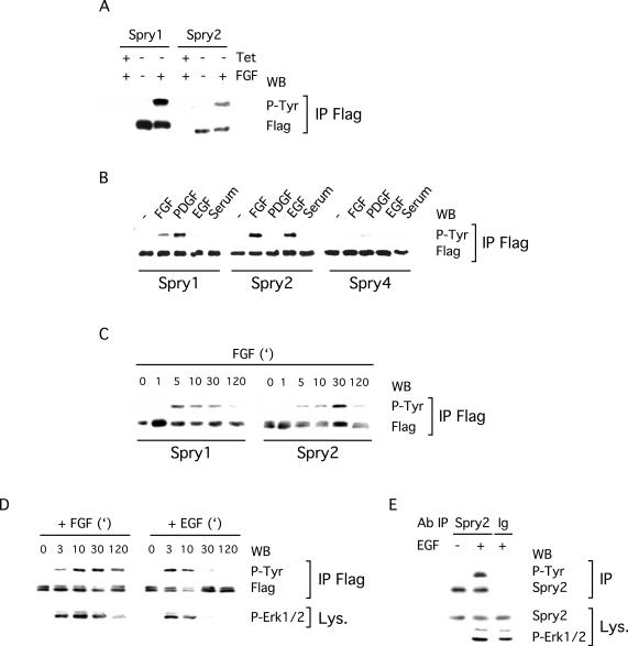
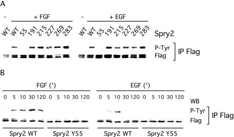
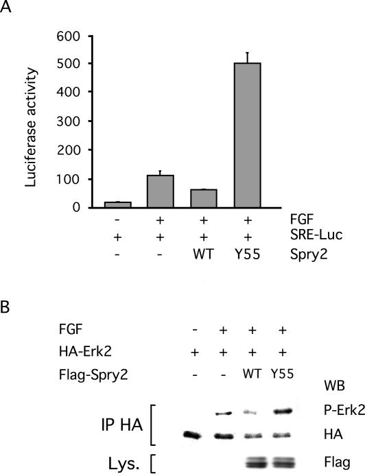
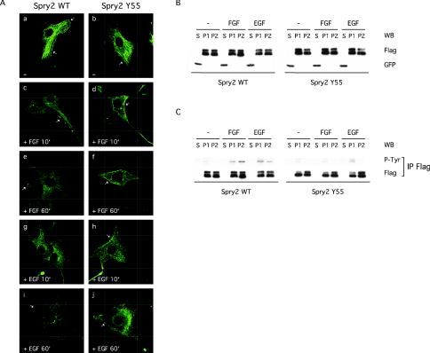
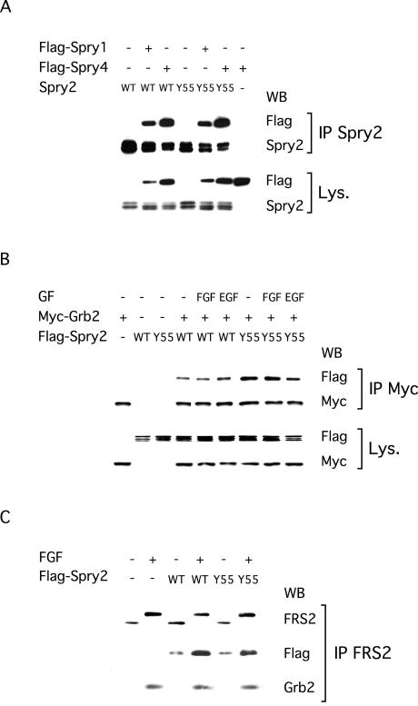
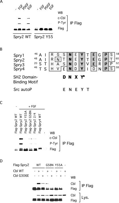
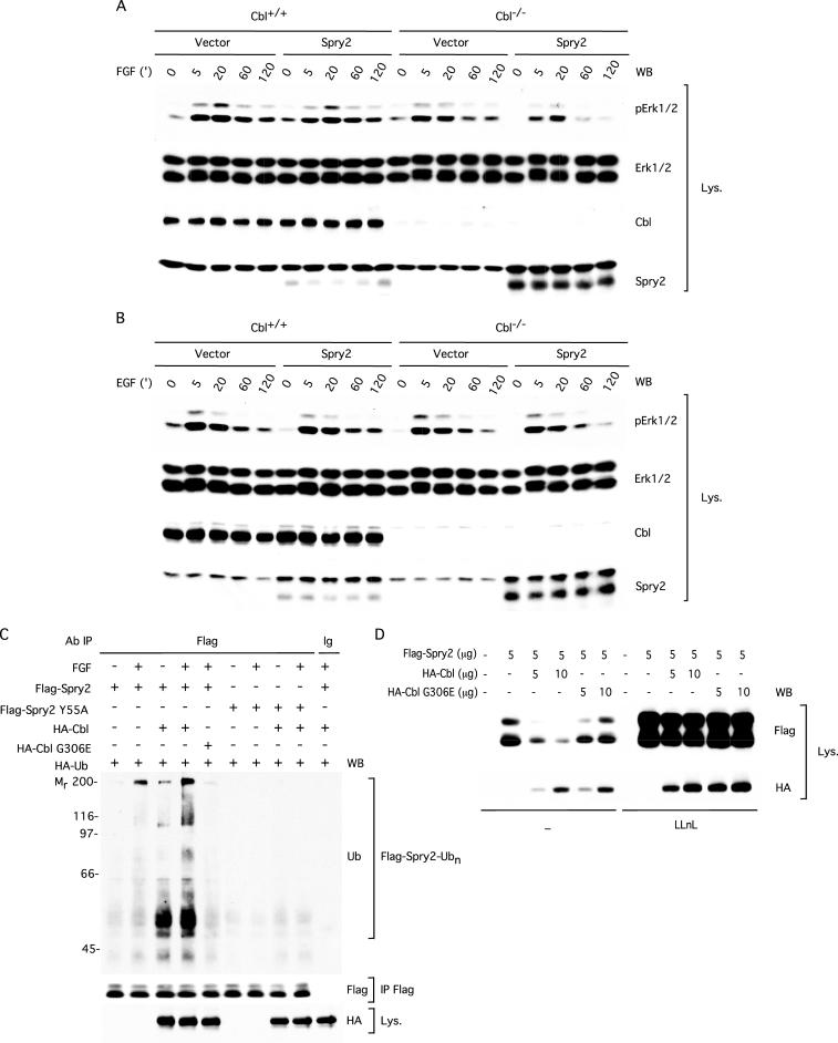
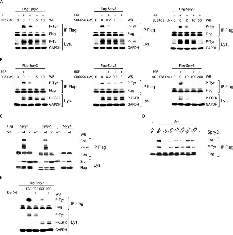
Sprouty proteins are recently identified receptor tyrosine kinase (RTK) inhibitors potentially involved in many developmental processes. Here, we report that Sprouty proteins become tyrosine phosphorylated after growth factor treatment. We identified Tyr55 as a key residue for Sprouty2 phosphorylation and showed that phosphorylation was required for Sprouty2 to inhibit RTK signaling, because a mutant Sprouty2 lacking Tyr55 augmented signaling. We found that tyrosine phosphorylation of Sprouty2 affected neither its subcellular localization nor its interaction with Grb2, FRS2/SNT, or other Sprouty proteins. In contrast, Sprouty2 tyrosine phosphorylation was necessary for its binding to the Src homology 2-like domain of c-Cbl after fibroblast growth factor (FGF) stimulation. To determine whether c-Cbl was required for Sprouty2-dependent cellular events, Sprouty2 was introduced into c-Cbl-wild-type and -null fibroblasts. Sprouty2 efficiently inhibited FGF-induced phosphorylation of extracellular signal-regulated kinase 1/2 in c-Cbl-null fibroblasts, thus indicating that the FGF-dependent binding of c-Cbl to Sprouty2 was dispensable for its inhibitory activity. However, c-Cbl mediates polyubiquitylation/proteasomal degradation of Sprouty2 in response to FGF. Last, using Src-family pharmacological inhibitors and dominant-negative Src, we showed that a Src-like kinase was required for tyrosine phosphorylation of Sprouty2 by growth factors. Thus, these data highlight a novel negative and positive regulatory loop that allows for the controlled, homeostatic inhibition of RTK signaling.


Figures

Figure 1.
Figure 1.
Spry proteins are tyrosine phosphorylated upon growth factor treatment. (A) Spry1 or Spry2 NIH3T3 Tet-repressible cells were serum starved in the presence (+Tet) or absence (–Tet) of tetracycline and stimulated for 5 min with FGF (40 ng/ml) before lysis. Proteins were incubated with a FLAG antibody, and the immunoprecipitates (IP FLAG) were analyzed by immunoblotting by using an antibody directed against phosphotyrosine (P-Tyr) and the FLAG antibody. (B–D) NIH3T3 cells, transfected with FLAG-tagged Spry expression vectors, were serum starved and treated with the indicated growth factor (40 ng/ml) before lysis. Phosphorylation and expression of Spry proteins were detected as described above. (B) Spry1 and Spry2 but not Spry4 are tyrosine phosphorylated after 10 min of growth factor treatment. (C) Time course of FGF treatment showing that tyrosine phosphorylation of Spry1 and Spry2 exhibit different kinetics. (D) Time course indicating that tyrosine phosphorylation of Spry2 by FGF and EGF exhibits different kinetics. Erk1/2 activation was detected in lysates (Lys.) by using an antibody directed against phosphorylated Erk1/2 (P-Erk1/2). (E) C2C12 cells were serum starved and stimulated for 5 min with EGF (40 ng/ml) before lysis. Proteins were incubated with a Spry2 antibody or rabbit immunoglobulins as a negative control. The immunoprecipitates were analyzed by immunoblotting with an antibody directed against phosphotyrosine (P-Tyr) and the Spry2 antibody. Expression of endogenous Spry2 in the lysates was detected with the same Spry2 antibody, and stimulation was confirmed with a phospho-Erk1/2 antibody. Each experiment was repeated at least twice with similar results obtained.
Figure 2.
Figure 2.
Tyrosine 55 is crucial for tyrosine phosphorylation of Spry2 upon growth factor treatment. Transfected NIH3T3 cells were serum starved, treated for 10 min with FGF or EGF (40 ng/ml), and lysed. The proteins were incubated with a FLAG antibody, and the immunoprecipitates (IP FLAG) were analyzed by immunoblotting by using antibodies directed against phosphotyrosine (P-Tyr) and the FLAG tag. (A) With the exception of Spry2 Y55A, all Spry2 mutants analyzed were tyrosine phosphorylated to a similar extent as wild-type Spry2 (WT) upon FGF or EGF treatment. (B) Time course of growth factor treatment indicating that tyrosine phosphorylation of Spry2 Y55A was not delayed but dramatically reduced. Both experiments were repeated twice with similar results observed.
Figure 3.
Figure 3.
Tyrosine phosphorylation is required for Spry2 activity. (A) NIH3T3 cells were cotransfected with a SRE-Luciferase reporter gene (150 ng) along with an empty expression vector (–, 1 μg), a wild-type Spry2 expression vector (WT, 1 μg), or a Spry2 Y55A mutant expression vector (Y55A, 1 μg) for 48 h. After serum starvation, the cells were stimulated with FGF (20 ng/ml) for 4 h before lysis and Luciferase assay. The values correspond to the average Luciferase units (RLUs) derived from triplicates of a representative experiment. Transfection efficiency was monitored with a Renilla control plasmid and found to be comparable in all samples. (B) NIH3T3 cells were cotransfected with a HA-tagged Erk2 expression vector along with an empty expression vector (–), a wild-type Spry2 expression vector (WT), or a Spry2 Y55A mutant expression vector (Y55) for 48 h. After serum starvation, the cells were left unstimulated (–) or treated for 2 h with 40 ng/ml FGF (+). Cell lysates were incubated with an antibody directed against the HA tag, and immunoprecipitates (IP HA) were analyzed by immunoblotting sequentially with an antibody directed against phosphorylated Erk1/2 (P-Erk2) and an antibody directed against HA. Expression of Spry2 Y55A was detected in the lysates (Lys.) by using an antibody directed against the FLAG tag.
Figure 4.
Figure 4.
Tyrosine phosphorylation does not affect the subcellular localization of Spry2. (A) Transfected NIH3T3 cells on coverslips were serum starved, left unstimulated (–), or treated with growth factor as indicated, and fixed. After permeabilization and blocking, Spry2 localization was observed by confocal microscopy by using a primary antibody directed against the FLAG tag and a fluorescein isothiocyanate-conjugated secondary antibody. For each experimental condition, a representative cell is shown. Arrows: plasma membrane. (B) NIH3T3 cells transfected with GFP and FLAG-tagged wild-type Spry2 or Spry2 Y55A were serum starved and stimulated for 10 min with the indicated growth factor (40 ng/ml). Lysates were submitted to subcellular fractionation (S, soluble fraction; P1, particulate fraction, containing endosomes, peroxisomes, and mitochondria membranes; P2, particulate fraction, containing endoplasmic reticulum, Golgi, and plasma membranes), and 1.5% of each fraction (S, 20 μg; P1, 4 μg; and P2, 16 μg) were analyzed by immunoblotting by using the FLAG antibody to detect Spry2 and an antibody directed against the GFP to check the fractionation process. (C) Seventy-five percent of each fraction (S, 880 μg; P1, 180 μg; and P2, 700 μg) were incubated with an antibody directed against the FLAG tag, and immunoprecipitates (IP FLAG) were analyzed by immunoblotting sequentially with an antibody directed against phosphorylated tyrosine (P-Tyr) and an antibody directed against the FLAG tag.
Figure 5.
Figure 5.
Tyrosine phosphorylation does not affect Spry2 interaction with Grb2, FRS2/SNT, or other Spry proteins. After serum starvation, transfected NIH3T3 cells were left unstimulated (–) or treated for 10 min with the indicated growth factor (40 ng/ml). (A) Cells were transfected with plasmids expressing FLAG-tagged Spry1 or Spry4 and nontagged wild-type Spry2 or Spry2 Y55A. Cell extracts were incubated with an antibody specific to Spry2, and immunoprecipitates (IP Spry2) were analyzed by immunoblotting by using an antibody directed against the FLAG tag to visualize Spry1 or Spry4, and an antibody against Spry2 to verify the immunoprecipitation. Expression of all proteins was confirmed in the lysates (Lys.) by using the same antibodies. (B) Cell lysates were incubated with an antibody directed against the Myc tag and immunoprecipitates (IP Myc) were analyzed by immunoblotting with an antibody directed against the Myc tag to visualize Grb2 and an antibody directed against the FLAG tag to visualize Spry2. Expression of both proteins in the lysates (Lys.) was confirmed with the same antibodies. (C) Cell extracts were incubated with an antibody directed against FRS2/SNT and immunoprecipitates (IP FRS2) were analyzed by immunoblotting with antibodies directed against FRS2, the FLAG tag and Grb2. FGF stimulation caused a mobility shift in FRS2 (serine phosphorylation), induced coimmunoprecipitation of endogenous Grb2 with endogenous FRS2, and increased coimmunoprecipitation of overexpressed wild-type Spry2 and Spry2 Y55A with endogenous FRS2. All experiments were performed at least twice with the same results obtained.
Figure 6.
Figure 6.
Tyrosine phosphorylation regulates Spry2 interaction with c-Cbl. After serum starvation, transfected NIH3T3 cells were left unstimulated (–) or treated for 10 min with the indicated growth factor (40 ng/ml). (A) Cell lysates were incubated with an antibody directed against the FLAG tag, and immunoprecipitates (IP FLAG) were analyzed by immunoblotting with an antibody directed against c-Cbl (endogenous), an antibody specific to phosphorylated tyrosine (P-Tyr), and an antibody against the FLAG tag. (B) Amino acid sequence alignment of the N-terminal regions containing the crucial tyrosines of the various mouse Spry proteins. Identical residues are indicated in bold and boxed. Spry1 and Spry2 share a stretch of seven identical residues, which exhibit some similarities to the c-Cbl SH2 domain-binding motif (SH2 domain-binding motif) and the c-Src autophosphorylation site (Src autoP). (C) Cell extracts were treated as in A, and the immunoprecipitates (IP FLAG) were analyzed by immunoblotting with an antibody directed against c-Cbl (endogenous), an antibody specific to phosphorylated tyrosine (P-Tyr), and an antibody against the FLAG tag. (D) Cell extracts were treated as in A, and the immunoprecipitates (IP FLAG) were analyzed by immunoblotting with an antibody against c-Cbl (a short exposure allowing only detection of transfected c-Cbl is shown) and an antibody against the FLAG tag. Expression of transfected wild-type and mutant c-Cbl was confirmed in the lysates with an antibody against c-Cbl and a short exposure of the film. All experiments were repeated at least twice with similar results obtained.
Figure 7.
Figure 7.
c-Cbl is not required for Spry2 inhibition of growth factor-dependent Erk1/2 phosphorylation. (A) c-Cbl+/+ and c-Cbl–/– MEF cells were infected with either the pMSCV-MIGR1 retroviral vector (vector) or a vector harboring FLAG-tagged wild-type Spry2 (Spry2) and sorted for GFP-positive cells. These cells were then serum starved for 18 h followed by FGF stimulation (20 ng/ml) for the indicated amount of time. Lysates (50 μg) were analyzed by immunoblotting sequentially with an antibody against phosphorylated Erk1/2 (P-Erk1/2), an antibody against total Erk1/2, an antibody against c-Cbl, and an antibody against Spry2. In addition to Spry2, the Spry2 antibody cross-reacted with a slower-migrating, nonspecific band. B as in A but with EGF stimulation (20 ng/ml) for the indicated amount of time. (C) Flg22 cells, transfected with plasmids expressing HA-tagged ubiquitin (2 μg), FLAG-tagged wild-type Spry2 (5 μg) or Spry2 Y55A (5 μg), and HA-tagged wild-type c-Cbl (2 μg) or c-Cbl G306E (2 μg) were serum starved for 12 h in the presence of LLnL (50 μM), stimulated with FGF (10 min, 40 ng/ml), and lysed in radioimmunoprecipitation assay buffer. Anti-FLAG immunoprecipitates (IP FLAG) from 1-mg aliquots of lysate were analyzed by sequentially immunoblotting with an antibody against ubiquitin (Ub) and an antibody against the FLAG tag. Equal aliquots (50 μg) of cell lysates were immunoblotted with an antibody against HA. (D) NIH3T3 cells, transfected with plasmids expressing FLAG-tagged Spry2 and HA-tagged wild-type c-Cbl or c-Cbl G306E, were left untreated (–) or treated for 12 h with LLnL (50 μM). Lysates (50 μg) were analyzed by immunoblotting sequentially with an antibody against the FLAG tag and an antibody against the HA tag. All experiments were repeated three times with similar results obtained.
Figure 8.
Figure 8.
Tyrosine phosphorylation of Spry2 depends on a Src-like kinase activity. (A) NIH3T3 cells, transfected with FLAG-tagged Spry2, were serum starved for 12 h. Before FGF stimulation (10 min, 40 ng/ml), the cells were left untreated (0) or treated for 2 h with the indicated amount of PP2, SU6656, or SU5402. Lysates were incubated with the FLAG antibody, and immunoprecipitates (IP FLAG) were analyzed by immunoblotting sequentially with an antibody specific to phosphorylated tyrosine (P-Tyr) and an antibody against the FLAG tag. Equal aliquots (50 μg) of cell lysates were immunoblotted sequentially with an antibody against phosphotyrosine to detect the activated FGFR (p-Tyr) and an antibody against GAPDH. (B) NIH3T3 cells, transfected with FLAG-tagged Spry2, were serum starved for 12 h. Before EGF stimulation (10 min, 40 ng/ml), the cells were left untreated (0) or treated for 2 h with the indicated amount of PP2, SU6656, or AG1478. Lysates were incubated with the FLAG antibody and immunoprecipitates (IP FLAG) were analyzed by immunoblotting sequentially with an antibody specific to phosphorylated tyrosine (P-Tyr) and an antibody against the FLAG tag. Equal aliquots (50 μg) of cell lysates were immunoblotted sequentially with an antibody against the activated EGFR (P-EGFR) and an antibody against GAPDH. (C and D) NIH3T3 cells, transfected with the indicated plasmids, were serum starved and lysed. The cell lysates were incubated with the FLAG antibody, and immunoprecipitates (IP FLAG) were analyzed by immunoblotting with an antibody specific to phosphorylated tyrosine (P-Tyr), an antibody against c-Cbl (endogenous), and an antibody against the FLAG tag. Src k– is a mutated form of c-Src devoid of kinase activity (Src K297R). (E) NIH3T3 cells, transfected with the indicated plasmids and serum starved, were left untreated (–) or stimulated for 10 min with FGF (40 ng/ml) or EGF (40 ng/ml) before lysis. Immunoprecipitation and immunoblotting were performed as in A and B. Src DN is a dominant-negative form of c-Src (Src K296R/Y528F).

Similar articles

Cited by

References

    1. Blake, R.A., Broome, M.A., Liu, X., Wu, J., Gishizky, M., Sun, L., and Courtneidge, S.A. (2000). SU6656, a selective src family kinase inhibitor, used to probe growth factor signaling. Mol. Cell. Biol. 20, 9018–9027. - PMC - PubMed
    1. Blom, N., Gammeltoft, S., and Brunak, S. (1999). Sequence and structure-based prediction of eukaryotic protein phosphorylation sites. J. Mol. Biol. 294, 1351–1362. - PubMed
    1. Brown, M.T., and Cooper, J.A. (1996). Regulation, substrates and functions of src. Biochim. Biophys. Acta 1287, 121–149. - PubMed
    1. Campbell, S.L., Khosravi-Far, R., Rossman, K.L., Clark, G.J., and Der, C.J. (1998). Increasing complexity of Ras signaling. Oncogene 17, 1395–1413. - PubMed
    1. Casci, T., Vinos, J., and Freeman, M. (1999). Sprouty, an intracellular inhibitor of Ras signaling. Cell 96, 655–665. - PubMed

Publication types

MeSH terms

Substances