. 2004 Mar 5;32(4):1584-90.
doi: 10.1093/nar/gkh335. Print 2004.

DNA dynamically directs its own transcription initiation

Affiliations

DNA dynamically directs its own transcription initiation

Chu H Choi et al. Nucleic Acids Res. .

Abstract

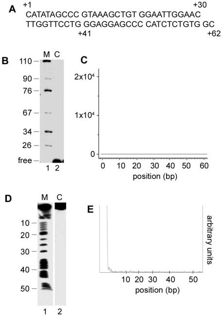
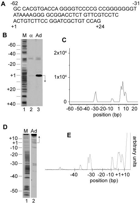
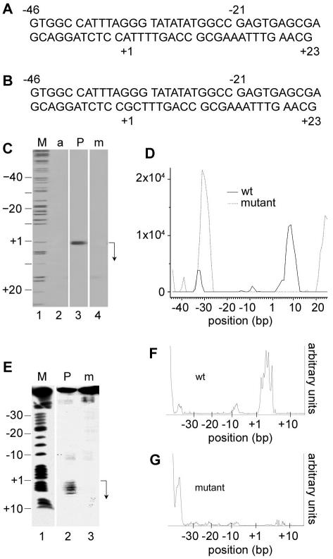
It has long been known that double-stranded DNA is subject to temporary, localized openings of its two strands. Particular regions along a DNA polymer are destabilized structurally by available thermal energy in the system. The localized sequence of DNA determines the physical properties of a stretch of DNA, and that in turn determines the opening profile of that DNA fragment. We show that the Peyrard-Bishop nonlinear dynamical model of DNA, which has been used to simulate denaturation of short DNA fragments, gives an accurate representation of the instability profile of a defined sequence of DNA, as verified using S1 nuclease cleavage assays. By comparing results for a non-promoter DNA fragment, the adenovirus major late promoter, the adeno-associated viral P5 promoter and a known P5 mutant promoter that is inactive for transcription, we show that the predicted openings correlate almost exactly with the promoter transcriptional start sites and major regulatory sites. Physicists have speculated that localized melting of DNA might play a role in gene transcription and other processes. Our data link sequence-dependent opening behavior in DNA to transcriptional activity for the first time.


Figures

Figure 1
Figure 1
Analysis of non-promoter control DNA fragment. (A) Upper strand sequence of the 62 bp non-promoter DNA. (B) Run-off transcription assay on a 105 bp linear fragment containing the non-promoter sequence in nuclear extract. The migration position of marker in base pairs and the position of free [32P]UTP is shown to the left of the panel. Lane 1, DNA marker (M); lane 2, reaction products (C). (C) PB simulation of the non-promoter sequence, plotting simulated instances of 2.1 Å-separated openings of 10 bp or more versus base position in the sequence. (D) S1 nuclease cleavage assay of the non-promoter DNA fragment. The corresponding sequence position is indicated to the left of the panel. Lane 1, lower strand-labeled P5 promoter GA sequencing reaction was used as a marker (M); lane 2, non-promoter DNA S1 digestion reaction (C). (E) Cleavage density profile of the non-promoter DNA sequence in the S1 nuclease experiment.
Figure 2
Figure 2
Analysis of adenovirus major late promoter. (A) Upper strand sequence of the 86 bp AdMLP. (B) Run-off transcription assay in nuclear extract using a 120 bp linear template containing the 86 bp fragment of the AdMLP. The arrow on the right indicates the TSS and the direction of transcription. The corresponding sequence position is indicated to the left of the marker. Lane 1, GA DNA sequencing reaction was used as a marker (M); lane 2, transcription with α-amanitin to specifically arrest polymerase II transcription (α); lane 3, RNA transcription product (Ad). (C) PB simulation of the AdMLP sequence, plotting simulated instances of 2.1 Å-separated openings of 10 bp or more versus base position in the sequence. (D) S1 nuclease cleavage assay of the AdMLP. The corresponding sequence position is indicated to the left of the marker. Lane 1, upper strand-labeled AdMLP fragment sequencing reaction was used as a marker (M); lane 2, S1 digestion of the 32P-labeled AdMLP fragment (Ad). (E) Cleavage density profile of the AdMLP fragment in the S1 nuclease experiment.
Figure 3
Figure 3
Analysis of AAV P5 promoter and P5 mutant promoter. (A) Upper strand sequence of the 69 bp P5 core promoter. (B) Upper strand sequence of the 69 bp mutant P5 promoter. (C) Transcription assay on a 120 bp fragment containing the P5 promoter and P5 mutant fragment in nuclear extract. The arrow on the right indicates the TSS and the direction of transcription. The corresponding sequence position is indicated to the left of the marker. Lane 1, GA DNA sequencing reaction was used as a marker (M); lane 2, transcription from the P5 promoter with α-amanitin (a); lane 3, RNA transcription products with P5 promoter (P); lane 4, RNA transcription products from the P5 mutant template (m). (D) PB simulation of the AAV P5 sequence, plotting simulated instances of 2.1 Å-separated openings of 10 bp or more versus base position in the sequence. The solid line represents the results of the wild-type (wt) P5 promoter, and the broken line represents results with the mutant P5 promoter sequence. (E) S1 nuclease cleavage of the P5 promoter and the P5 mutant promoter. The corresponding sequence position is indicated to the left of the panel. Lane 1, lower strand-labeled P5 promoter GA sequencing reaction (M); lane 2, AAV P5 promoter S1 cleavage reaction (P); lane 3, AAV P5 mutant promoter cleavage reaction (m). (F) Cleavage density profile of the wild-type (wt) AAV P5 promoter DNA in the S1 nuclease experiment. (G) Cleavage density profile of the mutant AAV P5 promoter DNA in the S1 nuclease experiment.

References

    1. Benham C.J. (1992) Energetics of the strand separation transition in superhelical DNA. J. Mol. Biol., 225, 835–847. - PubMed
    1. Benham C.J. (1993) Sites of predicted stress-induced DNA duplex destabilization occur preferentially at regulatory loci. Proc. Natl Acad. Sci. USA, 90, 2999–3003. - PMC - PubMed
    1. Benham C.J. (1996) Duplex destabilization in superhelical DNA is predicted to occur at specific transcriptional regulatory regions. J. Mol. Biol., 255, 425–434. - PubMed
    1. Breslauer K.J., Frank,R., Blocker,H. and Marky,L.A. (1986) Predicting DNA duplex stability from the base sequence. Proc. Natl Acad. Sci. USA, 83, 3746–3750. - PMC - PubMed
    1. Packer M.J., Dauncey,M.P. and Hunter,C.A. (2000) Sequence-dependent DNA structure: tetranucleotide conformational maps. J. Mol. Biol., 295, 85–103. - PubMed

Publication types