. 2004 Mar 16;101(11):3880-5.
doi: 10.1073/pnas.0400339101. Epub 2004 Mar 5.

Interleukin 2 plays a central role in Th2 differentiation

Affiliations

Interleukin 2 plays a central role in Th2 differentiation

Javier Cote-Sierra et al. Proc Natl Acad Sci U S A. .

Abstract

Differentiation of naïve CD4 T cells into T helper (Th) 2 cells requires signaling through the T cell receptor and an appropriate cytokine environment. IL-4 is critical for such Th2 differentiation. We show that IL-2 plays a central role in this process. The effect of IL-2 on Th2 generation does not depend on its cell growth or survival effects. Stat5a(-/-) cells show diminished differentiation to IL-4 production, and forced expression of a constitutively active form of Stat5a replaces the need for IL-2. In vivo IL-2 neutralization inhibits IL-4 production in two models. Studies of restriction enzyme accessibility and binding of Stat5 to chromatin indicate that IL-2 mediates its effect by stabilizing the accessibility of the Il4 gene. Thus, IL-2 plays a critical role in the polarization of naive CD4 T cells to the Th2 phenotype.


Figures
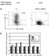
Fig. 1.
Fig. 1.
Effect of blocking IL-2 on cytokine production. (a) 5C.C7 CD4 T cells were cultured for 86 h with APC and 0.5 μM PCC peptide under Th2 conditions with anti-CD28. At the end of the priming culture, viable cell numbers, cell division history, and intracellular IL-4 expression were analyzed. (b) DO11.10 cells were cultured under Th2 conditions with immobilized anti-CD3 plus anti-CD28. mRNA levels for various cytokines were measured at 84 h of culture by real-time PCR analysis. GM-CSF, granulocyte/macrophage colony-stimulating factor.
Fig. 2.
Fig. 2.
Priming for IL-4 production in IL-2-/- CD4 T cells. IL-2-/- and IL-2+/+ 5C.C7 CD4 T cells were cultured for 86 h with APC and 0.5 μM PCC peptide under Th2 or Th1 conditions plus anti-CD28 with or without 100 units/ml human IL-2 (hIL-2). At the end of the priming culture, viable cell numbers, cell division history, and intracellular IL-4 expression were analyzed.
Fig. 3.
Fig. 3.
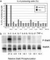
Effect of different cytokines on IL-4 production and Stat5 phosphorylation. (a) 5C.C7 CD4 T cells were cultured with APC and 0.2 μM PCC peptide under Th2 conditions for 94 h, with human (hIL-2) (20 units/ml) or with anti-mouse IL-2 (anti-mIL-2) without or with other cytokines. Intracellular IL-4 expression was analyzed after restimulation. (b) DO11.10 CD4 T cells were cultured under Th2 conditions with immobilized anti-CD3 plus anti-CD28 for 48 h. Cells were washed, cultured overnight in 0.5% FCS containing RPMI medium 1640, washed again, and cultured in serum-free medium for 2 h before cytokine stimulation for 15 min. Phospho-Stat5 and Stat5 were analyzed by immunoblotting. TNF, tumor necrosis factor; TGF, transforming growth factor.
Fig. 4.
Fig. 4.
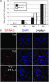
Effect of IL-2 on GATA-3 induction. (a) DO11.10 CD4 T cells were cultured under Th2 conditions with immobilized anti-CD3 plus anti-CD28. GATA-3 mRNA levels were measured by real-time PCR at 0, 12, 48, and 84 h in the presence or absence of anti-IL-2. (b) DO11.10 CD4 T cells were cultured under Th1 and Th2 conditions. Cells were fixed and permeabilized at 72 h, stained with a monoclonal anti-GATA-3, and analyzed by confocal microscopy. DAPI, 4′,6-diamidino-2-phenylindole.
Fig. 5.
Fig. 5.
Il4 gene accessibility. (a) 5C.C7 CD4 T cells stimulated under Th2 conditions with APC and 0.2 μM PCC peptide. Either IL-2 or anti-IL-2, anti-IL-2Rα, and anti-IL-2Rβ (anti-IL-2) were added. REA at HSII was analyzed by PCR at 86 h. (b) DO11.10 CD4 T cells stimulated with immobilized anti-CD3 plus anti-CD28 under Th2 conditions with either IL-2 or anti IL-2 for 48 h. Cells were washed and cultured under Th2-priming conditions for another 48 h, in the presence of either IL-2 or anti-IL-2, anti- IL-2Rα, and anti-IL-2Rβ. REA was assessed in HSII and HSIII at the outset of culture and at 48 and 96 h of Th2 priming. IL-4-producing capacity was assessed after restimulation. (c) DO11.10 CD4 T cells primed for 48 h under Th2 conditions without exogenous IL-2. Cells were cultured under Th2 conditions for the next 48 h in the presence of IL-2 (IL-4/2), IL-2 plus cyclosporin A (50 ng/ml) (IL-4/2 CsA) or anti-IL-2, anti-IL-2Rα, and anti-IL-2Rβ (IL-4/a2). REA at HS II and IL-4-producing capacity were analyzed at 92 h. (d) DO11.10 cells were cultured under Th2 conditions with immobilized anti-CD3 plus anti-CD28 for 86 h. IL-4-producing cells, identified by cytokine capture, were purified by cell sorting. IL-4+ cells were stimulated for 3 days under Th2 conditions in the presence of IL-2 (IL-4/IL-2) or anti-IL-2, anti-IL-2Rα, and anti-IL-2Rβ (IL-4/a2). REA at HSII and HSIII and IL-4-producing capacity were analyzed at 72 h.
Fig. 6.
Fig. 6.
Stat5a and IL-4 production. (a) C57BL/6 Stat5a-/- and WT CD4 T cells primed for 86 h with soluble anti-CD3 (0.5 μg/ml) and anti-CD28 (3 μg/ml) under Th-neutral conditions with either anti-IL-2 and anti-IL-2Rα or with added IL-2. KO, knockout. (b and c) 5C.C7 CD4 T cells were activated for 40 h before retrovirus infection. Cells were infected with a GATA3- (b) or a STAT5A1*6- (c) encoding retrovirus and cultured for an additional 3 days under Th2 conditions with IL-2 or anti-IL-2, anti-IL-2Rα, and anti-IL-2Rβ. Intracellular IL-4 expression was analyzed after stimulation in CD4 T cells (a) or STAT5A1*6-, GATA-3-, or control retrovirus-infected cells (b and c). NGFR, nerve growth factor receptor. (d) 5C.C7 CD4 T cells were stimulated under Th1 or Th2 conditions with APC and 1 μM PCC peptide for three rounds. Each round consisted of 4 days priming and 3 days resting in IL-2. Chromatin immunoprecipitation (ChIP) assays were carried out by using anti-Stat5a and control sera. Amounts of immunoprecipitated DNA sequences from HSII, HSIII, and SOCS-3 promoter Stat-binding motif (S3PS) were assessed by real-time PCR. Relative enrichment of the sequences were normalized by using a 3′-UTR t-bet sequence, which does not have a Stat-binding site.
Fig. 7.
Fig. 7.
In vivo IL-2 neutralization. (a) BALB/c mice implanted with miniosmotic pumps containing 1 μg of ovalbumin in PBS received 5 × 10⁶ purified DO11.10 CD4 T cells. Mice were injected i.p. with anti-IL-2 (S4B6), isotype control, or PBS. Draining lymph node cells were restimulated with ovalbumin peptide and soluble anti-CD28. IL-4 expression, cell division, and number of KJ1-26+ cells were analyzed. Mean IL-4+ KJ1-26 cells in control Ig and anti-IL-2 groups were significantly different (P < 0.0002).

References

    1. Zheng, W. & Flavell, R. A. (1997) Cell 89, 587-596. - PubMed
    1. Ouyang, W., Ranganath, S. H., Weindel, K., Bhattacharya, D., Murphy, T. L., Sha, W. C. & Murphy, K. M. (1998) Immunity 9, 745-755. - PubMed
    1. Henkel, G., Weiss, D. L., McCoy, R., Deloughery, T., Tara, D. & Brown, M. A. (1992) J. Immunol. 149, 3239-3246. - PubMed
    1. Bird, J. J., Brown, D. R., Mullen, A. C., Moskowitz, N. H., Mahowald, M. A., Sider, J. R., Gajewski, T. F., Wang, C. R. & Reiner, S. L. (1998) Immunity 9, 229-237. - PubMed
    1. Takemoto, N., Koyano-Nakagawa, N., Yokota, T., Arai, N., Miyatake, S. & Arai, K. (1998) Int. Immunol. 10, 1981-1985. - PubMed