

Magnetosome vesicles are present before magnetite formation, and MamA is required for their activation
- PMID: 15004275
- PMCID: PMC374331
- DOI: 10.1073/pnas.0400391101
Magnetosome vesicles are present before magnetite formation, and MamA is required for their activation
Abstract
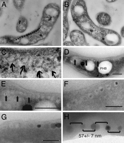
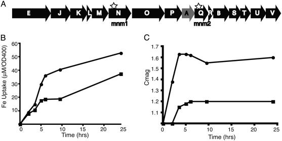
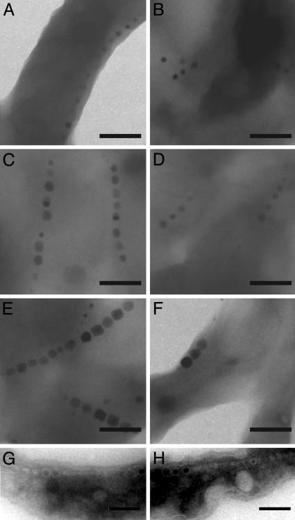
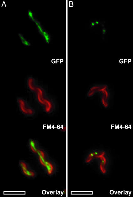
Bacterial magnetosomes are intracellular compartments that house highly ordered magnetite crystals. By using Magnetospirillum sp. AMB-1 as a model system, we show that magnetosome vesicles exist in the absence of magnetite, biomineralization of magnetite proceeds simultaneously in multiple vesicles, and biomineralization proceeds from the same location in each vesicle. The magnetosome-associated protein, MamA, is required for the formation of functional magnetosome vesicles and displays a dynamic subcellular localization throughout the growth cycle of magnetotactic bacteria. Together, these results suggest that the magnetosome precisely coordinates magnetite biomineralization and can serve as a model system for the study of organelle biogenesis in noneukaryotic cells.
Figures
Similar articles
-
Comparative Subcellular Localization Analysis of Magnetosome Proteins Reveals a Unique Localization Behavior of Mms6 Protein onto Magnetite Crystals.J Bacteriol. 2016 Sep 22;198(20):2794-802. doi: 10.1128/JB.00280-16. Print 2016 Oct 15. J Bacteriol. 2016. PMID: 27481925 Free PMC article.
-
The MagA protein of Magnetospirilla is not involved in bacterial magnetite biomineralization.J Bacteriol. 2012 Mar;194(5):1018-23. doi: 10.1128/JB.06356-11. Epub 2011 Dec 22. J Bacteriol. 2012. PMID: 22194451 Free PMC article.
-
The cation diffusion facilitator proteins MamB and MamM of Magnetospirillum gryphiswaldense have distinct and complex functions, and are involved in magnetite biomineralization and magnetosome membrane assembly.Mol Microbiol. 2011 Nov;82(4):818-35. doi: 10.1111/j.1365-2958.2011.07863.x. Epub 2011 Oct 18. Mol Microbiol. 2011. PMID: 22007638
-
Magnetosome biogenesis in magnetotactic bacteria.Nat Rev Microbiol. 2016 Sep 13;14(10):621-37. doi: 10.1038/nrmicro.2016.99. Nat Rev Microbiol. 2016. PMID: 27620945 Review.
-
Genetics and cell biology of magnetosome formation in magnetotactic bacteria.FEMS Microbiol Rev. 2008 Jul;32(4):654-72. doi: 10.1111/j.1574-6976.2008.00116.x. Epub 2008 Jun 2. FEMS Microbiol Rev. 2008. PMID: 18537832 Review.
Cited by
-
Comparative genome analysis of four magnetotactic bacteria reveals a complex set of group-specific genes implicated in magnetosome biomineralization and function.J Bacteriol. 2007 Jul;189(13):4899-910. doi: 10.1128/JB.00119-07. Epub 2007 Apr 20. J Bacteriol. 2007. PMID: 17449609 Free PMC article.
-
Magnetic Nanotweezers for Interrogating Biological Processes in Space and Time.Acc Chem Res. 2018 Apr 17;51(4):839-849. doi: 10.1021/acs.accounts.8b00004. Epub 2018 Mar 28. Acc Chem Res. 2018. PMID: 29589897 Free PMC article. Review.
-
Complete genome sequence of the chemolithoautotrophic marine magnetotactic coccus strain MC-1.Appl Environ Microbiol. 2009 Jul;75(14):4835-52. doi: 10.1128/AEM.02874-08. Epub 2009 May 22. Appl Environ Microbiol. 2009. PMID: 19465526 Free PMC article.
-
Nanotechnology for Targeted Detection and Removal of Bacteria: Opportunities and Challenges.Adv Sci (Weinh). 2021 Nov;8(21):e2100556. doi: 10.1002/advs.202100556. Epub 2021 Sep 23. Adv Sci (Weinh). 2021. PMID: 34558234 Free PMC article. Review.
-
Tethered Magnets Are the Key to Magnetotaxis: Direct Observations of Magnetospirillum magneticum AMB-1 Show that MamK Distributes Magnetosome Organelles Equally to Daughter Cells.mBio. 2017 Aug 8;8(4):e00679-17. doi: 10.1128/mBio.00679-17. mBio. 2017. PMID: 28790202 Free PMC article.
References
-
- Schuler, D. (1999) J. Mol. Microbiol. Biotechnol. 1, 79-86. - PubMed
-
- Kirschvink, J. L. & Hagadorn, J. W. (2000) in Biomineralization: From Biology to Biotechnology and Medical Application, ed. Baeuerlein, E. (Wiley, Weinheim, Germany), pp. 139-147.
-
- Mann, S., Sparks, N. H. & Board, R. G. (1990) Adv. Microb. Physiol. 31, 125-181. - PubMed
-
- Vali, H. & Kirschvink, J. L. (1990) Iron Biominerals (Plenum, New York).
Publication types
MeSH terms
Substances
Associated data
- Actions
LinkOut - more resources
Full Text Sources
Other Literature Sources
Medical
Molecular Biology Databases
