. 2004 Jan 27:5:3.
doi: 10.1186/1471-2202-5-3.

Pattern of distribution and cycling of SLOB, Slowpoke channel binding protein, in Drosophila

Affiliations

Pattern of distribution and cycling of SLOB, Slowpoke channel binding protein, in Drosophila

Angela M Jaramillo et al. BMC Neurosci. .

Abstract

Background: SLOB binds to and modulates the activity of the Drosophila Slowpoke (dSlo) calcium activated potassium channel. Recent microarray analyses demonstrated circadian cycling of slob mRNA.

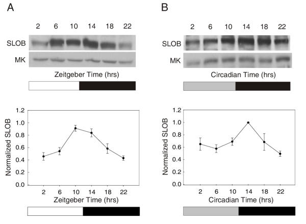
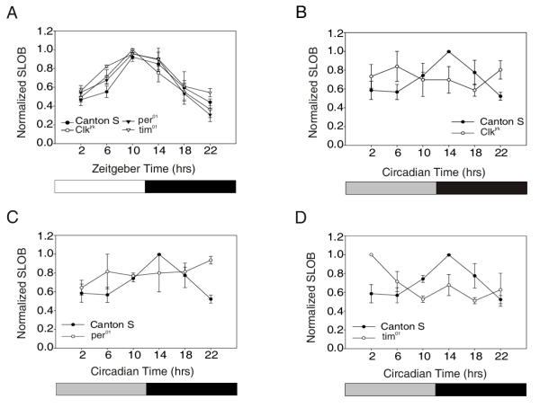
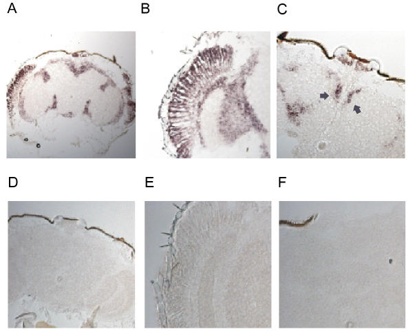
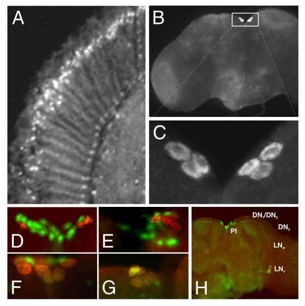
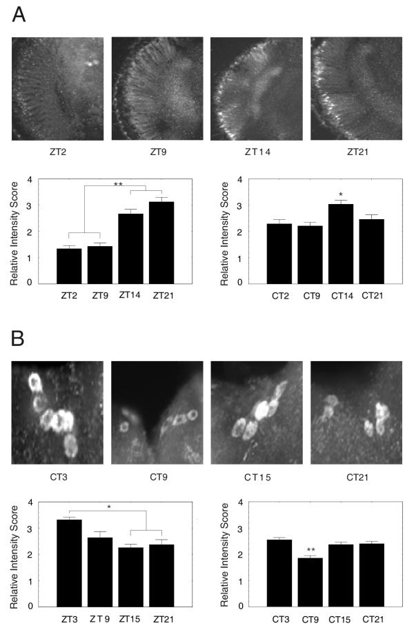
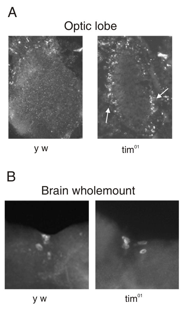
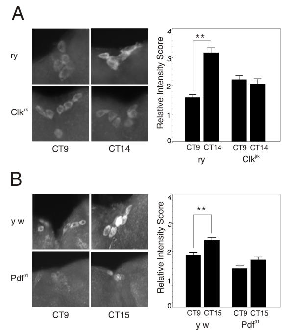
Results: We report the mRNA and protein expression pattern of slob in Drosophila heads. slob transcript is present in the photoreceptors, optic lobe, pars intercerebralis (PI) neurons and surrounding brain cortex. SLOB protein exhibits a similar distribution pattern, and we show that it cycles in Drosophila heads, in photoreceptor cells and in neurosecretory cells of the PI. The cycling of SLOB is altered in various clock gene mutants, and SLOB is expressed in ectopic locations in tim01 flies. We also demonstrate that SLOB no longer cycles in the PI neurons of Clkjrk flies, and that SLOB expression is reduced in the PI neurons of flies that lack pigment dispersing factor (PDF), a neuropeptide secreted by clock cells.

Conclusions: These data are consistent with the idea that SLOB may participate in one or more circadian pathways in Drosophila.


Figures

Figure 1
Figure 1
SLOB protein cycles in both LD and DD. (A) Canton S flies were entrained in LD for three days and collected at six time points over a 24 hour period. Fly head lysates were prepared and an anti-SLOB antibody was used to identify the 58 kDa band representing SLOB on the top western blot. The bottom blot was probed for MAPK, the protein level of which does not cycle [37] and was used as a loading control. SLOB oscillation has a trough at ZT 2 and a peak between ZT 10–14. The graph below represents the mean ± SEM for each time point from a minimum of three independent experiments. (B) Canton S flies were entrained in LD for three days and transferred to constant darkness. They were collected on the second day of DD at six time points, and SLOB cycling was determined as in (A).
Figure 2
Figure 2
SLOB cycling is altered under DD conditions in clock mutants. (A) Western blot analysis of SLOB cycling in Canton S, per01, tim01 and Clkjrk flies under LD conditions. Oscillations in all flies are in phase with one another. The graphs represent the mean ± SEM for each time point from a minimum of three experiments for each genotype. (B, C) Western blot analysis of Canton S, Clkjrk and per01 flies in DD conditions. There is no obvious cycling of SLOB in Clkjrk and per01 flies. (D) Western blot analysis of Canton S and tim01 flies in DD conditions. There is a shift in the phase of SLOB cycling in tim01 flies. The peak is no longer at CT 14 but at CT 2. The same Canton S data are illustrated in panels (B-D).
Figure 3
Figure 3
Distribution of slob mRNA. Frontal sections of Drosophila heads were probed with digoxigenin labeled RNA probes for slob. (A) Low magnification to survey entire head section. Antisense slob probes hybridized to the photoreceptors, optic lobe, PI neurons and the surrounding brain cortex. (B) High levels of expression detected in the photoreceptors, lamina and medulla. (C) slob message appears in the PI region. The arrows point to the dorsal medial PI neurons. (D-F) Digoxigenin labeled sense probes of slob reveal no significant labeling. The three panels correspond to the head sections of (A-C).
Figure 4
Figure 4
SLOB is expressed prominently in photoreceptor cells and PI neurons. (A) Head frontal section from y w (wild type) flies immunostained for SLOB protein. SLOB is expressed in the nuclei of the photoreceptor cells. (B) Brain wholemounts from y w flies immunostained for SLOB protein. SLOB is expressed prominently in the PI neurons of the protocerebrum (box), as well as elsewhere in the brain. (C) Enlargement of the boxed area in (B) to illustrate the cytoplasmic localization of SLOB in the PI neurons. Typically 6–8 PI neurons are SLOB positive. (D-G) GAL4 flies, specific for the three subsets of PI neurons, were crossed to UAS-GFP flies and immunostained for SLOB. (D) Mai 301 GAL4 is expressed in PI-1 neurons [22], shown by GFP expression. UAS-GFP tends to have a nuclear localization. SLOB positive PI neurons are immunostained with Texas Red, and SLOB has a cytoplasmic expression pattern. SLOB does not localize within PI-1 neurons. (E) Kurs 58 GAL4 is expressed in PI-2 neurons. SLOB is not found within the PI-2 neurons. (F) Mai 281 GAL4 is expressed in two of the three subsets of PI neurons (PI-2 and PI-3). SLOB is localized within some of these PI neurons and because SLOB was not found within PI-2 this suggests that the SLOB positive subset is PI-3. (G) Kurs 45 GAL4 is expressed only in PI-3 neurons. SLOB positive cells colocalize with the GFP expressing PI-3 cells. (H) Brain wholemount from y w flies highlighting components of the circadian system – the small ventral lateral neurons (LNv), the dorsal lateral neurons (LNd), the dorsal neurons (DN1–3) and the pars intercerebralis neurons (PI). The wholemount was immunostained for SLOB, PER and PDF. The red component is PER staining; the green staining in the PI neurons corresponds to SLOB, and the green staining elsewhere is PDF. Previous staining in Pdf01 flies reveals no PDF in the PI neurons (data not shown). PDF filled projections end close to, but appear to stop short of, the PI neurons. The projections of the DN (not visible) may synapse on the PI neurons.
Figure 5
Figure 5
SLOB cycles in the photoreceptor cell and the PI neurons. y w flies were entrained in LD for three days and collected at four time points over a 24 hour period. Some flies, after three days entrainment, were transferred to DD for two additional days. At least 5 heads were assayed per time point. (A) Head frontal sections were immunostained for SLOB. The bars depict mean ± SEM staining intensity scores (*p < 0.05, ** p < 0.001). The left bar graph is for LD and the right is for DD. SLOB cycling in the photoreceptors closely follows that of the western blots for both LD and DD. (B) Brain wholemounts from y w flies immunostained for SLOB. SLOB cycles in the PI neurons with an altered phase compared to the photoreceptors. The left bar graph is for LD and the right is for DD. Statistical analysis was done as in (A).
Figure 6
Figure 6
Elevated or ectopic expression of SLOB in tim01 flies. (A) Head sections from y w and tim01 flies immunostained for SLOB. There is greater expression of SLOB in the optic lobe in tim01 flies. The arrowheads point to the outer edges of the lamina. (B) Brain wholemounts from y w and tim01 flies immunostained for SLOB. There is no change in SLOB expression within the PI neurons of the tim01 flies.
Figure 7
Figure 7
SLOB expression is altered in Clkjrk and Pdf01 flies. (A) Brain wholemount from wild type rosy (ry) and Clkjrk flies. Two time points, taken on the second day of DD, show that SLOB does not cycle in the PI neurons of the Clkjrk flies. The accompanying bar graph illustrates relative intensity scores. Data were analyzed as described in Figure 5. (B) Brain wholemount from wild type y w and Pdf01 flies. Two time points, taken on the second day of DD, demonstrate the decrease in SLOB expression and dampened cycling in the PI neurons of Pdf01 flies. PDF appears to positively regulate SLOB protein. The bar graph represents relative intensity scores.
Figure 8
Figure 8
Overexpression of SLOB alters behavioral rhythms. Elavc155, GMR and Kurs 45 GAL4 flies were crossed to UAS-slob, UAS-mtslob and Df(1)w flies. Progeny were entrained for three days in LD cycles and then transferred to a locomotor monitoring device for 14 days in DD. (A) Actograms representing control, UAS-slob and UAS-mtslob lines crossed to elavc155. There is an apparent breakdown of rhythmicity after 7 days in flies overexpressing wild type and mutant SLOBs (middle and right panels). (B) Summary of behavioral data for elavc155 flies crossed to control and UAS lines. The FFT value is a measure of rhythmic strength and is plotted in the bar graph for the first and second week periods per fly line. Using Student's t test, all overexpressing SLOB lines exhibit significant breakdown of rhythms during the second seven days (*p < 0.05 and **p < 0.001, respectively). Controls show no significant breakdown except for one line, UAS-slob1/+. However, as demonstrated by the inserted western blot, this line expresses transgenic SLOB in the absence of a GAL4 driver, whereas the UAS-mtslob1/+ line does not. The blot was probed with an anti-HA antibody that detects only transgenic SLOB. Levels of MAPK were used as loading controls (data not shown). (C) Summary of behavioral data for GMR and Kurs 45 GAL4 flies crossed to various lines. No change in rhythmicity is evident when SLOB is overexpressed only in the Drosophila eye or PI neurons.

References

    1. Ceriani MF, Hogenesch JB, Yanovsky M, Panda S, Straume M, Kay SA. Genome-wide expression analysis in Drosophila reveals genes controlling circadian behavior. J Neurosci. 2002;22:9305–9319. - PMC - PubMed
    1. Claridge-Chang A, Wijnen H, Naef F, Boothroyd C, Rajewsky N, Young MW. Circadian regulation of gene expression systems in the Drosophila head. Neuron. 2001;32:657–671. - PubMed
    1. McDonald MJ, Rosbash M. Microarray analysis and organization of circadian gene expression in Drosophila. Cell. 2001;107:567–578. - PubMed
    1. Lin Y, Han M, Shimada B, Wang L, Gibler TM, Amarakone A, Awad TA, Stormo GD, Van Gelder RN, Taghert PH. Influence of the period-dependent circadian clock on diurnal, circadian, and aperiodic gene expression in Drosophila melanogaster. Proc Natl Acad Sci U S A. 2002;99:9562–9567. doi: 10.1073/pnas.132269699. - DOI - PMC - PubMed
    1. Ueda HR, Matsumoto A, Kawamura M, Iino M, Tanimura T, Hashimoto S. Genome-wide transcriptional orchestration of circadian rhythms in Drosophila. J Biol Chem. 2002;277:14048–14052. doi: 10.1074/jbc.C100765200. - DOI - PubMed

Publication types

MeSH terms

LinkOut - more resources