. 2004 Jan 30:5:4.
doi: 10.1186/1471-2202-5-4.

Efficient delivery of Cre-recombinase to neurons in vivo and stable transduction of neurons using adeno-associated and lentiviral vectors

Affiliations

Efficient delivery of Cre-recombinase to neurons in vivo and stable transduction of neurons using adeno-associated and lentiviral vectors

Bushra Y Ahmed et al. BMC Neurosci. .

Abstract

Background: Inactivating genes in vivo is an important technique for establishing their function in the adult nervous system. Unfortunately, conventional knockout mice may suffer from several limitations including embryonic or perinatal lethality and the compensatory regulation of other genes. One approach to producing conditional activation or inactivation of genes involves the use of Cre recombinase to remove loxP-flanked segments of DNA. We have studied the effects of delivering Cre to the hippocampus and neocortex of adult mice by injecting replication-deficient adeno-associated virus (AAV) and lentiviral (LV) vectors into discrete regions of the forebrain.

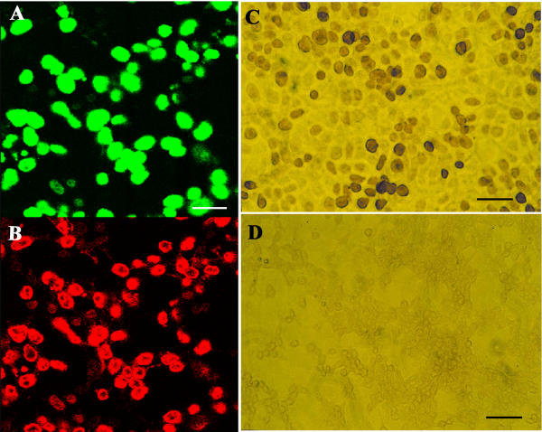
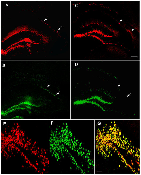
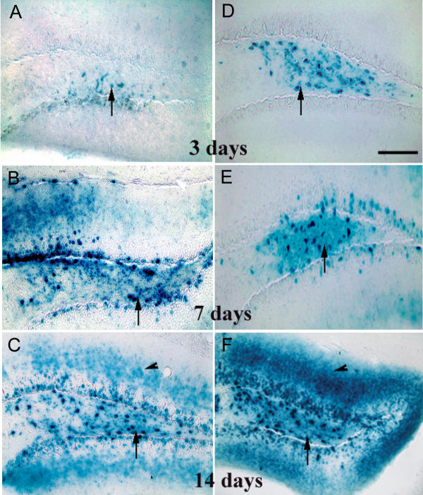
Results: Recombinant AAV-Cre, AAV-GFP (green fluorescent protein) and LV-Cre-EGFP (enhanced GFP) were made with the transgene controlled by the cytomegalovirus promoter. Infecting 293T cells in vitro with AAV-Cre and LV-Cre-EGFP resulted in transduction of most cells as shown by GFP fluorescence and Cre immunoreactivity. Injections of submicrolitre quantities of LV-Cre-EGFP and mixtures of AAV-Cre with AAV-GFP into the neocortex and hippocampus of adult Rosa26 reporter mice resulted in strong Cre and GFP expression in the dentate gyrus and moderate to strong labelling in specific regions of the hippocampus and in the neocortex, mainly in neurons. The pattern of expression of Cre and GFP obtained with AAV and LV vectors was very similar. X-gal staining showed that Cre-mediated recombination had occurred in neurons in the same regions of the brain, starting at 3 days post-injection. No obvious toxic effects of Cre expression were detected even after four weeks post-injection.

Conclusion: AAV and LV vectors are capable of delivering Cre to neurons in discrete regions of the adult mouse brain and producing recombination.


Figures

Figure 1
Figure 1
(A, B) EGFP fluorescence (A) and Cre immunoreactivity (B) in HEK 293T cells infected with LV-Cre-EGFP recombinant virus in vitro. All the cells which express EGFP also express Cre. (C, D) Immunohistochemistry for Cre, in HEK 293T cells 16 hrs after exposure to the AAV-Cre virus (1C) and in uninfected HEK 293T cells (1D). Cre is present in the nucleus of large numbers of cells infected with the virus but is absent from uninfected cells. Scale bars A and B = 25 μm, C = 50 μm, D = 100 μm.
Figure 2
Figure 2
Immunohistochemistry for GFP after infusing LV-Cre-EGFP virus (A, B) and AAV-Cre/AAV-GFP mixture viruses (C, D) into the neocortex and hippocampus of Rosa26 mouse brain. Very strong GFP expression is present in the molecular layer and polymorphic layer (arrows in B and D) of the dentate gyrus. GFP positive cells are present in CA1, CA2 and terminal part of the CA3 region of the hippocampus (A, C). Strong staining was also observed in presumptive glial cells in the subcortical white matter and corpus callosum (arrows in A and C). Fig. 2B and 2D are enlargements of Fig. 2A and 2C. Scale bars, A and C = 200 μm, B and D = 100 μm.
Figure 3
Figure 3
Double immunofluorescence for Cre and GFP using Cy3 and FITC labelled secondary antibodies respectively with single excitations of 543 nm to detect Cre (A, C) or 488 nm to detect GFP (B, D). LV-Cre-EGFP virus (A-B) and AAV-Cre/AAV-GFP mixture viruses (C-D) were injected into the hippocampus and neocortex of Rosa26 mice 4 weeks before perfusion. The patterns of expression of the two transgenes are almost identical (allowing for the slightly stronger staining obtained with the tyramide-enhanced Cre immunohistochemistry) irrespective of the vector used. Fig. 3E,3F, and 3G show the extent of colocalization of GFP and Cre in the dentate gyrus of an AAV-Cre/AAV-GFP mixture injected Rosa26 mouse brain. In the original micrographs, more cells were Cre positive: 373 cells were Cre positive and 256 cells were GFP positive. However the few green cells in 3G show that some cells expressed GFP alone. The arrowheads and arrows in A-D indicate immunofluorescence in pyramidal cells of CA1 and CA2 respectively. Scale bars, A-D = 200 μm, E-G = 400 μm.
Figure 4
Figure 4
Cre-mediated recombination following injection of LV-Cre-EGFP (A-C) and a mixture of AAV-Cre and AAV-GFP recombinant viruses (AAV-Cre/AAV-GFP mixture) (D-F) into the neocortex and hippocampus of Rosa26 reporter mice. Expression of β-galactosidase, indicating Cre-mediated excision of the loxP flanked stop signal, was detected by the X-gal staining. β-galactosidase activity was prominent in the dentate gyrus of animals at all postinjection intervals (arrows in A-F). Scale bars A= 100 μm (applies also to B-F).
Figure 5
Figure 5
Cre-mediated recombination in the neocortex and hippocampus of Rosa26 reporter mice, 4 weeks following injection of LV-Cre-EGFP recombinant virus (A-C) or a mixture of AAV-Cre and AAV-GFP recombinant viruses (AAV-Cre/AAV-GFP mixture) (D-F). β-galactosidase activity is apparent in and around the needle track in the neocortex (arrows in A and D), in parts of the CA1, CA2 and terminal portion of the CA3 region of the hippocampus and in the dentate gyrus. Cells in stratum lacunosum-moleculare (arrowheads in A and D), corpus callosum (arrow in B and E) were also observed. Parts of CA1 and CA2 from Fig. 5A and 5D are enlarged in Fig. 5B and 5E and the region of the dentate gyrus from Fig. 5A and 5D is enlarged in Fig. 5C and 5F. Scale bars in A = 200 μm (applies also to D); B = 100 μm (applies also to C, E and F).

References

    1. Tsien JZ, Chen DF, Gerber D, Tom C, Mercer EH, Anderson DJ, Mayford M, Kandel ER, Tonegawa S. Subregion- and cell type restricted gene knockout in mouse brain. Cell. 1996;87:1317–1326. - PubMed
    1. Cinato E, Mirotsou M, Sablitzky F. Cre-mediated transgene activation in the developing and adult mouse brain. Genesis. 2001;31:118–125. doi: 10.1002/gene.10014. - DOI - PubMed
    1. Silver DP, Livingston DM. Self-excising retroviral vectors encoding the Cre recombinase overcome Cre-mediated cellular toxicity. Mol Cell. 2001;8:233–243. - PubMed
    1. Bueler H. Adeno-associated viral vectors for gene transfer and gene therapy. Biol Chem. 1999;380:613–622. - PubMed
    1. Naldini L. Lentiviruses as gene transfer agents for delivery to non-dividing cells. Curr Opin Biotechnol. 1998;9:457–463. doi: 10.1016/S0958-1669(98)80029-3. - DOI - PubMed

Publication types

MeSH terms

LinkOut - more resources