. 2004 Mar;70(3):1633-40.
doi: 10.1128/AEM.70.3.1633-1640.2004.

Phage community dynamics in hot springs

Affiliations

Phage community dynamics in hot springs

Mya Breitbart et al. Appl Environ Microbiol. 2004 Mar.

Abstract

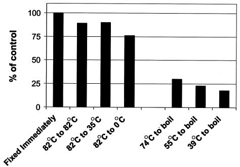
In extreme thermal environments such as hot springs, phages are the only known microbial predators. Here we present the first study of prokaryotic and phage community dynamics in these environments. Phages were abundant in hot springs, reaching concentrations of a million viruses per milliliter. Hot spring phage particles were resistant to shifts to lower temperatures, possibly facilitating DNA transfer out of these extreme environments. The phages were actively produced, with a population turnover time of 1 to 2 days. Phage-mediated microbial mortality was significant, making phage lysis an important component of hot spring microbial food webs. Together, these results show that phages exert an important influence on microbial community structure and energy flow in extreme thermal environments.


Figures

FIG. 1.
FIG. 1.
California hot springs sampled in the study: Travertine Springs (Bridgeport), several hot springs located in the Long Valley caldera (Little Hot Creek, Casa Diablo, and Shepherd's Pool), and Imperial Spa (part of the Desert Hot Springs geothermal field). The line drawing of the caldera was adapted from an image at http://lvo.wr.usgs.gov/images/maps/index.html.
FIG. 2.
FIG. 2.
Photographs of the hot springs sampled in this study. Direct counts of viruses and prokaryotes were performed at all sites (Imperial Spa, Travertine Springs, Casa Diablo, Shepherd's Pool, and Little Hot Creek). Different sampling sites along the Little Hot Creek system are indicated by numbers on the central overview map, with lines leading to expanded pictures detailing each site. Viral and microbial production measurements were performed at Little Hot Creek sites 3 and 4 and the spigot at the Imperial Spa where the water was emerging from the ground (not the pool).
FIG. 3.
FIG. 3.
Example of SYBR Gold staining of prokaryotic cells and VLP in the hot springs samples. SYBR Gold stained, typical sample from Little Hot Creek site 4, which was fixed with 2% paraformaldehyde, filtered onto a 0.02-μm Anodisc, stained with SYBR Gold, and viewed by epifluorescent microscopy. Unstained, aliquot of the same sample viewed under epifluorescent microscopy in the absence of SYBR Gold staining. No autofluorescence of the samples was observed. The phase-contrast panel was the same field of view as the unstained sample, viewed under phase contrast to show that the filter was in focus and contained cells.
FIG. 4.
FIG. 4.
Representative electron micrographs of VLP observed in the hot spring water from Little Hot Creek site 4.
FIG. 5.
FIG. 5.
Temperature shift experiments demonstrated that hot spring phage particles were relatively resistant to lower temperatures but sensitive to boiling. Water samples collected from Little Hot Creek site 3 (82°C) were incubated for ≈20 h at various temperatures to determine the stability of the phage particles at different temperatures. Similarly, samples from Little Hot Creek site 4 (74°C), Little Hot Creek site 8 (55°C), and Little Hot Creek site 9 (39°C) were incubated in a pot of boiling water (≈105°C) for ≈20 h. The number of intact phage particles observed by epifluorescent microscopy in the samples that were fixed immediately was set at 100%, and the number of intact VLP after the incubations is expressed as a percentage of that control value.

References

    1. Acheson, D. W. K., J. Reidl, X. Zhang, G. T. Keusch, J. J. Mekalanos, and M. K. Waldor. 1998. In vivo transduction with Shiga toxin 1-encoding phage. Infect. Immun. 66:4496-4498. - PMC - PubMed
    1. Anderson, N. G. (ed.). 1967. Isolation of viral particles from large volumes. Interscience Publishers, New York, N.Y.
    1. Anderson, N. G., and G. B. Cline. 1967. New centrifugal methods for virus isolation, p. 137-178. In M. K. Maramorosch and H. Koprowski (ed.), Methods in virology, vol. II. Academic Press, New York, N.Y.
    1. Arnold, H. P., W. Zillig, U. Ziese, I. Holz, M. Crosby, T. Utterback, J. F. Weidmann, J. K. Kristjanson, H. P. Klenk, K. E. Nelson, and C. M. Fraser. 2000. A novel lipothrixvirus, SIFV, of the extremely thermophilic crenarchaeon Sulfolobus. Virology 267:252-266. - PubMed
    1. Azam, F. 1998. Microbial control of oceanic carbon flux: the plot thickens. Science 280:694-696.

Publication types

LinkOut - more resources