. 2004 Mar 15;164(6):923-33.
doi: 10.1083/jcb.200312018. Epub 2004 Mar 8.

Misfolding diverts CFTR from recycling to degradation: quality control at early endosomes

Affiliations

Misfolding diverts CFTR from recycling to degradation: quality control at early endosomes

Manu Sharma et al. J Cell Biol. .

Abstract

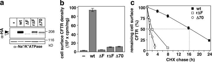
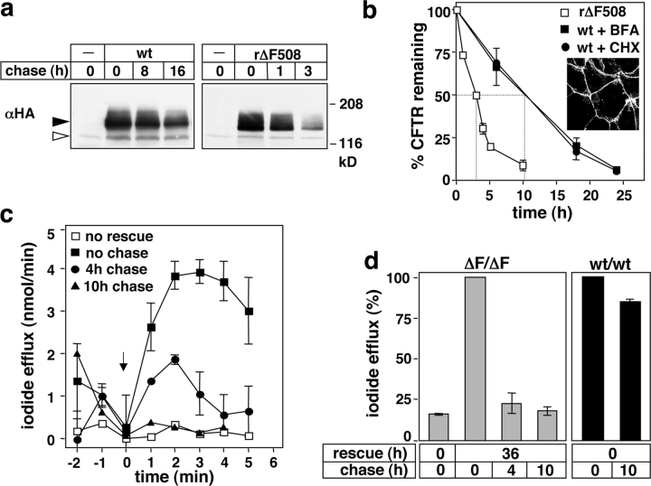
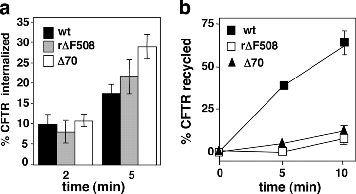
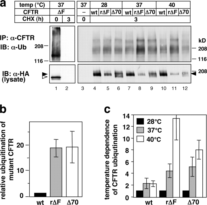
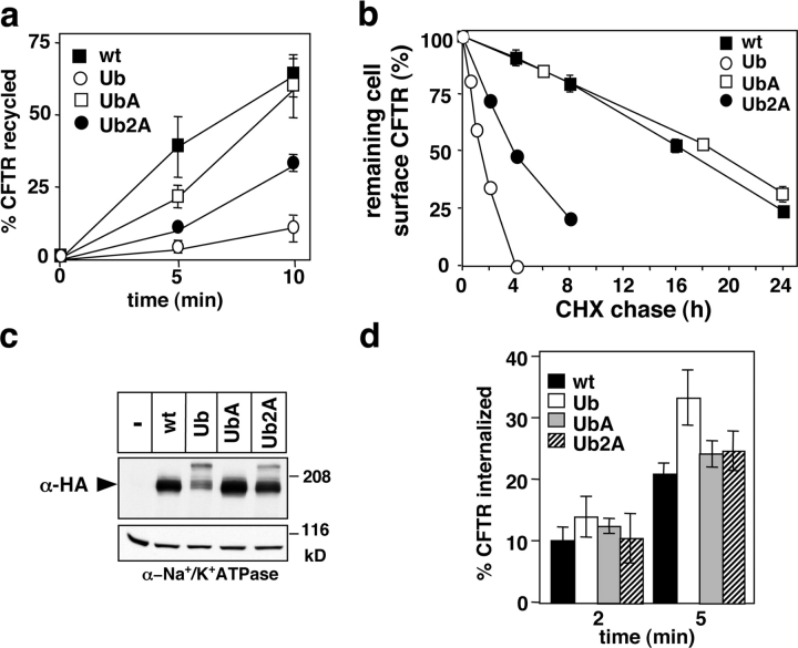
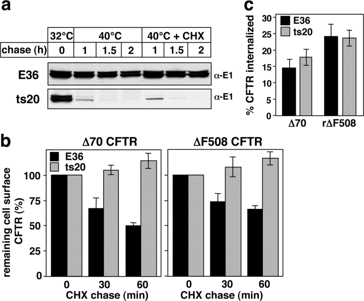
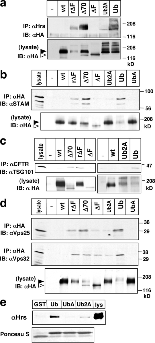
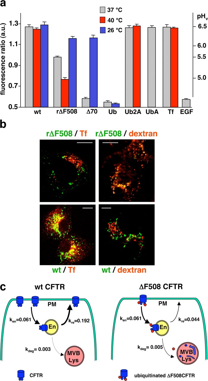
To investigate the degradation mechanism of misfolded membrane proteins from the cell surface, we used mutant cystic fibrosis transmembrane conductance regulators (CFTRs) exhibiting conformational defects in post-Golgi compartments. Here, we show that the folding state of CFTR determines the post-endocytic trafficking of the channel. Although native CFTR recycled from early endosomes back to the cell surface, misfolding prevented recycling and facilitated lysosomal targeting by promoting the ubiquitination of the channel. Rescuing the folding defect or down-regulating the E1 ubiquitin (Ub)-activating enzyme stabilized the mutant CFTR without interfering with its internalization. These observations with the preferential association of mutant CFTRs with Hrs, STAM-2, TSG101, hVps25, and hVps32, components of the Ub-dependent endosomal sorting machinery, establish a functional link between Ub modification and lysosomal degradation of misfolded CFTR from the cell surface. Our data provide evidence for a novel cellular mechanism of CF pathogenesis and suggest a paradigm for the quality control of plasma membrane proteins involving the coordinated function of ubiquitination and the Ub-dependent endosomal sorting machinery.


Figures

Figure 1.
Figure 1.
Destabilizing mutations down-regulate CFTR from the plasma membrane. For all experiments, rescued ΔF508 CFTR (rΔF508) was accumulated at 28°C for 24–36 h before the measurements. (a) Steady-state expression of CFTR variants. Equal amounts of cell lysates from BHK cells, expressing the indicated construct, were separated by SDS-PAGE. CFTR and Na+/K+-ATPase was visualized by immunoblotting with anti-HA and anti-Na+/K+-ATPase antibodies, respectively. Filled and empty arrowheads indicate the complex- and core-glycosylated CFTR, respectively. (b) The cell surface density of the CFTR variant was determined by anti-HA antibody and 125I-conjugated secondary antibody binding at 0°C in BHK cells, and was normalized for cellular protein. Nonspecific antibody binding to mock-transfected BHK cells is indicated (-). (c) The turnover of cell surface resident wt, rΔF508, and Δ70 CFTR harboring the 3HA tag was monitored by their disappearance kinetics in the presence of 100 μg/ml CHX by the radioactive anti-HA antibody-binding assay at 37°C. Data are means ± SEM, n = 2–4. Similar results were obtained by monitoring the disappearance kinetics of prebound anti-HA antibody in the absence of CHX (not depicted).
Figure 2.
Figure 2.
Rescued ΔF508 CFTR (rΔF508) is unstable in polarized epithelia. (a) Stably transfected PANC-1 cells were grown for >3 d at confluence, and then ΔF508 CFTR was rescued (at 26°C for 36 h). Disappearance of rΔF508 and wt CFTR was measured in the presence of 100 μg/ml CHX or 10 μg/ml brefeldin-A (not depicted) at 37°C by immunoblot analysis of equal amounts of cell lysates. Filled and empty arrowheads indicate the complex- and core-glycosylated CFTR, respectively. (b) Densitometric analysis of the disappearance of the complex-glycosylated wt and rΔF508 CFTR on immunoblots shown in a. Data are expressed as percentage of the initial amount and represent means ± SEM (n = 3–4). Inset: PANC-1 monolayer was immunostained for occludin to confirm polarization. (c) Functional stability of rΔF508 CFTR was measured by the iodide efflux assay in PANC-1 monolayers. The ΔF508 CFTR processing defect was rescued at 28°C (24 h) and the PKA-stimulated iodide release was measured after the indicated chase (0–10 h) at 37°C. (d) Functional turnover of rΔF508 and wt CFTR in differentiated respiratory epithelia derived from nasal polyps of homozygous (ΔF508/ΔF508) and healthy (wt/wt) individuals. The PKA-stimulated iodide release into the apical compartment on filter grown epithelia was monitored as a function of chase at 37°C after rescuing ΔF508 CFTR at 28°C for 36 h. Biosynthesis of wt CFTR was inhibited by 100 μg/ml CHX at 0 h.
Figure 3.
Figure 3.
Misfolding disrupts the constitutive recycling of CFTR. (a) Endocytosis rates of the wt, rescued ΔF508 (rΔF508), and Δ70 CFTR were measured by antibody-capture assay in stably transfected BHK cells. CFTR-3HA variants were labeled with anti-HA antibody at 0°C, and the complex was internalized at 37°C. Antibody remaining at the cell surface was measured with 125I-labeled secondary antibody. Data are expressed as percentage of the initial amount of specific antibody binding. (b) The recycling of wt, rΔF508, and Δ70 CFTR was measured after labeling the endosomal CFTR with anti-HA antibody at 37°C for 30 min, as described in the Materials and methods. After 5–10-min incubations at 37°C, recycled anti-HA–CFTR complexes were determined by biotinylated secondary antibody and 125I- labeled streptavidin at 4°C. Recycling efficiency of CFTR is expressed as percentage of internalized anti-HA antibody, measured in parallel samples. Data are obtained from 3–4 independent experiments.
Figure 4.
Figure 4.
Misfolding enhances the ubiquitination susceptibility of CFTR. (a) Ub modification of wt, Δ70, and rΔF508 CFTR in post-Golgi compartments. Ubiquitinated core-glycosylated CFTR was eliminated by CHX chase (100 μg/ml, 3 h; lanes 2–12) in BHK cells. For the 28°C incubation, the CHX chase was performed at 37°C for 1 h and then at 28°C for 2 h. When indicated, cells were incubated at 37°C for 2 h and then at 40°C for 1 h. Solubilized CFTR was denatured in 2% SDS to avoid the isolation of irrelevant proteins. Then, CFTR was precipitated with the L12B4 anti-CFTR antibody. Immunoprecipitates were probed with anti-Ub antibody (FK2) and with visualized ECL (top). CFTR was visualized in the lysate with anti-HA antibody (bottom). Immunoprecipitates of both rΔF508 and Δ70 CFTR were obtained from 50 and 150% more cells at 37 and 40°C, respectively, than used for wt CFTR. Filled and empty arrowheads indicate the complex- and core-glycosylated form of CFTR, respectively. (b) Ubiquitination susceptibility of the rΔF508 and Δ70 relative to wt CFTR in post-Golgi compartments. Data are derived from densitometry of ubiquitinated CFTR shown in a, with apparent molecular mass including and larger than the complex-glycosylated CFTR. The abundance of ubiquitinated CFTR adducts was normalized for the expression level of their complex-glycosylated form and was expressed as fold increase relative to that of the wt CFTR. Data are means ± SEM, n = 3–4. (c) The temperature-dependent ubiquitination level of Δ70, rΔF508, and wt CFTR variants was measured as described in a and b. The level of ubiquitination for each construct is expressed as fold increase relative to that measured at 28°C.
Figure 5.
Figure 5.
The CFTR-Ub chimera mimics the peripheral trafficking defect of the misfolded CFTR. (a) The recycling of CFTR (wt), CFTR-Ub (Ub), CFTR-UbA (UbA), and CFTR-Ub2A (Ub2A) was monitored by the biotin–streptavidin sandwich technique described in Fig. 3 b, in BHK cells stably expressing the respective construct. Data are means ± SEM, n = 3–4. (b) The cell surface stability of CFTR-Ub variants was determined by the anti-HA antibody-binding assay during the course of a CHX chase as in Fig. 1 c. (c) To determine the expression level of the CFTR variants, equal amount of lysates from BHK cells, expressing wt, CFTR-Ub, CFTR-UbA, and CFTR-Ub2A were separated by SDS-PAGE and probed with anti-HA antibody. The predominant complex-glycosylated forms are indicated by the filled arrowhead and have been verified by endoglycosidase H and peptide-N-glycanase F (not depicted). The origin of the lower mobility derivatives of CFTR is not known. (d) Endocytosis rates of CFTR, CFTR-Ub, CFTR-UbA, and CFTR-Ub2A chimeras were determined by the antibody-capture assay in BHK cells as described in Fig. 3 a.
Figure 6.
Figure 6.
The ubiquitination machinery is required for the disposal of misfolded CFTR by the endosomal sorting machinery. (a) Down-regulation of the thermosensitive form and the wt E1 Ub-activating enzyme was monitored by immunoblotting the ts20 and E36 cells lysates, respectively, using anti-E1 antibody. Cells were lysed after shifting the culture temperature from 32 to 40°C. CHX does not influence the down-regulation of E1. (b) Stably transfected ts20 and E36 cells were incubated at 40°C for 1.5 h to down-regulate the E1 enzyme. Then, the cell surface density of Δ70 and rΔF508 CFTR was monitored by anti-HA antibody-binding assay in the presence of CHX. Data are means ± SEM (n = 2), performed in triplicate. (c) Internalization rates of Δ70 and rΔF508 CFTR were measured as described in Fig. 3 a after the incubation of ts20 and E36 cells at 40°C for 2 h to down-regulate the E1 enzyme.
Figure 7.
Figure 7.
Association of destabilized CFTR with the components of the Ub-dependent endosomal sorting machinery. (a–c) Hrs, STAM-2, and TSG101 associate with destabilized CFTR in post-Golgi compartments. BHK cells expressing the indicated CFTR variant, except for Δ70 and ΔF508, were incubated in CHX for 3 h. Cells were lysed and Hrs was immunoprecipitated with anti-Hrs antibody. CFTR was visualized by anti-HA antibody. STAM-2 (b) and TSG101 (c) association was probed in the immunoprecipitates of CFTR with anti-TSG101 and anti-STAM-2 antibodies. (d) CFTR was immunoprecipitated with L12B4 and M3A7 anti-CFTR antibodies, and the complex was probed with anti-Vps25 and anti-Vps32 antibody. Considering the high abundance of wt CFTR, binding of the Vps is negligible to the wt form. (e) Hrs association with the indicated fusion protein; GST, GST-Ub (Ub), GST-UbF4A (UbA), and GST-UbF4A,I44A (Ub2A) was monitored by pull-down assay. Fusion proteins were incubated with HeLa cell lysate, protein complexes were isolated on glutathione-Sepharose, and the eluent was immunoblotted with rabbit anti-Hrs antibody (top). Fusion proteins were visualized by Ponceau S staining on the nitrocellulose membrane (bottom); lys, HeLa cell lysate. Filled and empty arrowheads indicate the complex- and core-glycosylated CFTR, respectively.
Figure 8.
Figure 8.
Conformation-dependent sorting of CFTR in early endosomes. (a) Monitoring the destination of internalized CFTR by organellar pH measurements. Ratiometric fluorescence video imaging of CFTR-containing vesicles in live cells was performed as described in the Materials and methods. Folding of mutants was facilitated by culturing the cells at 26°C for 1.5 h before internalization, whereas unfolding was promoted at 40°C. Internalization was performed for 3 h at 26 and 37°C and only for 1 h at 40°C. The pH of recycling endosome and lysosome was determined after the internalization of FITC-transferrin and FITC-EGF, respectively. The mean pH of vesicle populations was calculated by Origin software (Fig. S4). (b) BHK cells, expressing the 3HA-tagged rΔF508 or wt CFTR, were allowed to internalize monoclonal or polyclonal anti-HA antibody at 37°C for 3 h. Cells were fixed, permeabilized, and CFTR was colocalized with specific organellar markers. Recycling endosomes were labeled with Alexa Fluor® 598–transferrin (Tf). Lysosomes were visualized with TRITC-dextran by overnight labeling and subsequent chase for 3 h. Single optical sections of representative cells were obtained by fluorescence laser confocal microscopy. Bars, 10 μm. (c) Schematic model for the transport route of wt and rΔF508 CFTR from the cell surface. The recycling (kre) and degradation (kdeg) rate constants were calculated by the SAAM II program (see Materials and methods) using the two-compartmental model, and were expressed as fractional transfer of molecules/min.

Similar articles

Cited by

References

    1. Arvan, P., X. Zhao, J. Ramos-Castaneda, and A. Chang. 2002. Secretory pathway quality control operating in Golgi, plasmalemmal, and endosomal systems. Traffic. 3:771–780. - PubMed
    1. Benharouga, M., M. Haardt, N. Kartner, and G.L. Lukacs. 2001. COOH-terminal truncations promote proteasome-dependent degradation of mature cystic fibrosis transmembrane conductance regulator from post-Golgi compartments. J. Cell Biol. 153:957–970. - PMC - PubMed
    1. Benharouga, M., M. Sharma, J. So, M. Haardt, L. Drzymala, M. Popov, B. Schwapach, S. Grinstein, K. Du, and G.L. Lukacs. 2003. The role of the C terminus and Na+/H+ exchanger regulatory factor in the functional expression of cystic fibrosis transmembrane conductance regulator in nonpolarized cells and epithelia. J. Biol. Chem. 278:22079–22089. - PubMed
    1. Bilodeau, P., J. Urbanowski, S. Winistorfer, and R. Piper. 2002. The Vps27p Hse1p complex binds ubiquitin and mediates endosomal protein sorting. Nat. Cell Biol. 4:534–539. - PubMed
    1. Bilodeau, P., S. Winistorfer, W. Kearney, A. Robertson, and R. Piper. 2003. Vps27-Hse1 and ESCRT-I complexes cooperate to increase efficiency of sorting ubiquitinated proteins at the endosome. J. Cell Biol. 163:237–243. - PMC - PubMed

Publication types

MeSH terms

Substances