Comparative Study
. 2004 Mar 15;199(6):797-804.
doi: 10.1084/jem.20031639. Epub 2004 Mar 8.

Senescent impairment in synergistic cytokine pathways that provide rapid cardioprotection in the rat heart

Affiliations
Comparative Study

Senescent impairment in synergistic cytokine pathways that provide rapid cardioprotection in the rat heart

Munira Xaymardan et al. J Exp Med. .

Abstract

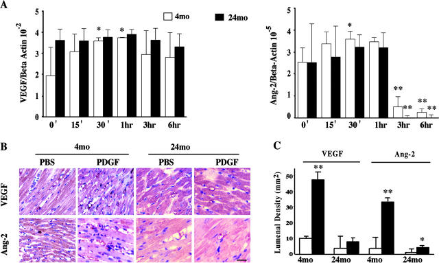
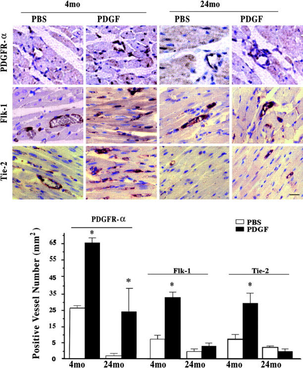
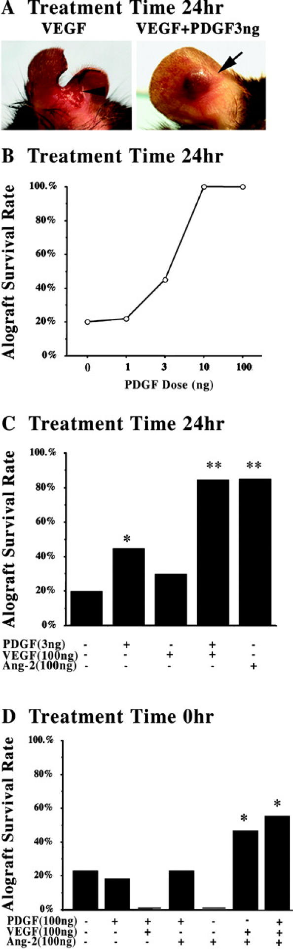
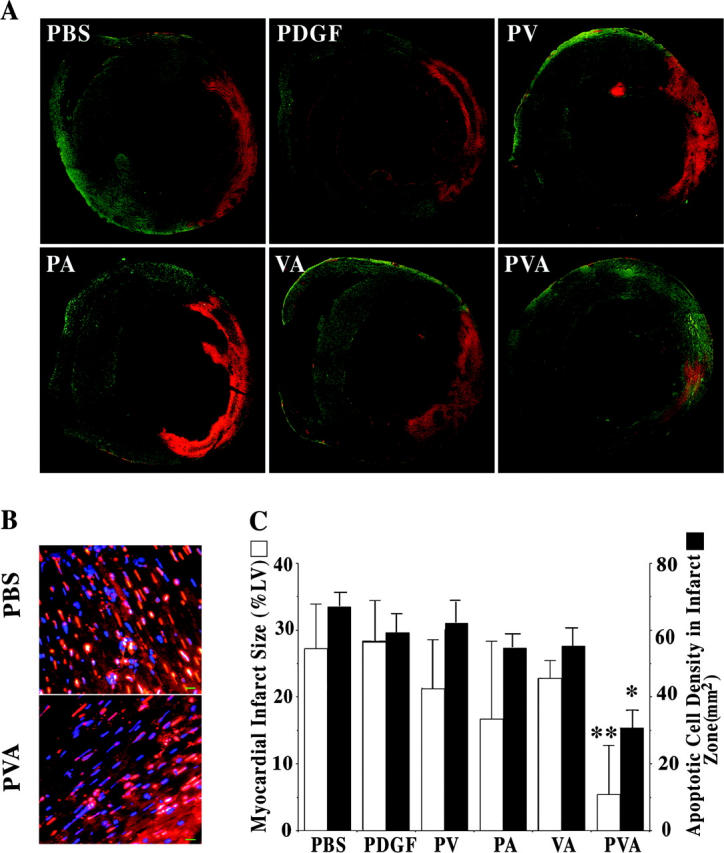
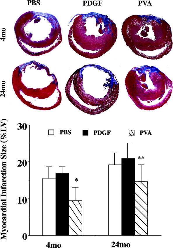
Pretreatment of rodent hearts with platelet-derived growth factor (PDGF)-AB decreases myocardial injury after coronary occlusion. However, PDGF-AB cardioprotection is diminished in older animals, suggesting that downstream elements mediating and/or synergizing the actions of PDGF-AB may be limited in aging cardiac vasculature. In vitro PDGF-AB induced vascular endothelial growth factor (VEGF) and angiopoietin (Ang)-2 expression in 4-mo-old rat cardiac endothelial cells, but not in 24-mo-old heart cells. In vivo injection of young hearts with PDGF-AB increased densities of microvessels staining for VEGF and its receptor, Flk-1, and Ang-2 and its receptor, Tie-2, as well as PDGF receptor (PDGFR)-alpha. In older hearts, PDGF-AB-mediated induction was primarily limited to PDGFR-alpha. Studies in a murine cardiac transplantation model demonstrated that synergist interactions of PDGF-AB plus VEGF plus Ang-2 (PVA) provided an immediate restoration of senescent cardiac vascular function. Moreover, PVA injection in young rat hearts, but not PDGF-AB alone or other cytokine combinations, at the time of coronary occlusion suppressed acute myocardial cell death by >50%. However, PVA also reduced the extent of myocardial infarction with an age-associated cardioprotective benefit (4-mo-old with 45% reduction vs. 24-mo-old with 24%; P < 0.05). These studies showed that synergistic cytokine pathways augmenting the actions of PDGF-AB are limited in older hearts, suggesting that strategies based on these interactions may provide age-dependent clinical cardiovascular benefit.


Figures

Figure 1.
Figure 1.
PDGF-AB induction of cardiac microvascular endothelial proangiogenic cytokines. (A) Real-time RT-PCR of VEGF and Ang-2 in PDGF-AB treated 4- and 24-mo-old rat CMECs in vitro. (B) Representative immunohistochemical staining of VEGF or Ang-2 in cardiac capillaries in rat hearts injected with vehicle (PBS) or PDGF-AB 24 h before euthanization (n = 3, each). Bar = 10 μm. (C) Graph shows the densities of VEGF and Ang-2 positive capillaries in PDGF-AB– or PBS-treated hearts. *, P < 0.05; **, P < 0.005. Treatment versus control.
Figure 2.
Figure 2.
Age-associated induction of cytokine receptors by intramyocardial injection of PDGF-AB. Representative immunohistochemical staining of PDGFR-α, Flk-1, and Tie-2 in cardiac capillaries in rat hearts injected with vehicle (PBS) or PDGF-AB 24 h before euthanization in 4- and 24-mo-old hearts (n = 3, each). Bar = 10 μm. Graph shows the densities of PDGFR-α, Flk-1, and Tie-2 positive capillaries (mm2) in PDGF-AB– or PBS-treated hearts. *, P < 0.05. PDGF-AB versus PBS.
Figure 3.
Figure 3.
Cytokine synergism in restoration of senescent cardiac allografts. (A) Representative examples of neonatal cardiac transplants into 18-mo-old senescent hosts with pretreatment of VEGF (100 ng) or VEGF (100 ng) and PDGF-AB (3 ng). The arrowhead (left) indicates the site of necrotic loss of both allograft and host pinnal tissue beyond the transplant site in the majority of the VEGF-pretreated transplants. The arrow (right) indicates viable/intact cardiac transplants in the host pinnal tissue. (B) Dose response curve of PDGF-AB pretreatment in the restoration of cardiac allograft viability in 18-mo-old mice (n ≥ 20, per group). 10 ng and 100 ng of PDGF-AB restored a significant number of allografts compared with 0- and 1-ng doses. P < 0.005. (C) Assessment of VEGF and Ang-2 pretreatment and combined VEGF and PDGF-AB (n ≥ 20, per group). *, P < 0.05. PDGF-AB (3 ng) versus PBS. **, P < 0.01. Treatment versus PDGF-AB (3 ng) or PBS. (D) Assessment of cytokine combinations in the peri-transplantation of pinnal tissue at the time of cardiac allograft transplantation (n > 10, per group). *, P < 0.05. PDGF-AB plus VEGF plus Ang-2 or VEGF plus Ang-2 versus PBS and all other treatment groups.
Figure 4.
Figure 4.
Synergistic suppression of cardiac apoptosis after coronary occlusion. TUNEL labeling of rat cardiac sections 24 h after injection with combinations of PDGF-AB, VEGF, and Ang-2 at the time of LAD ligation. (A) Reconstructed cross-section micrograph from lower magnification (4×) images for measurement of apoptotic area (percentage of left ventricular area). (B) High magnification (40×) TUNEL staining images used in the quantification of apoptotic cell density. Bar = 10 μm. (C) A graph is shown plotting mean apoptotic risk area size (white) and cell density (TUNEL+; black) in the sections of rat anterior left ventricular wall injected with PBS, PDGF-AB, PDGF-AB plus VEGF (PV), PDGF-AB plus Ang-2 (PA), VEGF plus Ang-2 (VA), or PDGF-AB plus VEGF and Ang-2 (PVA) (n = 3, each group) at the time of LAD ligation. *, P < 0.01; **, P < 0.005 PVA versus PBS and all other cytokine groups.
Figure 5.
Figure 5.
Age-associated cardioprotection by combined cytokine injection at the time of coronary occlusion. (A) Representative Masson's trichrome staining of rat hearts harvested 2 wk after peri-occlusion injections of PBS, PDGF-AB (P), or PDGF-AB plus VEGF and Ang-2 (PVA) and LAD ligation (n ≥ 5, each). Infarction area (blue stain) at the mid–papillary muscle level was significantly smaller in PVA peri-injection heart section than PBS- or PDGF-AB–injected sections. However, there is significant difference between 4- and 24-mo-old rats with PVA treatment. (B) Graph shows average myocardial infarction size (percentage of left ventricular area). *, P < 0.05. 4-mo-old PVA versus 4-mo-old PBS, 4-mo-old PDGF-AB, and 24-mo-old PVA. **, P < 0.05. 24-mo-old PVA versus 24-mo-old PBS and 24-mo-old PDGF-AB.

References

    1. Kass, D.A. 2002. Age-related changes in venticular-arterial coupling: pathophysiologic implications. Heart Fail. Rev. 7:51–62. - PubMed
    1. Lakatta, E.G., and D. Levy. 2003. Arterial and cardiac aging: major shareholders in cardiovascular disease enterprises: Part I: aging arteries: a “set up” for vascular disease. Circulation. 107:139–146. - PubMed
    1. Halcox, J.P., W.H. Schenke, G. Zalos, R. Mincemoyer, A. Prasad, M.A. Waclawiw, K.R. Nour, and A.A. Quyyumi. 2002. Prognostic value of coronary vascular endothelial dysfunction. Circulation. 106:653–658. - PubMed
    1. Gokce, N., J.F. Keaney, Jr., L.M. Hunter, M.T. Watkins, Z.S. Nedeljkovic, J.O. Menzoian, and J.A. Vita. 2003. Predictive value of noninvasively determined endothelial dysfunction for long-term cardiovascular events in patients with peripheral vascular disease. J. Am. Coll. Cardiol. 41:1769–1775. - PubMed
    1. Edelberg, J.M., S.H. Lee, M. Kaur, L. Tang, N.M. Feirt, S. McCabe, O. Bramwell, S.C. Wong, and M.K. Hong. 2002. Platelet-derived growth factor-AB limits the extent of myocardial infarction in a rat model: feasibility of restoring impaired angiogenic capacity in the aging heart. Circulation. 105:608–613. - PubMed

Publication types

MeSH terms

Substances