Comparative Study
. 2004 Mar 15;199(6):763-74.
doi: 10.1084/jem.20031619. Epub 2004 Mar 8.

Lysophosphatidic acid induces neointima formation through PPARgamma activation

Affiliations
Comparative Study

Lysophosphatidic acid induces neointima formation through PPARgamma activation

Chunxiang Zhang et al. J Exp Med. .

Abstract

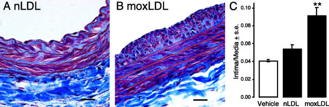
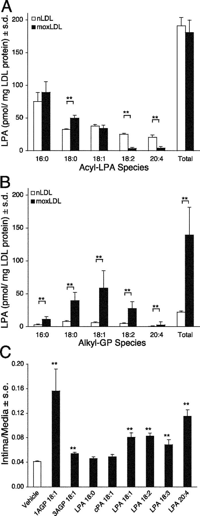
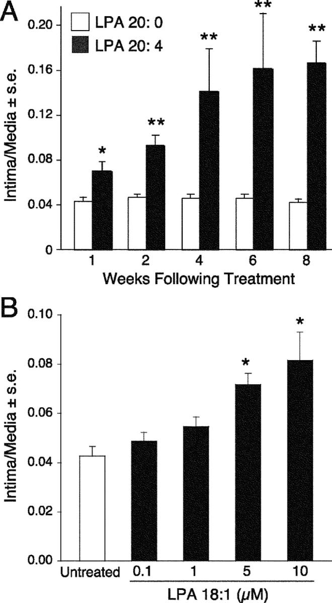
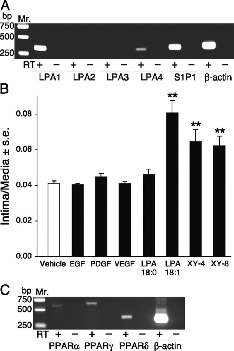
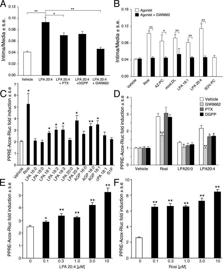
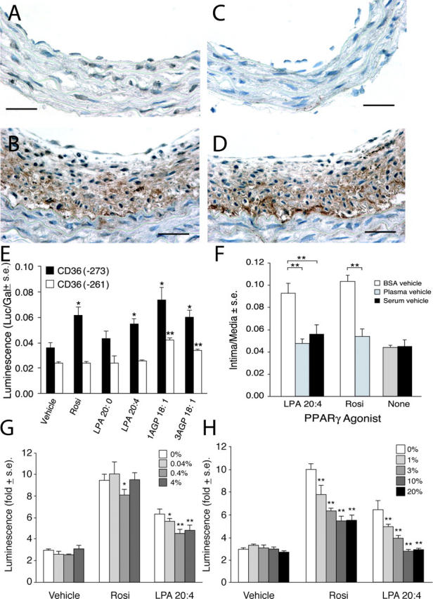
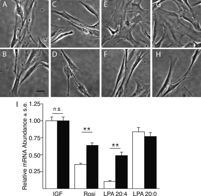
Neointimal lesions are characterized by accumulation of cells within the arterial wall and are a prelude to atherosclerotic disease. Here we report that a brief exposure to either alkyl ether analogs of the growth factor-like phospholipid lysophosphatidic acid (LPA), products generated during the oxidative modification of low density lipoprotein, or to unsaturated acyl forms of LPA induce progressive formation of neointima in vivo in a rat carotid artery model. This effect is completely inhibited by the peroxisome proliferator-activated receptor (PPAR)gamma antagonist GW9662 and mimicked by PPARgamma agonists Rosiglitazone and 1-O-hexadecyl-2-azeleoyl-phosphatidylcholine. In contrast, stearoyl-oxovaleryl phosphatidylcholine, a PPARalpha agonist and polypeptide epidermal growth factor, platelet-derived growth factor, and vascular endothelial growth factor failed to elicit neointima. The structure-activity relationship for neointima induction by LPA analogs in vivo is identical to that of PPARgamma activation in vitro and disparate from that of LPA G protein-coupled receptor activation. Neointima-inducing LPA analogs up-regulated the CD36 scavenger receptor in vitro and in vivo and elicited dedifferentiation of cultured vascular smooth muscle cells that was prevented by GW9662. These results suggest that selected LPA analogs are important novel endogenous PPARgamma ligands capable of mediating vascular remodeling and that activation of the nuclear transcription factor PPARgamma is both necessary and sufficient for neointima formation by components of oxidized low density lipoprotein.


Figures

Figure 1.
Figure 1.
Structural formulas of lipid mediators used in the study.
Figure 2.
Figure 2.
moxLDL treatment induces neointima formation in rat carotid arteries. Representative views of Masson's trichrome-stained, paraffin-embedded sections from animals treated with nLDL (A) or moxLDL (B) (5 mg LDL protein/ml) 2 wk after a 1-h treatment. Bar, 500 μm. Intima to media ratios were quantified (C, n = 5).
Figure 3.
Figure 3.
The five most abundant acyl-LPA (A) and alkyl-GP (B) species were quantified in nLDL and moxLDL using stable isotope dilution electrospray ionization mass spectrometry. The lack of difference in the total acyl-LPA content between nLDL and moxLDL is in sharp contrast to the sixfold increase in alkyl-GP levels in moxLDL (n = 4). In the batch of nLDL used in the experiments shown in Figs. 2 and 3, the alkyl-GP concentration was 0.1 μM, and the total concentration of unsaturated LPA plus alkyl-GP was 0.5 μM, whereas in moxLDL these concentrations were 0.7 and 0.9 μM, respectively. (C) Structure-activity relationship of neointimal lesion induction for various acyl-LPA (10 μM) and alkyl-GP (AGP) species (10 μM). Only select LPA species elicit neointima as the effect was stereoselective with a preference for 1AGP over 3AGP. LPA 18:0 and cPA 18:1 did not elicit detectable neointima (for structural formulas see Fig. 1).
Figure 4.
Figure 4.
(A) Exposure of rat carotid arteries for 1 h to 2.5 μM LPA 20:4, but not to LPA, 20:0 elicited progressive growth of neointima that continued for up to 8 wk posttreatment. Quantitative morphometric analysis for groups of 5 animals. (B) Dose–response curve for LPA 18:1-elicited neointimal response. Mean (± SE) intima to media ratios were determined for groups of five animals 2 wk after treatment.
Figure 5.
Figure 5.
(A) RT-PCR of LPA GPCR in the rat carotid tissue. LPA1, LPA4, and S1P1 were detected in RNA extracted from the whole carotid tissue. (B) Effect of polypeptide growth factors and non-LPA GPCR ligands fluorinated LPA analogs on neointima formation. Animals treated with 10 μM LPA 18:1, XY-4, and its regioisomer XY-8 but not those treated with EGF (50 ng/ml), VEGF (10 ng/ml), PDGF-BB (10 ng/ml), or LPA 18:0 (10 μM) showed neointima formation. Groups of five animals were treated with the compounds. (C) RT-PCR analysis detected PPARα, PPARδ, and PPARγ transcripts in the normal carotid tissue.
Figure 6.
Figure 6.
(A) PTX and DGPP, inhibitors of LPA GPCR signaling, partially attenuated neointima formation induced by LPA 20:4, whereas the PPARγ-specific antagonist GW9662 completely abolished this effect. (B) Rosi, AZ-PC, moxLDL, and unsaturated acyl forms of LPA all induced neointima formation that was completely abolished by GW9662. In contrast, SOV-PC, a PPARα-selective agonist, was ineffective in stimulating the development of neointima after 2 wk. (C) An in vitro assay using CV1 cells transfected with PPARγ and a PPRE-Acox-Rluc reporter gene showed an identical structure-activity relationship when exposed to different LPA species as found for the same set of ligands in the neointima assay in vivo (see Fig. 3 C). (D) The PPARγ antagonist GW9662 (10 μM) abolished, whereas PTX (100 ng/ml, 2 h) and DGPP (10 μM, 2 h) pretreatment and coapplication with LPA partially inhibited LPA 20:4-induced PPRE-Acox-Rluc reporter gene expression in vitro. Vehicle contained 1% DMSO, Rosi (10 μM), LPA20:0 (10 μM), or LPA 20:0 (10 μM) were applied for 20 h. Luciferase and β-galactosidase activities (mean ± SEM) were measured in the cell lysate (n = 4). (E and F) Dose–response relationship of LPA 20:4- and Rosi-induced activation of PPRE-Acox-Rluc reporter gene expression in vitro. *P < 0.05 and **P < 0.01, significant differences over vehicle control.
Figure 7.
Figure 7.
(A) Only a few nuclei show PPARγ immunoreactivity (in a carotid artery 4 wk after treatment with 2.5 μM LPA 20:0). In contrast, the multilayered neointima elicited by LPA 20:4 (B) expresses high levels of PPARγ immunoreactivity. Bars, 250 μm. Activation of PPARγ within neointima in LPA 20:4-treated carotid arteries is indicated by the strong expression of CD36 in a distribution that overlaps that of PPARγ (D). Little immunoreactivity for CD36 was noted in LPA 20:0-treated animals (C). Anti-PPARγ and anti-CD36 were from Santa Cruz Biotechnology, Inc. (E) Stimulation of CD36(−271)-Rluc and CD36(−261)-Rluc reporter genes by Rosi, LPA, and AGP in CV-1 cells. Rosi and LPA 20:4 but not LPA 20:0 (all 10 μM) elicited significant stimulation of CD36(−273)-Rluc that contains a PPRE between bp −273 and −261. Neither compound caused stimulation of the CD36(−261)-Rluc. 1AGP showed higher stimulation compared with 3AGP of the expression of the Rluc reporter. (F) Plasma and serum factors inhibit Rosi- and LPA 20:4-induced neointimal lesion formation in rat carotid arteries. Rosi (10 μM) and LPA 20:4 (2.5 μM) elicited neointima formation 2 wk after treatment when delivered as BSA complexes. In contrast, when the compounds were delivered in rat plasma or serum no neointima formation was detected (n = 5). Effect of BSA (G) or serum (H) on PPARγ activation by CPA and Rosi.
Figure 8.
Figure 8.
PPARγ agonists elicit phenotypic modulation and dedifferentiation of VSMCs in vitro. VSMC cultures established in the presence of 2 ng/ml IGF-1 (A) were treated with 1 μM of each LPA 20:0 (C), LPA 20:4 (E), and Rosi (G) for 3 d. LPA 20:4 and Rosi treatments lead to a pronounced change in the morphology of VSMCs. Pretreatment of the cultures with 200 nM GW9662 for 30 min did not affect the spindle-like morphology of the IGF-1– (B) and LPA 20:0-treated cultures (D). In contrast, GW9662 reversed the flattened morphology into a spindle-like shape in cultures treated with LPA 20:4 (F) and Rosi (H, calibration bar 100 μm). Expression of hCAD mRNA decreased significantly by day 5 in VSMCs treated with Rosi and LPA (I, white bars) compared with the IGF-treated control cultures. This trend was reversed in cultures pretreated with 200 nM GW9662 (I, black bars) as the PPARγ antagonist caused a significant increase in the abundance of hCAD mRNA measured by quantitative RT-PCR (P < 0.01, ANOVA).

References

    1. Lusis, A.J. 2000. Atherosclerosis. Nature. 407:233–241. - PMC - PubMed
    1. Steinberg, D. 2002. Atherogenesis in perspective: hypercholesterolemia and inflammation as partners in crime. Nat. Med. 8:1211–1217. - PubMed
    1. Dzau, V.J., R.C. Braun-Dullaeus, and D.G. Sedding. 2002. Vascular proliferation and atherosclerosis: new perspectives and therapeutic strategies. Nat. Med. 8:1249–1256. - PubMed
    1. Ross, R. 1999. Atherosclerosis is an inflammatory disease. Am. Heart J. 138:S419–S420. - PubMed
    1. Libby, P. 2002. Inflammation in atherosclerosis. Nature. 420:868–874. - PubMed

Publication types

MeSH terms