

Cbf1p modulates chromatin structure, transcription and repair at the Saccharomyces cerevisiae MET16 locus
- PMID: 15007107
- PMCID: PMC390324
- DOI: 10.1093/nar/gkh324
Cbf1p modulates chromatin structure, transcription and repair at the Saccharomyces cerevisiae MET16 locus
Abstract
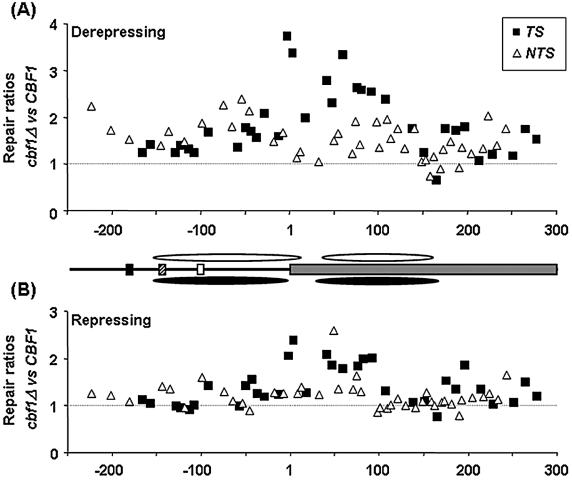
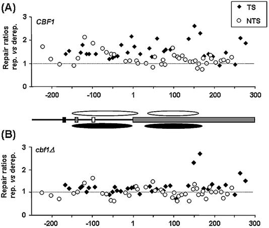
The presence of damage in the transcribed strand (TS) of active genes and its position in relation to nucleosomes influence nucleotide excision repair (NER) efficiency. We examined chromatin structure, transcription and repair at the MET16 gene of wild-type and cbf1Delta Saccharomyces cerevisiae cells under repressing or derepressing conditions. Cbf1p is a sequence-specific DNA binding protein required for MET16 chromatin remodelling. Irrespective of the level of transcription, repair at the MspI restriction fragment of MET16 exhibits periodicity in line with nucleosome positions in both strands of the regulatory region and the non-transcribed strand of the coding region. However, repair in the coding region of the TS is always faster, but exhibits periodicity only when MET16 is repressed. In general, absence of Cbf1p decreased repair in the sequences examined, although the effects were more dramatic in the Cbf1p remodelled area, with repair being reduced to the lowest levels within the nucleosome cores of this region. Our results indicate that repair at the promoter and coding regions of this lowly transcribed gene are dependent on both chromatin structure and the level of transcription. The data are discussed in light of current models relating NER and chromatin structure.
Figures
Similar articles
-
Roles for Gcn5p and Ada2p in transcription and nucleotide excision repair at the Saccharomyces cerevisiae MET16 gene.Nucleic Acids Res. 2006 Feb 9;34(3):976-85. doi: 10.1093/nar/gkj501. Print 2006. Nucleic Acids Res. 2006. PMID: 16473851 Free PMC article.
-
Histone acetylation, chromatin remodelling, transcription and nucleotide excision repair in S. cerevisiae: studies with two model genes.DNA Repair (Amst). 2005 Jul 28;4(8):870-83. doi: 10.1016/j.dnarep.2005.04.006. DNA Repair (Amst). 2005. PMID: 15950549
-
Nucleosome structure and positioning modulate nucleotide excision repair in the non-transcribed strand of an active gene.EMBO J. 1997 Aug 15;16(16):5046-56. doi: 10.1093/emboj/16.16.5046. EMBO J. 1997. PMID: 9305646 Free PMC article.
-
Nucleosome positioning, nucleotide excision repair and photoreactivation in Saccharomyces cerevisiae.DNA Repair (Amst). 2015 Dec;36:98-104. doi: 10.1016/j.dnarep.2015.09.012. Epub 2015 Sep 16. DNA Repair (Amst). 2015. PMID: 26429065 Review.
-
Transcription coupled nucleotide excision repair in the yeast Saccharomyces cerevisiae: The ambiguous role of Rad26.DNA Repair (Amst). 2015 Dec;36:43-48. doi: 10.1016/j.dnarep.2015.09.006. Epub 2015 Sep 10. DNA Repair (Amst). 2015. PMID: 26429063 Review.
Cited by
-
Histone variant Htz1 promotes histone H3 acetylation to enhance nucleotide excision repair in Htz1 nucleosomes.Nucleic Acids Res. 2013 Oct;41(19):9006-19. doi: 10.1093/nar/gkt688. Epub 2013 Aug 7. Nucleic Acids Res. 2013. PMID: 23925126 Free PMC article.
-
Nucleotide excision repair in cellular chromatin: studies with yeast from nucleotide to gene to genome.Int J Mol Sci. 2012;13(9):11141-11164. doi: 10.3390/ijms130911141. Epub 2012 Sep 7. Int J Mol Sci. 2012. PMID: 23109843 Free PMC article. Review.
-
Evidence that the transcription elongation function of Rpb9 is involved in transcription-coupled DNA repair in Saccharomyces cerevisiae.Mol Cell Biol. 2006 Dec;26(24):9430-41. doi: 10.1128/MCB.01656-06. Epub 2006 Oct 9. Mol Cell Biol. 2006. PMID: 17030604 Free PMC article.
-
Repair-independent chromatin assembly onto active ribosomal genes in yeast after UV irradiation.Mol Cell Biol. 2005 Nov;25(22):9773-83. doi: 10.1128/MCB.25.22.9773-9783.2005. Mol Cell Biol. 2005. PMID: 16260595 Free PMC article.
-
Derepression of INO1 transcription requires cooperation between the Ino2p-Ino4p heterodimer and Cbf1p and recruitment of the ISW2 chromatin-remodeling complex.Eukaryot Cell. 2010 Dec;9(12):1845-55. doi: 10.1128/EC.00144-10. Epub 2010 Oct 8. Eukaryot Cell. 2010. PMID: 20935143 Free PMC article.
References
-
- Wolffe A. (1998) Chromatin: Structure and Function. Academic Press, San Diego, USA.
-
- Meijer M. and Smerdon,M.J. (1999) Accessing DNA damage in chromatin: insights from transcription. BioEssays, 21, 596–603. - PubMed
-
- Meyer P. (2001) Chromatin remodelling. Curr. Opin. Plant Biol., 4, 457–462. - PubMed
-
- Pfeifer G.P. (1997) Formation and processing of UV photoproducts: effects of DNA sequence and chromatin environment. Photochem. Photobiol., 65, 270–283. - PubMed
-
- Smerdon M.J. and Conconi,A. (1999) Modulation of DNA damage and DNA repair in chromatin. Prog. Nucleic Acid Res. Mol. Biol., 62, 227–255. - PubMed
Publication types
MeSH terms
Substances
Grants and funding
LinkOut - more resources
Full Text Sources
Molecular Biology Databases
