. 2004 Mar 8;32(5):1627-37.
doi: 10.1093/nar/gkh309. Print 2004.

Telomere-bound TRF1 and TRF2 stall the replication fork at telomeric repeats

Affiliations

Telomere-bound TRF1 and TRF2 stall the replication fork at telomeric repeats

Rieko Ohki et al. Nucleic Acids Res. .

Abstract

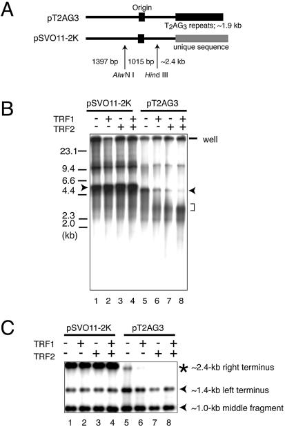
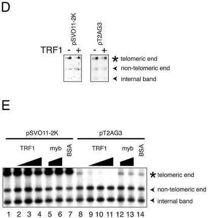
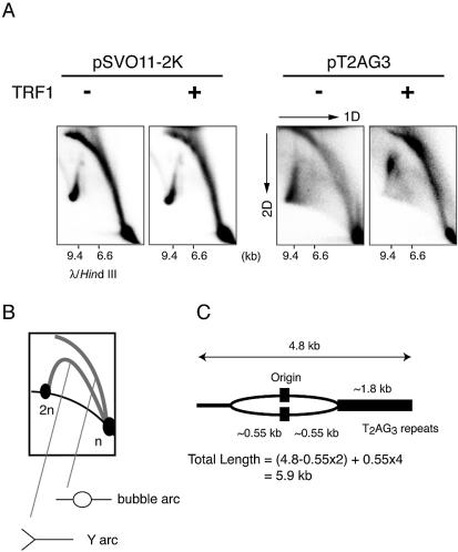
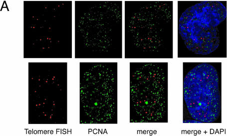
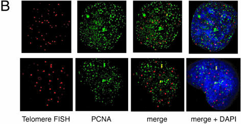
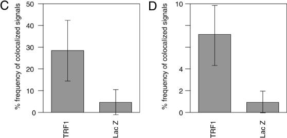
Vertebrate telomeres consist of tandem repeats of T2AG3 and associated proteins including the telomeric DNA-binding proteins, TRF1 and TRF2. It has been proposed that telomeres assume two interswitchable states, the open state that is accessible to various trans-acting factors and the closed state that excludes those factors. TRF1 and TRF2 are believed to promote the formation of the closed state. However, little is known about how those two states influence DNA replication. We analyzed the effects of TRF1 and TRF2 on telomeric replication both in vitro and in vivo. By exploiting the in vitro replication system of linear SV40 DNA, we found that telomeric repeats are a poor replication template. Moreover, the addition of recombinant TRF1 and TRF2 significantly stalled the replication fork progression at telomeric repeats. When TRF1 was overexpressed in HeLa cells, cells with 4N DNA content were accumulated. Furthermore, cytological analyses revealed that the replication focus overlapped with telomere signals at a significantly higher frequency in TRF1-overexpressing cells than in control cells. The results suggest that TRF1 and TRF2 exert inhibitory effects on replication fork progression.


Figures

Figure 1
Figure 1
SV40-based in vitro replication system of linear DNA with a stretch of T2AG3 repeats at one end. In vitro replication of linear DNA containing telomeric repeats at one end. pT2AG3 contains ∼1.9 kb telomeric repeats and a single restriction site of BbsI abutting the tract of telomeric repeats. BbsI leaves 4 nt 5′ protrusions at the digested sites and the 3′- recessive ends were filled-in with dATPs, dGTPs, biotinylated dUTPs and biotinylated dCTPs. Those biotin-labeled DNAs were captured on avidin-coated beads and subjected to replication reactions. After the reactions, bead-bound DNAs were collected and subjected to analysis. Only the replication products produced by a single round of replication from the original template DNAs retain the terminal biotin labels. Therefore, it is possible to analyze those products by analyzing the bead-bound DNAs. Note that one end of linearized pT2AG3 has a telomeric sequence to the extreme end in the same direction as that of the native telomeres (3′-OH of G-strand at termini). The BbsI recognition sequence is shadowed. Biotin-labeled nucleotides are shown by small filled circles.
Figure 2
Figure 2
In vitro replication products templating linear DNA molecules with or without telomeric repeats. (A) Schematic structures of linearized pT2AG3 and pSVO11-2K. Both pT2AG3 and pSVO11-2K were derived from pSVO11 that contains an SV40 replication origin (Origin) (55). The T2AG3-repeat in pT2AG3 and the corresponding region in pSVO11-2K derived from a cloning vector are shown by black and gray squares, respectively. The positions of AlwNI and HindIII sites used in experiments performed in panels C–E are shown and fragment lengths are indicated (not to scale). (B) Replication products of pSVO11-2K and pT2AG3. pSVO11-2K and pT2AG3 were linearized and captured on beads (pT2AG3-beads and pSVO11-2K-beads). The beads were incubated with T-antigen, 293 cell extracts with or without recombinant TRF1 and/or TRF2 for 10 min on ice. 250 ng of baculovirus-produced purified recombinant TRF1 (lanes 2 and 6) or TRF2 (lanes 3 and 7), or both (lanes 4 and 8) was added per 25 µl reaction containing 50 ng of DNA. Replication reactions were started by shifting the incubation temperature to 37°C and terminated after 2 h. DNA from each reaction was purified and separated on 0.7% agarose gel. Arrowheads indicate the full-length product from each plasmid. The bracket shows the signal that increased when TRF1 and/or TRF2 were added to the reaction. Due to the intrinsic DNA ligase activity present in S100 extracts, linear dimers were formed and detected (bands seen at ∼10 kb). (C) Telomeric repeats are replicated inefficiently in the presence of TRF1 or TRF2. Purified replication products were digested by AlwNI and HindIII. Restriction digests were run on 1% agarose gel and autoradiographed. The positions of the three digested fragments (the ∼2.4 kb telomeric end, the 1015 bp middle fragment and the 1397 bp non-telomeric fragment, see A) are shown. (D) Replication reaction was performed in the absence of SV40 T-antigen, and products were analyzed as in (C). Autoradiographs after long exposure are shown. Note that the fragments are labeled proportionally to their length. Signals appear stronger at the right side of each lane, because the samples were distributed and migrated unequally in the lane. (E) Experiments were performed as in (B) except that increasing amounts of the full-length TRF1, Myb-like domain-only TRF1 and BSA were added as indicated. The concentrations of the added protein were: lanes 2 and 9, 250 ng/25 µl reaction; lanes 3, 5, 10 and 12, 500 ng/25 µl reaction; and lanes 4, 6, 7, 11, 13 and 14, 1 µg/25 µl reaction.
Figure 2
Figure 2
In vitro replication products templating linear DNA molecules with or without telomeric repeats. (A) Schematic structures of linearized pT2AG3 and pSVO11-2K. Both pT2AG3 and pSVO11-2K were derived from pSVO11 that contains an SV40 replication origin (Origin) (55). The T2AG3-repeat in pT2AG3 and the corresponding region in pSVO11-2K derived from a cloning vector are shown by black and gray squares, respectively. The positions of AlwNI and HindIII sites used in experiments performed in panels C–E are shown and fragment lengths are indicated (not to scale). (B) Replication products of pSVO11-2K and pT2AG3. pSVO11-2K and pT2AG3 were linearized and captured on beads (pT2AG3-beads and pSVO11-2K-beads). The beads were incubated with T-antigen, 293 cell extracts with or without recombinant TRF1 and/or TRF2 for 10 min on ice. 250 ng of baculovirus-produced purified recombinant TRF1 (lanes 2 and 6) or TRF2 (lanes 3 and 7), or both (lanes 4 and 8) was added per 25 µl reaction containing 50 ng of DNA. Replication reactions were started by shifting the incubation temperature to 37°C and terminated after 2 h. DNA from each reaction was purified and separated on 0.7% agarose gel. Arrowheads indicate the full-length product from each plasmid. The bracket shows the signal that increased when TRF1 and/or TRF2 were added to the reaction. Due to the intrinsic DNA ligase activity present in S100 extracts, linear dimers were formed and detected (bands seen at ∼10 kb). (C) Telomeric repeats are replicated inefficiently in the presence of TRF1 or TRF2. Purified replication products were digested by AlwNI and HindIII. Restriction digests were run on 1% agarose gel and autoradiographed. The positions of the three digested fragments (the ∼2.4 kb telomeric end, the 1015 bp middle fragment and the 1397 bp non-telomeric fragment, see A) are shown. (D) Replication reaction was performed in the absence of SV40 T-antigen, and products were analyzed as in (C). Autoradiographs after long exposure are shown. Note that the fragments are labeled proportionally to their length. Signals appear stronger at the right side of each lane, because the samples were distributed and migrated unequally in the lane. (E) Experiments were performed as in (B) except that increasing amounts of the full-length TRF1, Myb-like domain-only TRF1 and BSA were added as indicated. The concentrations of the added protein were: lanes 2 and 9, 250 ng/25 µl reaction; lanes 3, 5, 10 and 12, 500 ng/25 µl reaction; and lanes 4, 6, 7, 11, 13 and 14, 1 µg/25 µl reaction.
Figure 3
Figure 3
Two-dimensional gel electrophoresis of replication products. (A) The replication fork is stalled in the replication reaction from pT2AG3-beads with TRF1. Labeled replicated DNAs obtained from pT2AG3- and pSVO11-2K-beads were analyzed by the neutral–neutral 2D gel electrophoresis assay (22,30). λ DNA digested with HindIII was also run in the first dimension to serve as a marker for the linear DNA species (migration positions are shown). (B) Schematic representation of the relative positions of linear DNA, bubble arcs and Y arcs. n and 2n represent the positions of full-sized and double-sized linear DNA molecules. The strong signal found at the 2n position of the linear species probably represents the linear dimer formed by intrinsic DNA ligase activity present in S100 extracts, in addition to replication intermediates. (C) Expected size of the replication intermediate that stalls the replication when the fork encounters telomeric repeats.
Figure 4
Figure 4
Colocalization of telomeres and the replication foci in HeLa cells overexpressing TRF1. (A and B) Cells infected with LacZ- (A) or TRF- expressing adenoviruses (B) were harvested 36 h post-infection and stained with anti-PCNA antibody together with telomeric DNA detected by Cy3-labeled (T2AG3)3 PNA probe. DNA was stained by DAPI. Two representative cells are indicated in both (A) and (B). Colocalized signals are represented in yellow. (C) Frequencies of telomeric FISH signals that colocalized with PCNA signals. Ten randomly chosen mid-to-late S phase cells were analyzed. For each cell, the number of telomeric signals and number of telomere–PCNA overlapping signals were counted. Subsequently, the percentage of telomere–PCNA overlapping signals among total telomeric signals was obtained. Average of the percentage of the overlapping signals were obtained for LacZ- and TRF-expressing cells, and shown as a graph. (D) Frequencies of PCNA signals that colocalized with telomeric FISH signals. Ten randomly chosen mid-to-late S phase cells were analyzed. For each cell, the number of PCNA signals and number of telomere–PCNA overlapping signals were counted. Subsequently, percentage of telomere–PCNA overlapping signals among total PCNA signals was obtained. Average of the percentage of the overlapping signals were obtained for LacZ- and TRF-expressing cells, and shown as a graph.
Figure 4
Figure 4
Colocalization of telomeres and the replication foci in HeLa cells overexpressing TRF1. (A and B) Cells infected with LacZ- (A) or TRF- expressing adenoviruses (B) were harvested 36 h post-infection and stained with anti-PCNA antibody together with telomeric DNA detected by Cy3-labeled (T2AG3)3 PNA probe. DNA was stained by DAPI. Two representative cells are indicated in both (A) and (B). Colocalized signals are represented in yellow. (C) Frequencies of telomeric FISH signals that colocalized with PCNA signals. Ten randomly chosen mid-to-late S phase cells were analyzed. For each cell, the number of telomeric signals and number of telomere–PCNA overlapping signals were counted. Subsequently, the percentage of telomere–PCNA overlapping signals among total telomeric signals was obtained. Average of the percentage of the overlapping signals were obtained for LacZ- and TRF-expressing cells, and shown as a graph. (D) Frequencies of PCNA signals that colocalized with telomeric FISH signals. Ten randomly chosen mid-to-late S phase cells were analyzed. For each cell, the number of PCNA signals and number of telomere–PCNA overlapping signals were counted. Subsequently, percentage of telomere–PCNA overlapping signals among total PCNA signals was obtained. Average of the percentage of the overlapping signals were obtained for LacZ- and TRF-expressing cells, and shown as a graph.

Similar articles

Cited by

References

    1. Chong L., van Steensel,B., Broccoli,D., Erdjument,B.H., Hanish,J., Tempst,P. and de Lange,T. (1995) A human telomeric protein. Science, 270, 1663–1667. - PubMed
    1. Broccoli D., Smogorzewska,A., Chong,L. and de Lange,T. (1997) Human telomeres contain two distinct Myb-related proteins, TRF1 and TRF2. Nature Genet., 17, 231–235. - PubMed
    1. van Steensel B. and de Lange,T. (1997) Control of telomere length by the human telomeric protein TRF1. Nature, 385, 740–743. - PubMed
    1. Smogorzewska A., van Steensel,B., Bianchi,A., Oelmann,S., Schaefer,M.R., Schnapp,G. and de Lange,T. (2000) Control of human telomere length by TRF1 and TRF2. Mol. Cell. Biol., 20, 1659–1668. - PMC - PubMed
    1. Griffith J.D., Comeau,L., Rosenfield,S., Stansel,R.M., Bianchi,A., Moss,H. and de Lange,T. (1999) Mammalian telomeres end in a large duplex loop. Cell, 97, 503–514. - PubMed

Publication types

MeSH terms

Substances