

doi: 10.1073/pnas.0400481101.
Epub 2004 Mar 8.
Fhit is a physiological target of the protein kinase Src
Affiliations
- PMID: 15007172
- PMCID: PMC374320
- DOI: 10.1073/pnas.0400481101
Item in Clipboard
Fhit is a physiological target of the protein kinase Src
Proc Natl Acad Sci U S A.
.
Abstract
The FHIT gene is a tumor suppressor that is frequently inactivated by genomic alterations at chromosomal region 3p14.2. In the last few years, a considerable amount of data describing inactivation of FHIT in a variety of human malignancies and demonstrating the tumor suppressor potential of Fhit have been reported. Despite the demonstration that FHIT functions as a tumor suppressor, the pathway through which Fhit induces apoptosis and inhibits growth of cancer cells is not known. Our data demonstrate that Fhit is a target of tyrosine phosphorylation by the Src protein kinase. We show that Src phosphorylates Y114 of Fhit in vitro and in vivo, providing insight into a biochemical pathway involved in Fhit signaling.
Figures
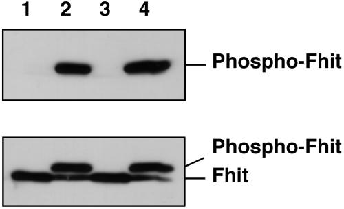
Src phosphorylates Fhit in vitro. One microgram of recombinant Fhit was incubated with 10 units of recombinant Src (lanes 1 and 2) or Lyn (lanes 3 and 4) at 30°C and immunoblotted with anti-phosphotyrosine (Upper) or anti-Fhit (Lower) antibodies. Incubations were carried out for 0 (lanes 1 and 3) or 30 (lanes 2 and 4) min.
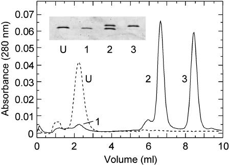
Chromatographic separation of unphosphorylated and phosphorylated forms of Fhit. Dashed line, the MonoQ column elution profile of Fhit incubated in buffer with ATP and without Src kinase; solid line, the MonoQ column elution profile of Fhit incubated in the presence of ATP and Src kinase. (Inset) SDS gel electrophoresis pattern of Fhit samples corresponding to the indicated column peaks: U, unphosphorylated Fhit incubated without Src kinase; 1, residual, unphosphorylated Fhit after incubation with Src kinase; 2, Fhit phosphorylated on one subunit; and 3, Fhit phosphorylated on both subunits. Approximately 0.2 μg of each sample was subjected to electrophoresis, and the gel was stained with Sypro Ruby.
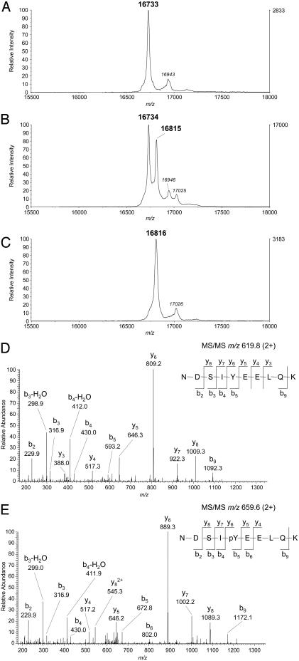
Mass spectral analysis of Fhit. (A-C) MALDI-TOF spectra of unphosphorylated, monophosphorylated, and diphosphorylated forms of Fhit, respectively, were acquired on an Applied Biosystems Voyager Elite as described in Materials and Methods. The m/z values shown in bold represent the average of three spectra obtained from the same sample spot on the MALDI target. The lower intensity peaks in each panel represent matrix adducts. (D and E) ESI tandem mass spectra were acquired on a Thermo Finnigan LCQ Classic spectrometer as described in Materials and Methods. (D) Tandem mass spectrum of the 2+ ion (m/z 619.8) of tryptic peptide 109-118 from unphosphorylated Fhit. (E) Tandem mass spectrum from the 2+ ion (m/z 659.6) of tryptic peptide 109-118 from phosphorylated Fhit. Spectra were obtained by targeted scans of the indicated ions. Peptide fragments are denoted on the spectra using the nomenclature of Roepstorff and Fohlman (23). Identification of the site of phosphorylation was obtained by comparison of the y-series ions in the two spectra.
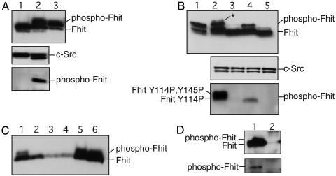
Src phosphorylates Fhit in vivo. (A) 293 cells were cotransfected with WT FHIT and SRCDN (lane 1) or activated SRC (lane 2) and immunoblotted with anti-Fhit (Top), anti-Src (Middle), or anti-phosphotyrosine (Bottom) antibodies. Lysates were immunoprecipitated with anti-Fhit before Western blotting with anti-phosphotyrosine. Lane 3, an aliquot of in vitro phosphorylated Fhit was loaded for comparison. (B) 293 cells were cotransfected with SRCDN and WT FHIT (lane 2), FHITY114F (lane 3), FHITY145F (lane 4), and FHITY114F, Y145F (lane 5). Lysates were immunoblotted as in A. Lane 1, an aliquot of in vitro phosphorylated Fhit was loaded for comparison. (C) Western blot analysis of normal human tissues with anti-Fhit antibody; 100 μg of each lysate were loaded in each lane: lane 1, fetal liver; lane 2, fetal brain; lane 3, fetal lung; lane 4, lung; lane 5, fetal kidney; and lane 6, kidney. (D) One milligram of normal human kidney lysate was immunoprecipitated with anti-Fhit antibody (lane 1) or with rabbit IgG (lane 2) and immunoblotted with anti-Fhit (Upper) or with anti-phosphotyrosine (Lower).
Similar articles
-
FHIT-proteasome degradation caused by mitogenic stimulation of the EGF receptor family in cancer cells.Proc Natl Acad Sci U S A. 2006 Dec 12;103(50):18981-6. doi: 10.1073/pnas.0605821103. Epub 2006 Dec 1. Proc Natl Acad Sci U S A. 2006. PMID: 17142325 Free PMC article.
-
Fhit modulation of the Akt-survivin pathway in lung cancer cells: Fhit-tyrosine 114 (Y114) is essential.Oncogene. 2006 May 11;25(20):2860-72. doi: 10.1038/sj.onc.1209323. Oncogene. 2006. PMID: 16407838
-
Activation state-dependent interaction between Gαq subunits and the Fhit tumor suppressor.Cell Commun Signal. 2013 Aug 15;11:59. doi: 10.1186/1478-811X-11-59. Cell Commun Signal. 2013. PMID: 23947369 Free PMC article.
-
FHIT as tumor suppressor: mechanisms and therapeutic opportunities.Cancer Biol Ther. 2002 May-Jun;1(3):232-6. doi: 10.4161/cbt.73. Cancer Biol Ther. 2002. PMID: 12432269 Review.
-
[FHIT--tumor suppressor protein involved in induction of apoptosis and cell cycle regulation].Postepy Biochem. 2009;55(1):66-75. Postepy Biochem. 2009. PMID: 19514467 Review. Polish.
Cited by
-
Chemical Proteomics of the Tumor Suppressor Fhit Covalently Bound to the Cofactor Ap3A Elucidates Its Inhibitory Action on Translation.J Am Chem Soc. 2022 May 18;144(19):8613-8623. doi: 10.1021/jacs.2c00815. Epub 2022 May 6. J Am Chem Soc. 2022. PMID: 35522782 Free PMC article.
-
FHIT-proteasome degradation caused by mitogenic stimulation of the EGF receptor family in cancer cells.Proc Natl Acad Sci U S A. 2006 Dec 12;103(50):18981-6. doi: 10.1073/pnas.0605821103. Epub 2006 Dec 1. Proc Natl Acad Sci U S A. 2006. PMID: 17142325 Free PMC article.
-
Comprehensive characterization of the genomic alterations in human gastric cancer.Int J Cancer. 2015 Jul 1;137(1):86-95. doi: 10.1002/ijc.29352. Epub 2014 Dec 3. Int J Cancer. 2015. PMID: 25422082 Free PMC article.
-
The expression of FHIT, PCNA and EGFR in benign and malignant breast lesions.Br J Cancer. 2007 Jan 15;96(1):110-7. doi: 10.1038/sj.bjc.6603512. Epub 2006 Dec 12. Br J Cancer. 2007. PMID: 17164758 Free PMC article.
-
Fhit modulation of the Akt-survivin pathway in lung cancer cells: Fhit-tyrosine 114 (Y114) is essential.Oncogene. 2006 May 11;25(20):2860-72. doi: 10.1038/sj.onc.1209323. Oncogene. 2006. PMID: 16407838
References
-
- Ohta, M., Inoue, H., Cotticelli, M. G., Kastury, K., Baffa, R., Palazzo, J., Siprashvili, Z., Mori, M., McCue, P., Druck, T. et al. (1996) Cell 84, 587-597. - PubMed
-
- Sozzi, G., Tornielli, S., Tagliabue, E., Sard, L., Pezzella, F., Pastorino, U., Minoletti, F., Pilotti, S., Ratcliffe, C., Veronese, M. L., et al. (1997) Cancer Res. 57, 5207-5712. - PubMed
-
- Baffa, R., Veronese, M. L., Santoro, R., Mandes, B., Palazzo, J. P., Rugge, M., Santoro, E., Croce, C. M. & Huebner, K. (1998) Cancer Res. 58, 4708-4714. - PubMed
-
- Campiglio, M., Pekarsky, Y., Menard, S., Tagliabue, E., Pilotti, S. & Croce, C. M. (1999) Cancer Res. 59, 3866-3869. - PubMed
Publication types
MeSH terms
Substances
Grants and funding
LinkOut - more resources
Full Text Sources
Molecular Biology Databases
Miscellaneous
