. 2004 Mar 16;101(11):3833-8.
doi: 10.1073/pnas.0308186101. Epub 2004 Mar 8.

Tandem MS analysis of brain clathrin-coated vesicles reveals their critical involvement in synaptic vesicle recycling

Affiliations

Tandem MS analysis of brain clathrin-coated vesicles reveals their critical involvement in synaptic vesicle recycling

Francois Blondeau et al. Proc Natl Acad Sci U S A. .

Abstract

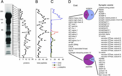
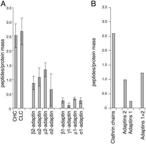
Tandem MS has identified 209 proteins of clathrin-coated vesicles (CCVs) isolated from rat brain. An overwhelming abundance of peptides were assigned to the clathrin coat with a 1:1 stoichiometry observed for clathrin heavy and light chains and a 2:1 stoichiometry of clathrin heavy chain with clathrin adaptor protein heterotetramers. Thirty-two proteins representing many of the known components of synaptic vesicles (SVs) were identified, supporting that a main function for brain CCVs is to recapture SVs after exocytosis. A ratio of vesicle-N-ethylmaleimide-sensitive factor attachment protein receptors to target-N-ethylmaleimide-sensitive factor attachment protein receptors, similar to that previously detected on SVs, supports a single-step model for SV sorting during CCV-mediated recycling of SVs. The uncovering of eight previously undescribed proteins, four of which have to date been linked to clathrin-mediated trafficking, further attests to the value of the current organelle-based proteomics strategy.


Figures

Fig. 2.
Fig. 2.
Distribution of proteins and peptides in CCVs. Proteins from one of the three independent CCV preparations were separated on SDS/PAGE, and 61 gel slices were cut as illustrated and processed for MS/MS. Molecular weight markers are indicated on the left. (A) Number of proteins identified in each gel slice. (B) Number of fragmented peptides assigned to proteins in each gel slice. (C) Numbers of peptides assigned to CHC (blue triangles, total assigned 398), CLC (black squares, total assigned 28), and μ-adaptin subunits of AP-1 and -2 (red circles, total assigned 84) are indicated. In slice 49 (arrow), 100% of the peptides are assigned to the CHC. In slice 32 (arrow), 63% of the total peptides are accounted for by the μ-subunits of AP-1 and -2, and in slice 21 (arrow), 39% of the total peptides are accounted for by CLC. (D) Known components of clathrin coats (coat) and SVs identified on CCVs by MS/MS are listed. The total number of proteins and peptides assigned to each category are presented in the pie graphs as a percentage of the total for the two categories.
Fig. 4.
Fig. 4.
Clathrin coat. (A) Normalization of the total number of peptides found in each preparation for each protein by dividing by the calculated mass of the protein (in kilodaltons) reveals a 1:1 relationship for CHC and CLCs as well as AP-2 and -1 subunits. The error bars represent the SD among the three preparations. (B) Peptide to protein mass of CHC and CLCs combined (clathrin) compared to the subunits of AP-2 combined, AP-1 combined, or the subunits of AP-2 and -1 combined.
Fig. 1.
Fig. 1.
Isolation of CCVs. (A) Coomassie blue staining of proteins separated by SDS/PAGE from each step of the enrichment protocol for CCVs. Aliquots of 40 μg of protein were loaded on each lane. H, P1, S2, P2, SGp, SGs, cpCCV, and CCV represent homogenate, first pellet, second supernatant, second pellet, sucrose/Ficoll gradient pellet, sucrose/Ficoll gradient supernatant, D2O cushion pellet, and CCVs, respectively. The predicted migratory positions of CHC, CLCs, and α-, β-, γ- and μ-adaptins are indicated. (B) Immunoblots detected the presence CHC, CLCs, and α-adaptin as indicated. (C) CCV fractions were evaluated by random sampling EM. Examples of CCVs are indicated by arrowheads, noncoated vesicles with comparable diameter to CCVs are indicated by short arrows, and noncoated profiles of diameter larger than CCVs are indicated by long arrows. (Bar = 200 nm.) (D) The proportion of coated profiles in the cpCCV and CCV preparations was evaluated from 64 and 66 independently and randomly sampled EM images, respectively. In total, 9,974 (cpCCV) and 10,457 (CCV) profiles were counted. The mean and SD from three independent experiments are 64.9 ± 1.2% and 72.7 ± 1.7% for cpCCV and CCV, respectively.
Fig. 3.
Fig. 3.
Western blot of selected proteins found in CCVs. Equal protein aliquots (40 μg) for cpCCV, CCV, and homogenate (H) fractions were prepared for Western blots with the indicated antibodies. For sucrose gradient fractions, an equal volume (30 μl) was applied to each lane.

References

    1. Conner, S. D. & Schmid, S. L. (2003) Nature 422, 37-44. - PubMed
    1. Brodsky, F. M., Chen, C. Y., Knuehl, C., Towler, M. C. & Wakenam, D. E. (2001) Annu. Rev. Cell Dev. Biol. 17, 517-568. - PubMed
    1. Ford, M. G., Mills, I. G., Peter, B. J., Vallis, Y., Praefcke, G. J., Evans, P. R. & McMahon, H. T. (2002) Nature 419, 361-366. - PubMed
    1. Conner, S. D. & Schmid, S. L. (2003) J. Cell Biol. 162, 773-779. - PMC - PubMed
    1. Motley, A., Bright, N. A., Seaman, M. N. & Robinson, M. S. (2003) J. Cell Biol. 162, 909-918. - PMC - PubMed

Publication types