

doi: 10.1073/pnas.0307877101.
Epub 2004 Mar 8.
The severe acute respiratory syndrome-coronavirus replicative protein nsp9 is a single-stranded RNA-binding subunit unique in the RNA virus world
Affiliations
- PMID: 15007178
- PMCID: PMC374323
- DOI: 10.1073/pnas.0307877101
Item in Clipboard
The severe acute respiratory syndrome-coronavirus replicative protein nsp9 is a single-stranded RNA-binding subunit unique in the RNA virus world
Proc Natl Acad Sci U S A.
.
Abstract
The recently identified etiological agent of the severe acute respiratory syndrome (SARS) belongs to Coronaviridae (CoV), a family of viruses replicating by a poorly understood mechanism. Here, we report the crystal structure at 2.7-A resolution of nsp9, a hitherto uncharacterized subunit of the SARS-CoV replicative polyproteins. We show that SARS-CoV nsp9 is a single-stranded RNA-binding protein displaying a previously unreported, oligosaccharide/oligonucleotide fold-like fold. The presence of this type of protein has not been detected in the replicative complexes of RNA viruses, and its presence may reflect the unique and complex CoV viral replication/transcription machinery.
Figures
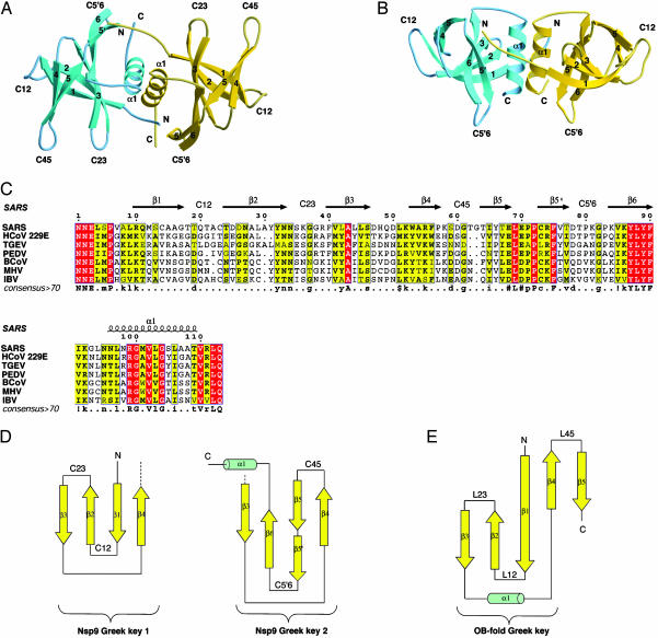
Crystal structure, sequence, and topology of SARS-CoV nsp9. (A) Ribbon representation of SARS-CoV nsp9. One molecule of the dimer is gold and the other is cyan. Loops between strands x and y are labeled C. (B) A 90° view of A. (C) Multiple alignment of nsp9 sequences from SARS-CoV National Center for Biotechnology Information (NCBI) accession no. AY291315, and several related CoVs: HCoV 229E, human CoV 229E, NCBI accession no. NP_073550; TGEV, transmissible gastroenteritis virus, NCBI accession no. NP_058423; PEDV, porcine epidemic diarrhea virus CV777, NCBI accession no. NP_598309, BCoV, bovine CoV, NCBI accession no. NP_150074; MHV, mouse hepatitis virus MHV-A59, NCBI accession no. NP_045298; and IBV, avian infectious bronchitis virus, NCBI accession no. NP_040829). The consensus sequence (identity cutoff >70%) is displayed under the multiple sequence alignment. Dots and residues in lowercase correspond to positions for which the residue conservation is under and above the cutoff value, respectively; positions marked by # correspond to either Asn, Asp, Glu, or Gln; positions marked by ! correspond to either Ile or Val, and $ corresponds to Leu or Met. Residues that are conserved in all sequences are boxed in red, and those for which conservation is >70% are boxed in yellow. For a given position, only residues homologous to the consensus are bold. The top numbers correspond to the amino acid sequence of SARS-CoV nsp9. Secondary structure elements and loops of nsp9 SARS-CoV are numbered according to Fig. 1 and are indicated above the alignment. (D) Schematic representation of nsp9 topology. nsp9 SARS-CoV β-barrel structure is a concatenation of two Greek key motifs, Greek key 1 having a g- topology and Greek key 2 a g+ topology (30), resulting in a six-stranded RH-g- to g+ topology. β-strands and α-helices are symbolized by arrows and cylinders, respectively, and they are numbered consistently with the sequence alignment. (E) Schematic representation of the typical Greek key (g- topology) motif found in the OB fold.

Surface analysis of SARS-CoV nsp9. (A) Electrostatic surface potential of nsp9 viewed from the same orientation as in Fig. 1 A. Potential values range from -5 kT (red) to 0 (white) and to +5 kT (blue), where k is the Boltzman constant and T is the temperature. Accessible Lys and Arg residues are indicated. (B) Back view from A, with the same color code. (C) Accessible surface colored according to the following: Lys or Arg are blue; Tyr, Phe, and Trp are yellow; and Val, Leu, and Ile are orange. The orientation is the same than in A. (D) Same coloring as in C with the same orientation as in B.

SARS-CoV nsp9 is an oligonucleotide-binding protein. (A) BIAcore analysis of nsp9 binding to immobilized DNA oligonucleotides. The protein (16 μM) was injected at a flow rate of 5 μl/min in HBS buffer on dextran layers containing 550 and 850 resonance units (RU) of the 25- and 45-mer oligonucleotides, respectively. The sensorgrams are the result of two independent experiments. RU values at 125 s (5 s after the end of the injection) are indicated. (B) Tryptophan fluorescence quenching study on SARS-CoV nsp9. The tryptophan fluorescence quenching at the plateau (in percent) is plotted versus the length of ssDNA probes. The KDapp values are extracted from the plot displayed in C and are discussed in the text. (C) The relative fluorescence of nsp9 at 340 nm is plotted as a function of the oligonucleotide concentration for ssDNA, ranging from 6- to 79-mer and for a 6, and 560-mer ssRNA. D, DNA; R, RNA. The KDapp values (discussed in the text), result from the fitting of the data to a single exponential (GraphPad).
References
-
- Drosten, C., Gunther, S., Preiser, W., van der Werf, S., Brodt, H. R., Becker, S., Rabenau, H., Panning, M., Kolesnikova, L., Fouchier, R. A., et al. (2003) N. Engl. J. Med. 348, 1967-1976. - PubMed
-
- Ksiazek, T. G., Erdman, D., Goldsmith, C. S., Zaki, S. R., Peret, T., Emery, S., Tong, S., Urbani, C., Comer, J. A., Lim, W., et al. (2003) N. Engl. J. Med. 348, 1953-1966. - PubMed
-
- Marra, M. A., Jones, S. J., Astell, C. R., Holt, R. A., Brooks-Wilson, A., Butterfield, Y. S., Khattra, J., Asano, J. K., Barber, S. A., Chan, S. Y., et al. (2003) Science 300, 1399-1404. - PubMed
-
- Rota, P. A., Oberste, M. S., Monroe, S. S., Nix, W. A., Campagnoli, R., Icenogle, J. P., Penaranda, S., Bankamp, B., Maher, K., Chen, M. H., et al. (2003) Science 300, 1394-1399. - PubMed
Publication types
MeSH terms
Substances
Associated data
- Actions
LinkOut - more resources
Full Text Sources
Other Literature Sources
Molecular Biology Databases
Miscellaneous
