Comparative Study
. 2004 Feb;19(4):898-906.
doi: 10.1111/j.0953-816x.2004.03117.x.

Olfactory bulb neurons express functional, entrainable circadian rhythms

Affiliations
Comparative Study

Olfactory bulb neurons express functional, entrainable circadian rhythms

Daniel Granados-Fuentes et al. Eur J Neurosci. 2004 Feb.

Abstract

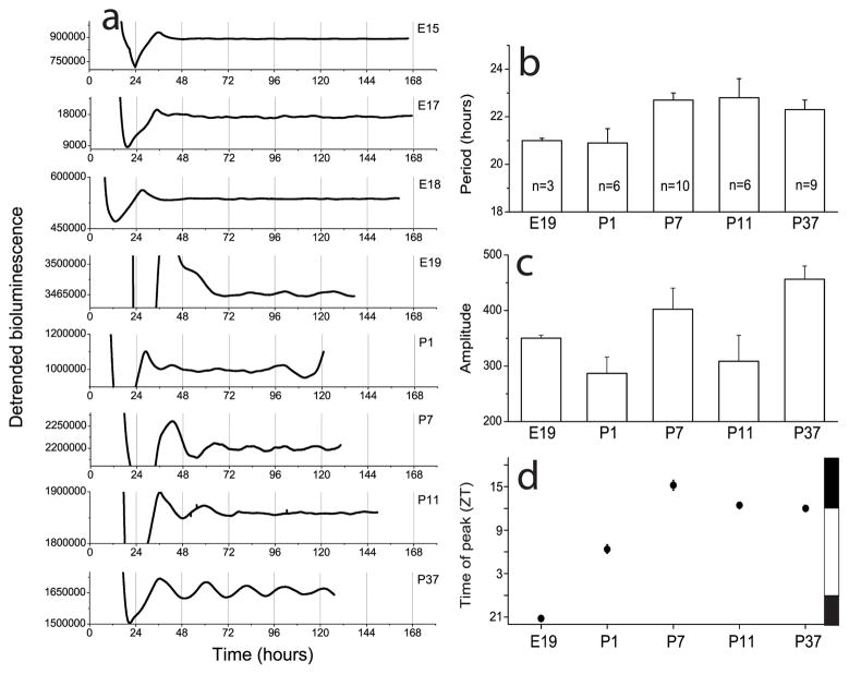
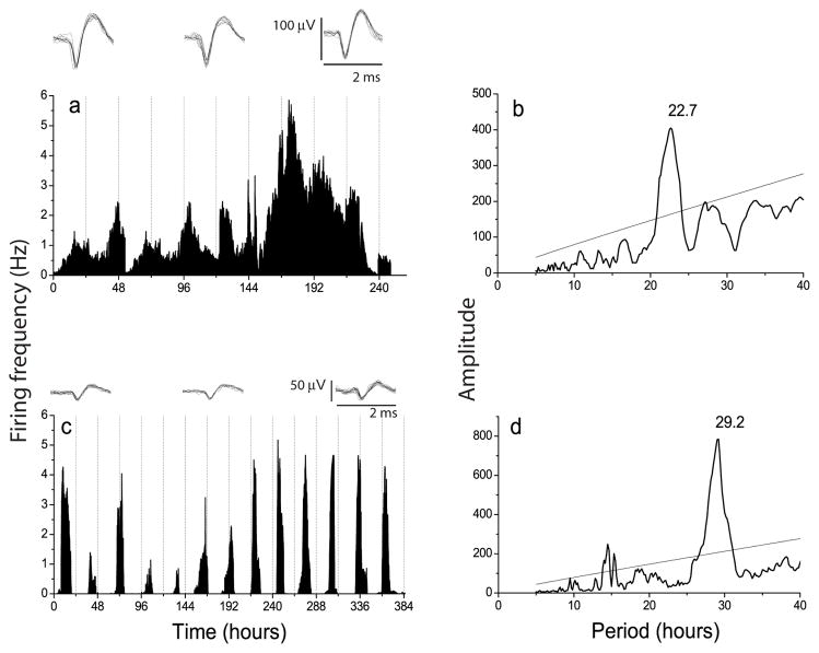
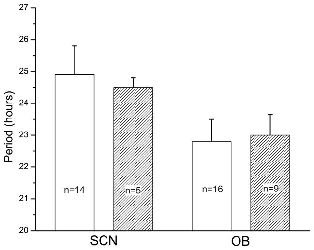
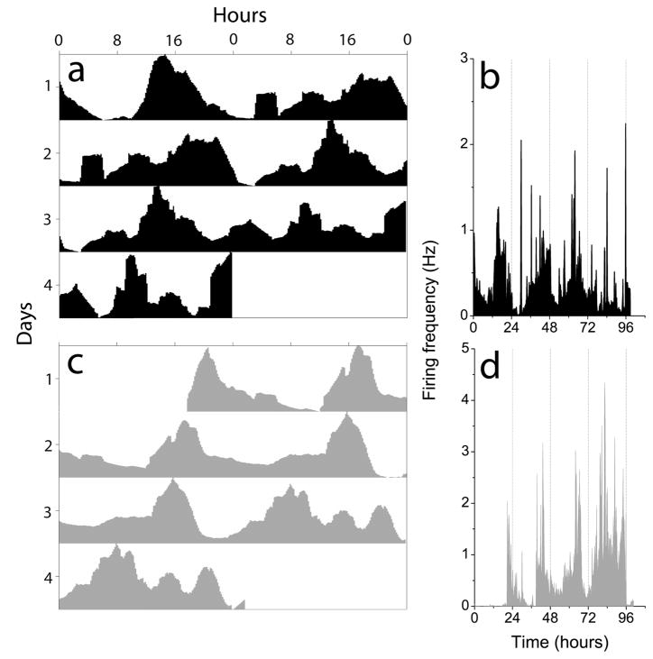
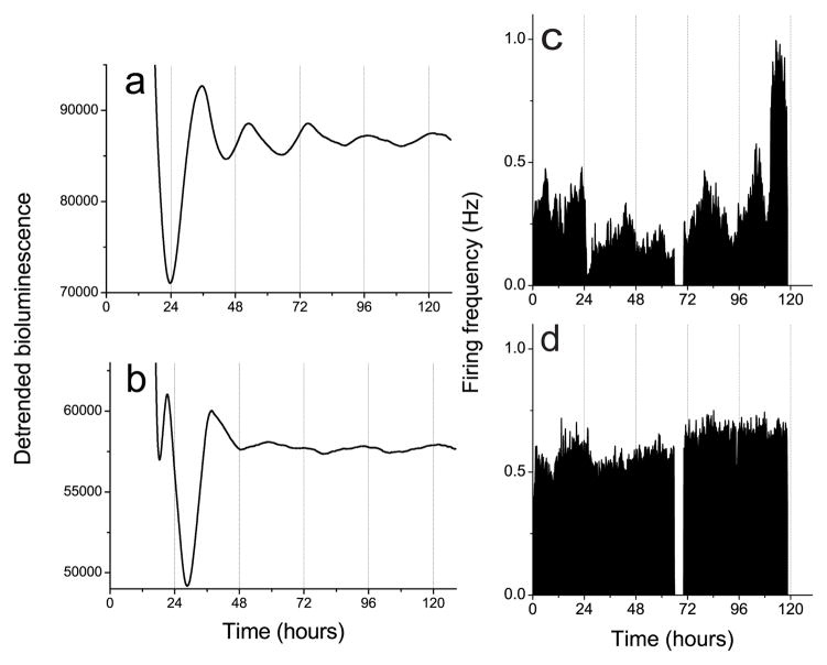
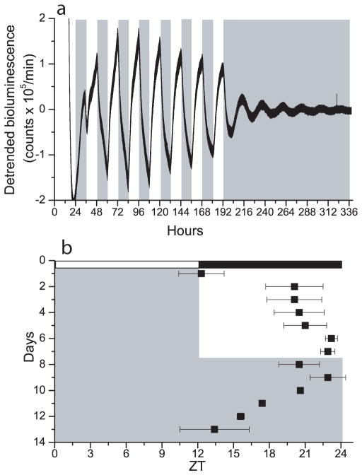
Circadian pacemakers drive many daily molecular, physiological and behavioural rhythms. We investigated whether the main olfactory bulb is a functional circadian pacemaker in rats. Long-term, multielectrode recordings revealed that individual, cultured bulb neurons expressed near 24-h oscillations in firing rate. Real-time recordings of Period1 gene activity showed that a population of cells within the bulb expressed synchronized rhythmicity starting on embryonic day 19. This rhythmicity was intrinsic to the mitral, and not the granule, cell layer, entrainable to physiological temperature cycles and temperature compensated in its period. However, removal of the olfactory bulbs had no effect on running wheel behaviour. These results indicate that individual mitral/tufted cells are competent circadian pacemakers which normally synchronize to each other. The daily rhythms in gene expression and firing rate intrinsic to the olfactory bulb are not required for circadian patterns of locomotion, indicating that they are involved in rhythms outside the canonical circadian system.


Figures

Figure 1
Figure 1
Circadian patterns of bioluminescence recorded from olfactory bulb explants taken from rats carrying a Period1-luciferase (Per1-luc) transgene. a, Daily oscillations of Per1 gene activity were present in the OB by E19. b–c, The average period and amplitude of rhythmic OB cultures did not depend upon developmental age. d, From E19 to P7, the Per1-luc expression in the OB peaked progressively later until it reached its mature phase around one week postnatal. Phase is given in Zeitgeber Time where ZT0 is dawn in the animal colony (7:00 a.m.) and ZT12 is dusk (7:00 p.m.). The time of lights on and off are shown on the right as white and black bars. Error bars show S.E.M.
Figure 2
Figure 2
a, Circadian rhythm in electrical activity of a representative OB neuron. Insets show 10 superimposed action potentials recorded at different times over 10 days to illustrate the reliable discrimination of the activity of a single OB cell. b, Chi-squared periodogram analysis of the firing pattern from the cell in a shows a dominant frequency at 22.7 h. c, Circadian firing pattern of an SCN neuron recorded for 16 days on a different multielectrode array. Insets show small action potentials, typical of SCN neurons. The period estimate for this cell, shown in d, was 29.2 h.
Figure 3
Figure 3
The average periods of firing rate (white bars) and Per1-luc activity (hatched bars) for cultured SCN and OB were similar. Period estimates were made from firing patterns of cells dispersed on P4-P6 and cultured for 16 days and from Per1-driven bioluminescence of tissues explanted and recorded beginning on postnatal day 37. While there was a tendency for the SCN to have a longer period than the OB, period differences were not significant (p=0.06, Student’s t-test). The periods of Per1-luc and electrical rhythms did not differ for either the SCN or OB (p>0.1, Student’s t-test). Error bars show S.E.M.
Figure 4
Figure 4
OB neurons in the same culture can express different circadian periods. a and c show actograms for the firing patterns shown in b and d, respectively. The two cells, recorded on electrodes 100 μm apart, had periods of 22.8 h (top) and 20.5 h (bottom).
Figure 5
Figure 5
Explant cultures of the mitral cell layer showed Per1-luc circadian rhythms (a) while the granule cell layer (b) did not. Similarly, cells dispersed from the mitral cell layer showed firing rate circadian rhythms (c) while cells from the granule cell layer did not (d). Data collection was briefly interrupted around 72 h for a medium change in c and d.
Figure 6
Figure 6
Temperature cycles entrain the OB in vitro. a, Bioluminescence recording from an OB explant in a temperature cycle of 35.3°C (white fill) to 36.8°C (gray fill). Per1 gene activity peaked near the cool-to-warm transitions during the 8 days of cyclic temperature. The rhythm free-ran from the last cool-to-warm transition in constant temperature. b, The peak of OB rhythmicity (black squares, mean ± SEM, n= 4) was tightly regulated by the temperature cycle (gray shows warm phases) so that after 8 days, the OB had delayed to peak approximately 1 h prior to the daily warm phase. Relative to the animals’ prior light cycle, the temperature cycle advanced the Per1-luc rhythm to around subjective dawn (white and black bars show the times of lights on and off in the animal colony, defined as ZT 0 and 12, respectively).

References

    1. Abe M, Herzog ED, Yamazaki S, Straume M, Tei H, Sakaki Y, Menaker M, Block GD. Circadian rhythms in isolated brain regions. J Neurosci. 2002;22:350–356. - PMC - PubMed
    1. Altman PL, Dittmer DS. Biological Handbooks. FASEB; Washington D.C: 1962. Growth, including reproduction and morphological development; pp. 304–313.
    1. Amir S, Cain S, Sullivan J, Robinson B, Stewart J. In rats, odor-induced Fos in the olfactory pathways depends on the phase of the circadian clock. Neurosci Lett. 1999;272:175–178. - PubMed
    1. Balsalobre A, Damiola F, Schibler U. A serum shock induces circadian gene expression in mammalian tissue culture cells. Cell. 1998;93:929–937. - PubMed
    1. Bayer SA. 3H-thymidine-radiographic studies of neurogenesis in the rat olfactory bulb. Exp Brain Res. 1983;50:329–340. - PubMed

Publication types