

Resetting the brain clock: time course and localization of mPER1 and mPER2 protein expression in suprachiasmatic nuclei during phase shifts
- PMID: 15009158
- PMCID: PMC3271804
- DOI: 10.1111/j.1460-9568.2004.03189.x
Resetting the brain clock: time course and localization of mPER1 and mPER2 protein expression in suprachiasmatic nuclei during phase shifts
Abstract
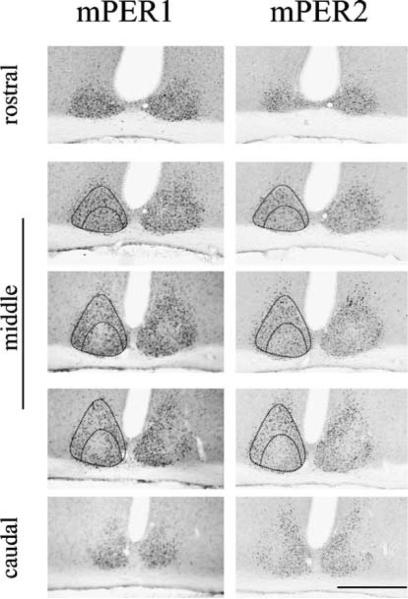
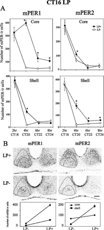
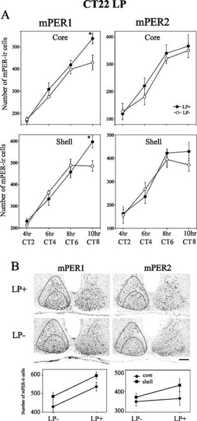
The mechanism whereby brief light pulses reset the mammalian circadian clock involves acute Per gene induction. In a previous study we investigated light-induced expression of mPer1 and mPer2 mRNA in the suprachiasmatic nuclei (SCN), with the aim of understanding the relationship between gene expression and behavioural phase shifts. In the present study, we examine the protein products of mPer1 and mPer2 genes in the core and shell region of SCN for 34 h following a phase-shifting light pulse, in order to further explore the molecular mechanism of photic entrainment. The results indicate that, during the delay zone of the phase response curve, while endogenous levels of mPER1 and mPER2 protein are falling, a light pulse produces an increase in the expression of both proteins. In contrast, during the advance zone of the phase response curve, while levels of endogenous mPER1 and mPER2 proteins are rising, a light pulse results in a further increase in mPER1 but not mPER2 protein. The regional distribution of mPER1 and mPER2 protein in the SCN follows the same pattern as their respective mRNAs, with mPER1 expression in the shell region of SCN correlated with phase advances and mPER2 in the shell region correlated with phase delays.
Figures
References
-
- Abrahamson EE, Moore RY. Suprachiasmatic nucleus in the mouse: retinal innervation, intrinsic organization and efferent projections. Brain Res. 2001;916:172–191. - PubMed
-
- Albrecht U, Zheng B, Larkin D, Sun ZS, Lee CC. MPer1 and mPer2 are essential for normal resetting of the circadian clock. J. Biol. Rhythms. 2001;16:100–104. - PubMed
-
- Bae K, Weaver DR. Light-induced phase shifts in mice lacking mPER1 or mPER2. J. Biol. Rhythms. 2003;18:123–133. - PubMed
-
- Dunlap JC. Molecular bases for circadian clocks. Cell. 1999;96:271–290. - PubMed
Publication types
MeSH terms
Substances
Grants and funding
LinkOut - more resources
Full Text Sources
