. 2004 Mar 1;18(5):512-27.
doi: 10.1101/gad.1177304. Epub 2004 Mar 10.

Phosphorylation of RCC1 in mitosis is essential for producing a high RanGTP concentration on chromosomes and for spindle assembly in mammalian cells

Affiliations

Phosphorylation of RCC1 in mitosis is essential for producing a high RanGTP concentration on chromosomes and for spindle assembly in mammalian cells

Hoi-Yeung Li et al. Genes Dev. .

Abstract

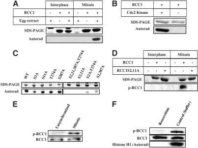
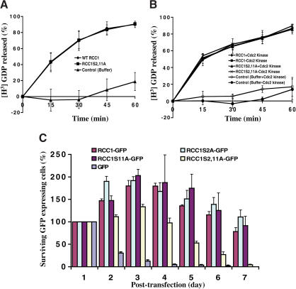
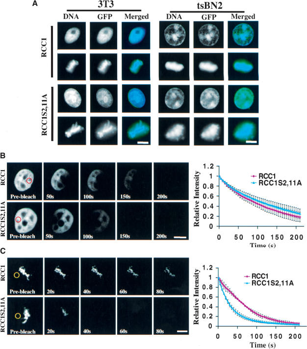
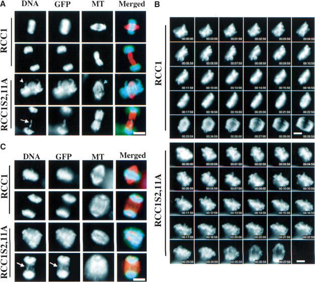
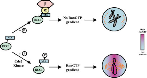
Spindle assembly is subject to the regulatory controls of both the cell-cycle machinery and the Ran-signaling pathway. An important question is how the two regulatory pathways communicate with each other to achieve coordinated regulation in mitosis. We show here that Cdc2 kinase phosphorylates the serines located in or near the nuclear localization signal (NLS) of human RCC1, the nucleotide exchange factor for Ran. This phosphorylation is necessary for RCC1 to generate RanGTP on mitotic chromosomes in mammalian cells, which in turn is required for spindle assembly and chromosome segregation. Moreover, phosphorylation of the NLS of RCC1 is required to prevent the binding of importin alpha and beta to RCC1, thereby allowing RCC1 to couple RanGTP production to chromosome binding. These findings reveal that the cell-cycle machinery directly regulates the Ran-signaling pathway by placing a high RanGTP concentration on the mitotic chromosome in mammalian cells.


Figures

Figure 1.
Figure 1.
RCC1 phosphorylation. (A) RCC1 is phosphorylated in mitosis. Purified human 6His-RCC1 or control buffer was incubated in interphase or mitotic egg extracts in the presence of γ-32p-ATP. RCC1 phosphorylation was analyzed by SDS-PAGE and autoradiography. (B) Cdc2 kinase phosphorylates 6His-RCC1. Purified 6His-RCC1 was incubated with Cdc2 kinase or control buffer in the presence of γ-32p-ATP, followed by SDS-PAGE and autoradiography. (C) S2 and S11 of RCC1 were phosphorylated in mitosis. Purified wild-type or mutant 6His-RCC1 was incubated in mitotic egg extracts in the presence of γ-32p-ATP and analyzed by SDS-PAGE and autoradiography. (D) Phosphospecific RCC1 antibody recognizes RCC1 treated with the mitotic, but not the interphase, egg extracts. (E) RCC1 is phosphorylated on S2 and/or S11 in mitotic HeLa cells. RCC1 isolated from unsynchronized or mitotic-arrested HeLa cells was probed with phosphospecific antibody (p-RCC1) or antibody recognizing both phosphorylated and unphosphorylated RCC1 (RCC1). (F) RCC1 is phosphorylated by Cdc2 kinase in HeLa cells. RCC1 isolated from mitotic-arrested HeLa cells treated with roscovitine or control buffer was probed with antibodies against RCC1 as in E. Cdc2 kinase activity in cell lysates was assayed using histone H1 as a substrate.
Figure 2.
Figure 2.
Mitotic phosphorylation of RCC1 is essential in vivo. (A) RCC1 and RCC1S2,11A have the same GEF activity in vitro. (B) Phosphorylation of RCC1 does not affect its GEF activity in vitro. Purified 6His-RCC1 or 6His-RCC1S2,11A was phosphorylated by Cdc2 kinase and then used in GEF assays. (C) Phosphorylation of S2 or S11 is essential for RCC1 function in vivo. RCC1-GFP, RCC1S2A-GFP, RCC1S11A-GFP, RCC1S2,11A-GFP, or GFP vector was used to transfect tsBN2 cells. One day posttransfection, the cells were shifted to 39.5°C to inactivate endogenous RCC1. The surviving GFP-expressing cells were determined as a percentage of the initial GFP-expressing cells at day 1.
Figure 3.
Figure 3.
Phosphorylation of RCC1 regulates its interaction with mitotic chromosomes. (A) Nonphosphorylatable RCC1 is properly localized. Swiss 3T3 and tsBN2 cells expressing wild-type RCC1-GFP or RCC1S2,11A-GFP were localized by fixation and fluorescence microscopy. FLIP of RCC1 in interphase (B) and mitosis (C). A spot marked by red or yellow circles in interphase nuclei or mitotic cytosol, respectively, was repeatedly photobleached in 3T3 cells expressing RCC1-GFP or RCC1S2,11A-GFP. Representative FLIP images are shown. Fluorescence intensity of the RCC1-GFP or RCC1S2,11A-GFP was quantified after each bleach pulse and plotted as relative intensity versus time. Error bars show S.D. of three to five independent experiments from different cells. Bars, 10μm.
Figure 4.
Figure 4.
Phosphorylation of RCC1 is required for spindle assembly and chromosome segregation. (A) Swiss 3T3 cells overexpressing either RCC1-GFP or RCC1S2,11A-GFP were fixed and stained with an anti-α-tubulin antibody (DM1-α) and DAPI. Examples of cells expressing RCC1-GFP with normal spindles and chromosomes or expressing RCC1S2,11A-GFP with defects in chromosome arm congression (white arrowhead), metaphase spindle (blue arrowhead), and chromosome segregation are shown (white arrow pointing to a lagging chromosome). (B) Time-lapse microscopy of Swiss 3T3 cells overexpressing RCC1-GFP or RCC1S2,11A-GFP from prometaphase to anaphase. Chromosomes in RCC1S2,11A-GFP-expressing cells failed to congress properly before segregation. (C) Temperature-shift experiments. tsBN2 cells stably expressing RCC1-GFP or RCC1S2,11A-GFP were arrested with nocodazole at the permissive temperature (37°C). After a further incubation at 39.5°C (to inactivate the endogenous RCC1), nocodazole was washed out and spindle assembly and chromosome segregation were analyzed by immunofluorescence microscopy. Representative metaphase, anaphase, and telophase cells fixed 40min after nocodazole washout are shown (arrow pointing to lagging chromosomes). Bars, 10μm.
Figure 5.
Figure 5.
Phosphorylation of RCC1 is essential for production of the RanGTP gradient. (A) YIC FRET reports the existence of RanGTP in the interphase nucleus. tsBN2 cells expressing YIC were incubated at permissive (33.5°C or 37°C) or restrictive (39.5°C) temperatures for 2–3 h before FRET. Images of interphase cells in CFP and YFP before and after photobleaching are shown. White dashed circles outline the interphase nuclei. Histogram shows quantification of FRET: the increase in CFP fluorescence intensity in bleached (black columns) and control unbleached (white columns) cells. (B) A typical quantification curve of FRET. The cell was scanned five times before and after bleaching YFP. Fluorescence intensities (I) of CFP from the last scan before bleaching (I5) and the first scan after bleaching (I6) were used to quantify FRET shown in the histograms. (C) RanGTP is concentrated on mitotic chromosomes in vivo. Images of pro-metaphase and anaphase cells before and after photobleaching are shown. White dashed circles outline mitotic cells. A higher FRET signal was detected on mitotic chromosomes than in the mitotic cytosol (see the histogram below the images). (D) Both RCC1-GFP and RCC1S2,11A-GFP support RanGTP production in interphase nuclei. FRET was carried out in tsBN2 cells expressing RCC1-GFP or RCC1S2,11A-GFP grown at 39.5°C for 2–3 h. Similar FRET signals were detected. (E) Phosphorylation of RCC1 is required for the RanGTP gradient production in mitosis. tsNB2 cells expressing RCC1-GFP or RCC1S2,11A-GFP were arrested in mitosis at the permissive temperature by nocodazole, and then shifted to 39.5°C for 2–3 h before FRET. Significantly stronger FRET was detected on the mitotic chromosomes in cells expressing RCC1-GFP than cells expressing RCC1S2,11A-GFP.
Figure 6.
Figure 6.
Importin α and β negatively regulate RanGTP production in mitosis. (A) The chromosome-coupled exchange mechanism. Core histones, Ran, and RCC1 are shown. As individual proteins, Ran and RCC1 weakly interact with chromatin via histones H3/H4 and histones H2A/H2B, respectively. However, formation of the binary complex of RCC1–Ran during nucleotide exchange allows the binary complex to bind to chromatin stably and irreversibly. Nucleotide exchange on Ran dissociates the binary complex into RanGTP and RCC1, thereby coupling RanGTP production to mitotic chromosomes. (B) FRAP analysis of RCC1 mobility in vivo. RanT24N was injected into the mitotic cytosol of cells expressing RCC1-GFP or RCC1S2,11-GFP followed by FRAP of RCC1 on mitotic chromosomes. RCC1-GFP, but not RCC1S2,11A-GFP, was immobilized on mitotic chromosomes. Error bars show S.D. from at least five independent experiments. (C) Phosphorylation of RCC1 in mitosis prevents its interaction with importin α and β. Purified 6His-RCC1 or 6His-RCC1S2,11A was incubated with interphase or mitotic egg extracts. Both forms of RCC1 bound to similar amounts of Ran. The 95-kD and 50-kD proteins that specifically interacted with RCC1S2,11A in mitosis (see arrowheads) were identified by microsequencing as importin α and β, respectively. Western blotting further confirmed the identity of importin β and Ran. (D) Unphosphorylated RCC1 binds to importin β in mitotic HeLa cells. Roscovitine or control buffer was used to inhibit Cdc2 kinase activity in the mitotic HeLa cells. Beads alone or beads bound to 6His-RanT24N were used to pull down RCC1 from the HeLa cell lysates. Western blotting revealed that roscovitine blocked RCC1 phosphorylation (as revealed by phosphospecific antibody, p-RCC1), which allowed RCC1 to bind importin β. (E) Competition experiments. The binding of RCC1-GFP or RCC1S2,11A-GFP to mitotic chromosomes in the presence of RanT24N was completed using an increasing concentration of unlabeled 6His-RCC1 or 6His-RCC1S2,11A, respectively. Histograms show the relative fluorescence intensity of GFP-labeled RCC1 on mitotic chromosomes under different conditions. RCC1S2,11A-GFP exhibits a weaker binding than RCC1-GFP. (F) Importin β inhibits the binding of RCC1S2,11A to chromosomes. Egg extracts depleted of importin β with or without add-back of importin β along with mock-depletion controls were used to assemble mitotic chromosomes in the presence of RanT24N. The binding of RCC1-GFP or RCC1S2,11A-GFP to the chromatin in the presence of unlabeled RCC1 competitors is quantified. Depletion of importin β allows binding of RCC1S2,11A-GFP to chromatin as strongly as RCC1-GFP.
Figure 7.
Figure 7.
A model. The N terminus of RCC1 (green) contains an NLS and is required for RCC1 to bind to chromatin. In mitosis, binding of importin α (yellow) and β (pink) to the NLS interferes with RCC1 binding to mitotic chromosomes. Therefore, phosphorylation of RCC1 is required to prevent the binding of importin α and β, thereby allowing RCC1 to bind to mitotic chromosomes with sufficient affinity to produce RanGTP on the chromosome. A high RanGTP concentration (purple) established on mitotic chromosomes (gray) is required for spindle assembly in mitosis.

Similar articles

Cited by

References

    1. Antonio C., Ferby, I., Wilhelm, H., Jones, M., Karsenti, E., Nebreda, A.R., and Vernos, I. 2000. Xkid, a chromokinesin required for chromosome alignment on the metaphase plate. Cell 102: 425–435. - PubMed
    1. Arnaoutov A. and Dasso, M. 2003. The Ran GTPase regulates kinetochore function. Dev Cell. 5: 99–111. - PubMed
    1. Bastiaens P.I.H. and Jovin, T.M. 1996. Microspectroscopic imaging tracks the intracellular processing of a signal transduction protein: Fluorescent-labeled protein kinase CβI. Proc. Natl. Acad. Sci. 93: 8407–8412. - PMC - PubMed
    1. Bilbao-Cortes D., Hetzer, M., Langst, G., Becker, P.B., and Mattij, I.W. 2002. Ran binds to chromatin by two distinct mechanisms. Curr. Biol. 12: 1151–1156. - PubMed
    1. Carazo-Salas R.E., Guarguaglini, G., Gruss, O.J., Segref, A., Karsenti, E., and Mattaj, I.W. 1999. Generation of GTP-bound Ran by RCC1 is required for chromatin-induced mitotic spindle formation. Nature 400: 178–181. - PubMed

Publication types

MeSH terms