. 2004 Mar 10;24(10):2366-74.
doi: 10.1523/JNEUROSCI.4711-03.2004.

Abnormal hippocampal axon bundling in EphB receptor mutant mice

Affiliations

Abnormal hippocampal axon bundling in EphB receptor mutant mice

Zhi-Yong Chen et al. J Neurosci. .

Abstract

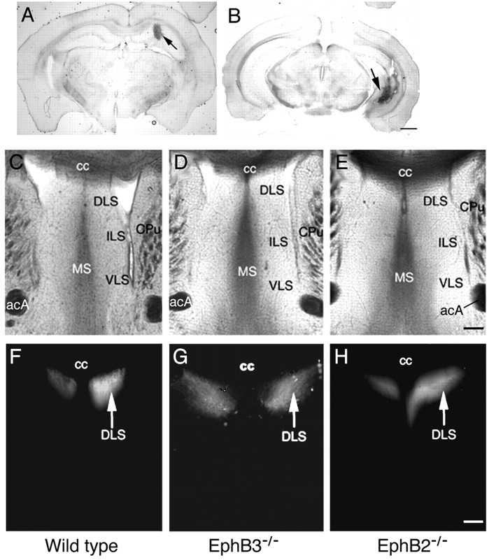
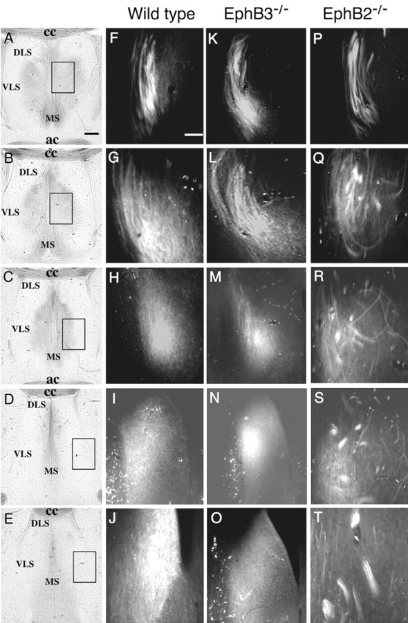
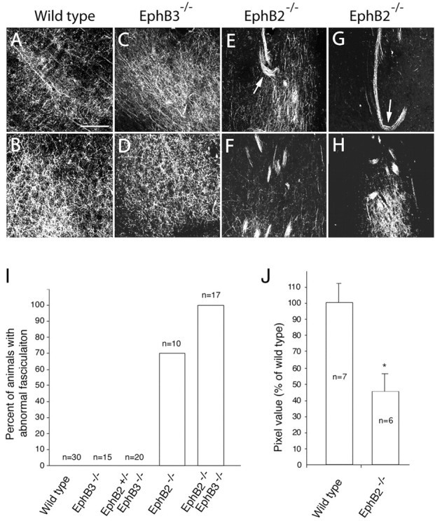
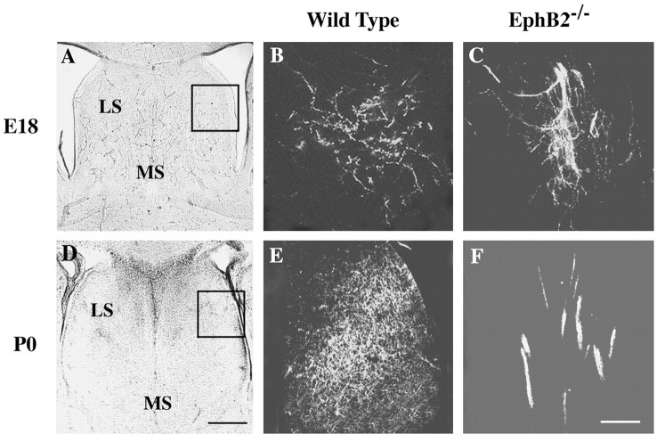
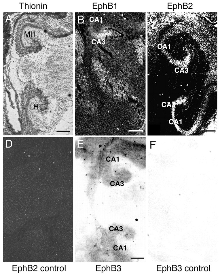
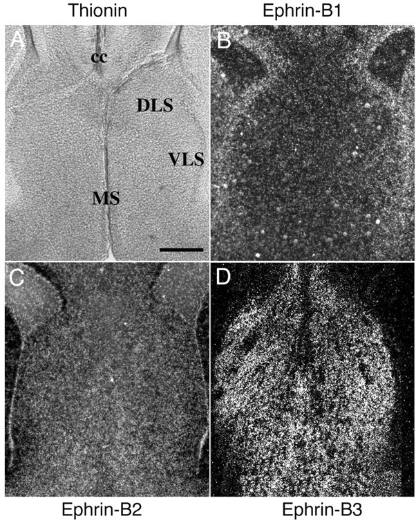
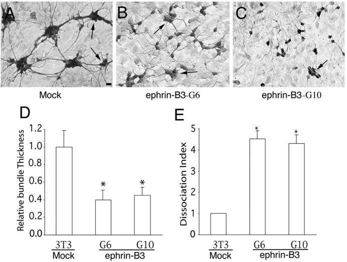
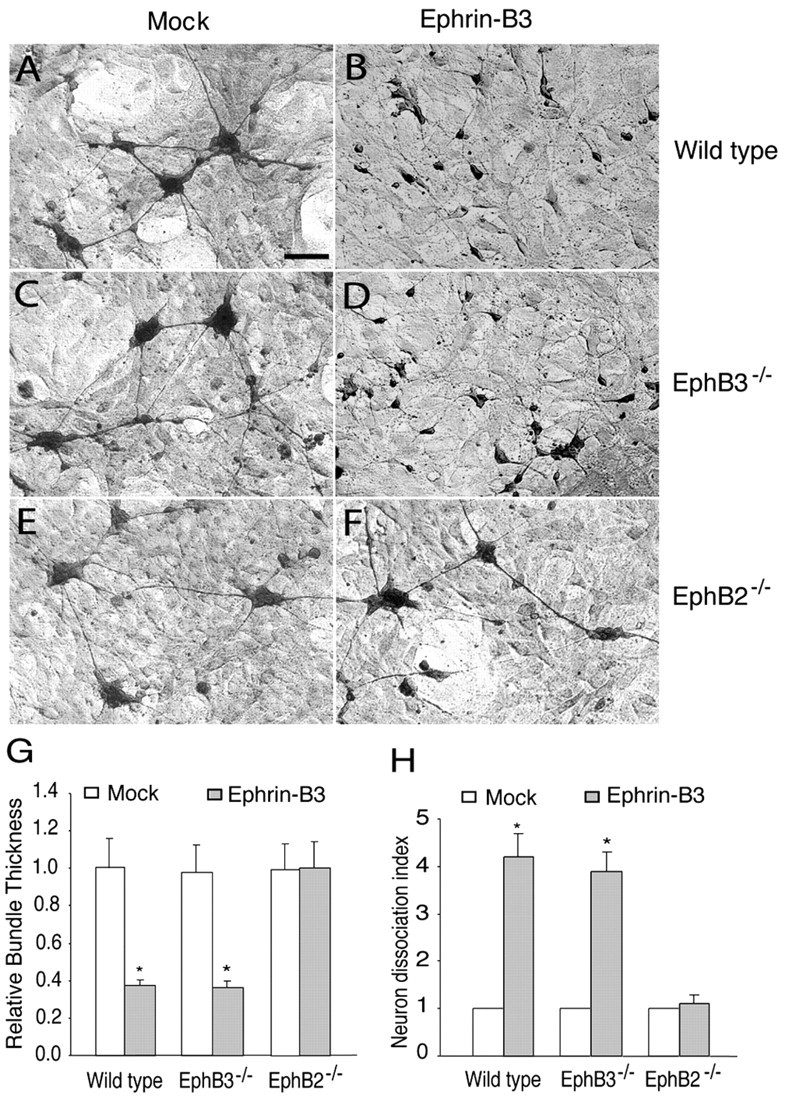
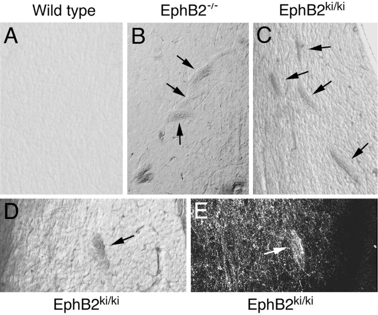
Axons travel frequently in bundles to reach their target. After arriving at the target, axon terminals defasciculate, migrate to topographically defined positions, and form synapses with appropriate target neurons. Here we present evidence that the B-type receptors of the erythropoietin-producing hepatocellular (Eph) family and a ligand, ephrin-B3, influence hippocampal axon defasciculation. The EphB receptors are expressed in the hippocampus, and the ligand, ephrin-B3, is transcribed in the lateral septum, the major subcortical target of hippocampal neurons. Ephrin-B3 promotes adhesion of hippocampal neurons to the ligand-expressing substrates in vitro, and the loss of the receptor EphB2 abrogates the effects of ephrin-B3. In mice deficient in EphB2 and EphB3, many hippocampal axons remain in bundles. This phenotype was also observed in mice that were specifically deleted for the cytoplasmic domain of EphB2. These observations indicate that the EphB receptors and their ligand regulate hippocampal axon defasciculation at the septal target, possibly through a receptor-mediated forward signaling mechanism.


Figures

Figure 1.
Figure 1.
Normal medial hippocampal projections to the lateral septum in EphB2- and EphB3-null mice. A, B, Typical Fluoro-ruby injection sites for hippocampal axon tracing in the medial hippocampus (A) and the lateral hippocampus (B). Injections were done according to stereotaxic coordinates by Franklin and Paxinos (1997). Arrows indicate injection sites. Scale bar, 1 mm. C-H, Tracing of the medial hippocampal axon terminal field in the lateral septum. C-E, Bright-field photomicrographs of sections of the traced brains containing the septal area from the wild-type (C), EphB3-/- (D), and EphB2-/- (E) mice. Major cytoarchitectural features are indicated. F-H, Dark-field photomicrographs depicting positions of the medial hippocampal axon terminals in wild-type (F), Eph B3-/- (G), and Eph B2-/- (H) mice. acA, Anterior commissure, anterior tract; cc, corpus callosum; CPu, caudate-putamen; DLS, dorsal lateral septum; ILS, intermediate lateral septum; MS, medial septum; VLS, ventral lateral septum. Scale bar, 0.2 mm.
Figure 2.
Figure 2.
Increased axon bundling in the terminal area of lateral hippocampal axons in the lateral septum in EphB receptor-null mice. Fluoro-ruby was injected into the lateral hippocampus to anterogradely label the lateral hippocampal axons. A-E, Bright-field photomicrographs of five different posterior to anterior coronal sections of a wild-type traced brain showing the major cytoarchitectural features of the septal region. These sections are ∼100 μm apart. Boxes in A-E indicate positions of dark-field panels on the right. F-T, Fluorescence photomicrographs of lateral hippocampal axon terminals at different posterior to anterior positions of wild-type (F-J), EphB3-/- single mutant (K-O), and EphB2-/- single mutant (P-T) mice. ac, Anterior commissure; cc, corpus callosum; DLS, dorsal lateral septum; MS, medial septum; VLS, ventral lateral septum. Scale bars: A, 0.2 mm; F, 0.02 mm.
Figure 3.
Figure 3.
Density of lateral hippocampal axon terminals in the lateral septum is reduced in EphB2-/- knock-out mice. A-H, High-magnification confocal images of hippocampal axon terminals in wild-type (A, B), EphB3-/- (C, D), and two different Eph B2-/- single mutant (E-H) mice. Positions of top panels correspond to Figure 2C, and bottom panels correspond to Figure 2 E. Arrows in E and G indicate axon bundles turning abnormally. I, Percentage of EphB knock-out animals with defasciculation defects. J, The density of the unbundled axon terminals in the EphB2-/- septum is significantly less than that of the wild-type septum. The pixel values of the unbundled axon terminals were measured using ImagePro image analysis software. Axon bundles were excluded from analysis. Asterisk indicates significant differences (p < 0.05; t test). Scale bar, 0.05 mm.
Figure 4.
Figure 4.
Hippocampal axon defasciculation defects in embryonic and newborn EphB-null mice. DiI was placed in the lateral hippocampal regions. A, D, Bright-field photomicrographs of wild-type brain sections showing major cytoarchitectural features of the septal region. B, E, Defasciculated hippocampal axons in the lateral septum of wild-type mouse brains. C, F, Abnormal hippocampal axon bundles in the lateral septum of the EphB2-/- mutant mice. Images in B, C, E, and F are from the boxed area in A and D. Scale bars: B, 0.25 mm; F, 0.05 mm.
Figure 5.
Figure 5.
Expression of EphB receptors in the hippocampus. Sagittal hippocampal sections of E18 mouse embryos were hybridized with 35S- or digoxigenin-labeled RNA probes. A, Bright-field photomicrograph of a hybridized sagittal mouse hippocampal section stained with thionin for cytoarchitecture reference. B, C, Dark-field photomicrographs of mouse hippocampal sections hybridized with 35S-labeled antisense riboprobes of EphB1 (B) and EphB2 (C). D, Dark-field photomicrographs of a similar section as shown in C hybridized with a control EphB2 sense riboprobe. E, F, Bright-field photomicrographs of hippocampal sections hybridized with digoxigenin-labeled antisense (E) or control sense (F) riboprobes of EphB3. MH and LH, Medial and lateral hippocampus. Scale bars, 0.5 mm.
Figure 6.
Figure 6.
Expression of the B-ephrins in the septum. Coronal sections of E18 mouse embryos were hybridized with 35S-labeled riboprobes of the B-ephrins. A, Bright-field photomicrograph of a thionin-stained section of the mouse septum for cytoarchitecture reference. B-D, Dark-field photomicrographs of mouse brain sections containing the septum hybridized with riboprobes of ephrin-B1 (B), ephrin-B2 (C), and ephrin-B3 (D). Sense control probes showed no specific hybridization signals. cc, Corpus callosum; DLS, dorsal lateral septum; VLS, ventral lateral septum; MS, medial septum. Scale bar, 0.2 mm.
Figure 7.
Figure 7.
Effect of ephrin-B ligands on the aggregation and axon bundling of rat hippocampal neurons. A, Hippocampal neurons of E18 embryos (105 per well of 12-well dish) were cocultured with control NIH-3T3 cells. B, C, E18 hippocampal neurons were cocultured with ephrin-B3-G6 (B) or ephrin-B3-G10 (C), two lines of ephrin-B3-expressing cells, respectively. The cocultures were maintained for 2 d and then stained with anti-neuron-specific enolase antibody. Arrows indicate aggregates with similar sizes from different cultures that were chosen for quantification. Scale bar, 20 μm. D, Relative bundle thickness. The diameter of all axonal bundles was measured from aggregates that had similar size in both control and ephrin-B3-expressing cell lines. E, Neuron dissociation index. Dissociation index is the ratio of the number of individual unaggregated neurons in cocultures of ephrin-B3-expressing and control cells. Because the total number of neurons seeded on each well is the same (105 per well of 12-well Corning tissue culture dishes), the number of unaggregated individual cells is a measure of how aggregated the neurons are in the presence of ephrin-B3 compared with the controls. The dissociation index on control cells was regarded as 1. Asterisk indicates statistical significance (p < 0.05; t test).
Figure 8.
Figure 8.
Effects of inactivation of EphB2 and EphB3 on the response of hippocampal neurons to ephrin-B3. A, C, E, E16 hippocampal neurons from the wild-type (A), EphB3-/- (C), and EphB2-/- (E) embryos were cocultured with a mock-transfected NIH-3T3 cell line. B, D, F, E16 hippocampal neurons from the wild-type (B), EphB3-/- (D), and EphB2-/- (F) mouse embryos were cocultured with an ephrin-B3-expressing cell line (G6). The cultures were maintained for 2 d and then stained with anti-neurofilament H antibody. Scale bar, 0.1 mm. G, Relative axon bundle diameter. H, Dissociation index. The diameter of axon bundles and the number of unaggregated neurons were quantified as described in Figure 7 and in Materials and Methods. Asterisk indicates statistical difference from the control (p < 0.05; t test).
Figure 9.
Figure 9.
Proper hippocampal axon defasciculation is dependent on the cytoplasmic domain of EphB2. Fluoro-ruby was injected into the lateral hippocampus of adult mice that were allowed to survive for 1 week. The traced brains were then sectioned with a vibratome, and the sections were examined for the presence of axon bundles and the morphology of axon terminals in the lateral septum. A, Bright-field photomicrograph of a vibratome section from an adult wild-type mouse. B, Bright-field photomicrograph of a vibratome section from an adult EphB2-/- mouse. C, D, Bright-field photomicrographs of vibratome sections from two different adult EphB2ki/ki homozygous mice. E, The axon bundle shown in D labeled with Fluoro-ruby anterograde tracing.

References

    1. Adams RH, Wilkinson GA, Weiss C, Diella F, Gale NW, Deutsch U, Risau W, Klein R (1999) Roles of ephrinB ligands and EphB receptors in cardiovascular development: demarcation of arterial/venous domains, vascular morphogenesis, and sprouting angiogenesis. Genes Dev 13: 295-306. - PMC - PubMed
    1. Bergemann AD, Zhang L, Chiang MK, Brambilla R, Klein R, Flanagan JG (1998) Ephrin-B3, a ligand for the receptor EphB3, expressed at the midline of the developing neural tube. Oncogene 16: 471-480. - PubMed
    1. Bohme B, VandenBos T, Cerretti DP, Park LS, Holtrich U, Rubsamen-Waigmann H, Strebhardt K (1996) Cell-cell adhesion mediated by binding of membrane-anchored ligand LERK-2 to the EPH-related receptor human embryonal kinase 2 promotes tyrosine kinase activity. J Biol Chem 271: 24747-24752. - PubMed
    1. Cowan CA, Henkemeyer M (2002) Ephrins in reverse, park and drive. Trends Cell Biol 12: 339-346. - PubMed
    1. Cowan CA, Yokoyama N, Bianchi LM, Henkemeyer M, Fritzsch B (2000) EphB2 guides axons at the midline and is necessary for normal vestibular function. Neuron 26: 417-430. - PubMed

Publication types

MeSH terms