. 2004 Mar 10;24(10):2440-8.
doi: 10.1523/JNEUROSCI.0783-03.2004.

Oscillating Purkinje neuron activity causing involuntary eye movement in a mutant mouse deficient in the glutamate receptor delta2 subunit

Affiliations

Oscillating Purkinje neuron activity causing involuntary eye movement in a mutant mouse deficient in the glutamate receptor delta2 subunit

Takashi Yoshida et al. J Neurosci. .

Abstract

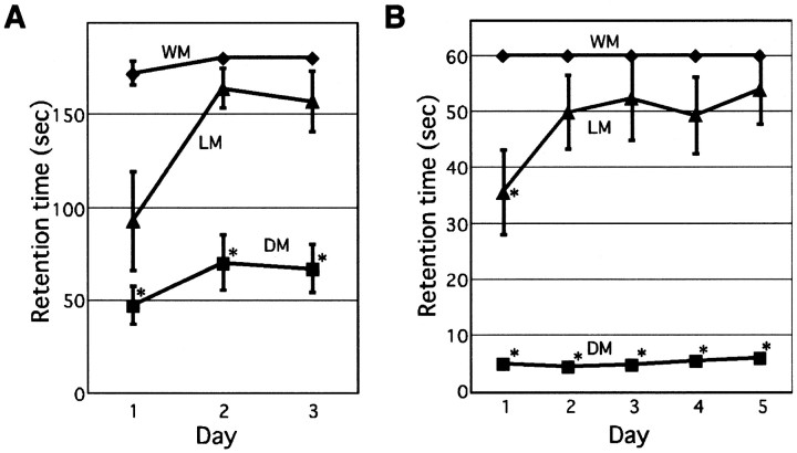
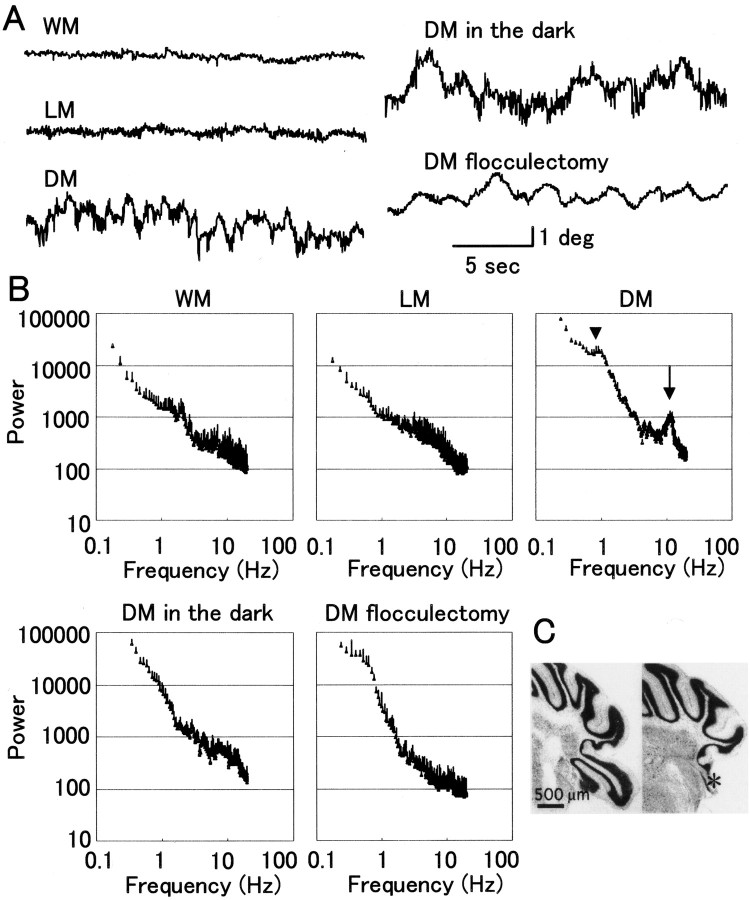
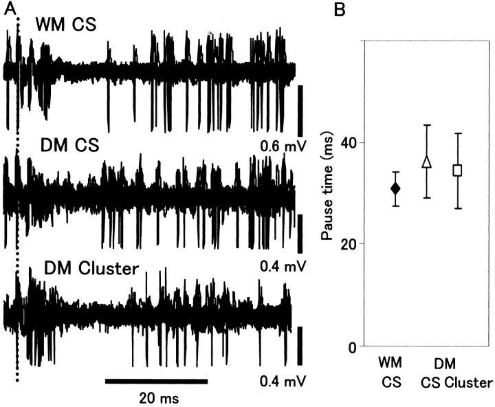
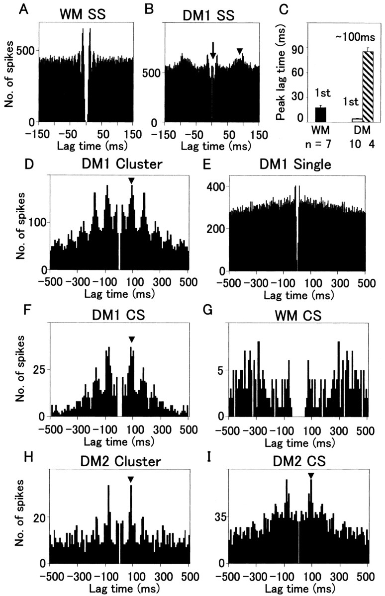
How failures in regulation of synaptic transmission in the mammalian CNS affect neuronal activity and disturb motor coordination is addressed. The mutant mouse deficient in the glutamate receptor delta2 subunit, specifically expressed in cerebellar Purkinje neurons, has defects in synaptic regulations such as synaptic plasticity, stabilization, and elimination of synaptic connections and shows failures in motor coordination and learning. In this study, the cause of motor discoordination of the delta2 mutant mouse was analyzed by comparing its motor control ability with those of the wild-type mouse and the lurcher mutant mouse, which loses all Purkinje neurons, the sole output neurons in the cerebellar cortex. Unexpectedly, the delta2 mutant mouse showed severer motor discoordination than the lurcher mouse without any cerebellar cortical outputs. The delta2 mutant mouse showed involuntary spontaneous eye movement with characteristic 10 Hz oscillation, which disappeared by ablation of the cerebellar flocculus, suggesting that the delta2 mutant cerebellar cortex outputs an abnormal signal. In vivo extracellular recordings of neuronal activity revealed that Purkinje neurons tended to fire clustered action potentials and complex spikes at approximately 10 Hz in the delta2 mutant mouse. A whole-cell patch-clamp recording from Purkinje neurons in cerebellar slices indicated that the clustered action potentials could be induced by climbing fiber activation. Taken together, our results suggest that the delta2 subunit deficiency produces the oscillating activity in Purkinje neurons by enhancing climbing fiber inputs, causing surplus movement and affecting motor control worse than no signal at all.


Figures

Figure 1.
Figure 1.
The cerebellar morphology of WMs, DMs, and LMs. A-C, Dorsal views of whole brains (A) and saggital slices of cerebella (B, C). Each arrow indicates the cerebellum. The cerebellum of an LM was the smallest, and that of a DM was slightly smaller than that of a WM. The cerebellar slices were stained with cresyl violet (B) or with anti-calbindin antibody (C). Calbindin was specifically expressed in PNs within the cerebellar cortex.
Figure 2.
Figure 2.
Motor control of WMs, DMs, and LMs. The retention times during which a mouse stayed on a stationary rod (A) or on a rotating rod (B) are shown (mean ± SEM). Each trial for a stationary rod test was 180 sec, and that for a rotating rod test was for 60 sec. The rotating rod test was started 1 d after the last day of the stationary rod test. Three rotating rod trials were performed each day, and the longest retention time was used to calculate the mean. n = 7 (WM); n = 11 (DM); n = 7 (LM). *Significantly lower than WMs (p < 0.05; Steel Dwass's test).
Figure 3.
Figure 3.
The spontaneous horizontal eye movement of mice. A, Traces show the horizontal eye position. B, The power specrtra calculated from eye position recordings. The spectra from 9-12 mice were averaged and then smoothed (Microsoft Excel). An arrowhead indicates the peak ∼1 Hz, and an arrow indicates that at ∼10 Hz. The mean ± SEM is shown at each frequency. C, The cerebella of the DM without (left) and after (right) ablation of the floccular complex (denoted by an asterisk).
Figure 4.
Figure 4.
Action potential firing of floccular PNs in a WM and DMs. A, Spontaneous activities of PNs in a WM and DMs (DM1 and DM2). Simple spikes, complex spikes (arrows), and clustered firings (arrowheads) are shown. B, Interval histograms of apparent simple spikes in a WM and a DM (DM1). C, The mean ± SEM of peak intervals of apparent simple spikes is shown. D, E, Complex spikes in a WM (D) and a DM (E; DM2). F, G, Clustered spikes (F) and simple spikes (G) in the PN of a DM (DM2). Fifteen traces were superimposed in D-F, and 100 traces were superimposed in G. In the PN of a WM, the time courses of complex spikes were constant. In contrast, multiple time courses of both complex spikes and clustered firings were recorded in the PN of a DM (DM2), although the time courses of simple spikes were constant.
Figure 5.
Figure 5.
The pose of simple spikes after a complex spike or a clustered firing. A, Fifteen single-unit recording traces after a complex spike or a clustered firing are superimposed for each. The dotted line indicates the onset of a complex spike or a clustered firing. B, The mean ± SEM of pose times is shown.
Figure 6.
Figure 6.
Autocorrelation of simple spikes, complex spikes, and clustered firings. A, B, Autocorrelation histograms of apparent simple spikes in PNs of a WM (A) and a DM (DM1) (B). The arrows indicate the shortest time lag, and the arrowheads indicate the peak ∼100 msec. Bin width is 1 msec. C, The mean ± SEM of peak lag times is shown. D, The autocorrelation histogram of clustered firings in the PN of a DM. E, The autocorrelation histogram of isolated simple spikes that does not constitute clustered firings in a DM. F, G, The autocorrelation histogram of complex spikes in the PNs of a DM (F) and a WM (G). The histograms shown in B and D-F were calculated from the data obtained from the same PN (DM1). H, I, The autocorrelation histograms of clustered firings (H) and complex spikes (I) obtained from a PN of another DM (DM2). The bin width is 10 msec in D-I. Each histogram was based on the 3 min recording, except for G (6 min).
Figure 7.
Figure 7.
Patch-clamp recording in slice preparations. A, Interval histograms of action potentials recorded extracellularly from PNs in slices prepared from a WM and a DM. B, The mean ± SD of peak intervals is shown. The larger variation in the interval histograms was noticed in DMs. C, EPSCs induced by activation of a climbing fiber were identified by the paired-pulse depression. D, Climbing fiber responses in a current-clamp condition in a WM and a DM. E, The numbers of complex spikes, clustered firings, and single spikes induced by climbing fiber activation are shown.
Figure 8.
Figure 8.
Simultaneous recording of the eye movement and the single PN activity. A, The top trace shows the eye position. The bottom vertical bars indicate the timings of complex spikes, clustered firings, and isolated simple spikes that do not constitute clustered firings. B, The averaged traces of eye position triggered by a complex spike or by a clustered firing occurring at time 0. Cells 1-3 were correlated to the horizontal eye movement, and cell 4 to the vertical movement. Cell 3 did not show a clear correlation. The eye position at 0 msec in each trace was set at 0° before averaging. Seventy to 120 traces were averaged. The SEM is also shown.
Figure 9.
Figure 9.
A scheme explaining the cause of 10 Hz oscillation in the PN activity and the motor discoordination. PF, Parallel fiber; CF, climbing fiber; ION, inferior olivary neuron.

References

    1. Aiba A, Kano M, Chen C, Stanton ME, Fox GD, Herrup K, Zwingman TA, Tonegawa S (1994) Deficient cerebellar long-term depression and impaired motor learning in mGluR1 mutant mice. Cell 7: 377-388. - PubMed
    1. Aizenman CD, Linden DJ (2000) Rapid, synaptically driven increases in the intrinsic excitability of cerebellar deep nuclear neurons. Nat Neurosci 3: 109-111. - PubMed
    1. Araki K, Meguro H, Kushiya E, Takayama C, Inoue Y, Mishina M (1993) Selective expression of the glutamate receptor channel δ2 subunit in cerebellar Purkinje cells. Biochem Biophys Res Commun 197: 1267-1276. - PubMed
    1. Chen C, Kano M, Abeliovich A, Chen L, Bao S, Kim JJ, Hashimoto K, Thompson RF, Tonegawa S (1995) Impaired motor coordination correlates with persistent multiple climbing fiber innervation in PKCγ mutant mice. Cell 83: 1233-1242. - PubMed
    1. Conquet F, Bashir ZI, Davies CH, Daniel H, Ferraguti F, Bordi F, Franz-Bacon K, Reggiani A, Matarese V, Conde F, Collingridge GL, Crepel F (1994) Motor deficit and impairment of synaptic plasticity in mice lacking mGluR1. Nature 372: 237-243. - PubMed

Publication types

MeSH terms