. 2004 Mar 10;24(10):2465-74.
doi: 10.1523/JNEUROSCI.4331-03.2004.

The two regulatory subunits of aplysia cAMP-dependent protein kinase mediate distinct functions in producing synaptic plasticity

Affiliations

The two regulatory subunits of aplysia cAMP-dependent protein kinase mediate distinct functions in producing synaptic plasticity

Jinming Liu et al. J Neurosci. .

Abstract

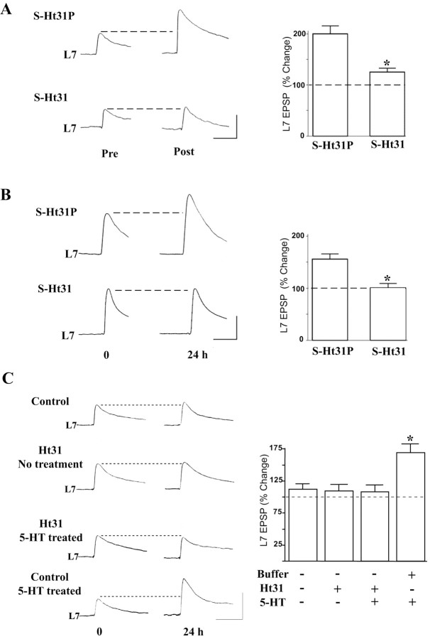
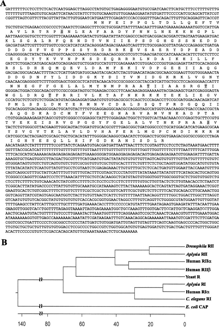
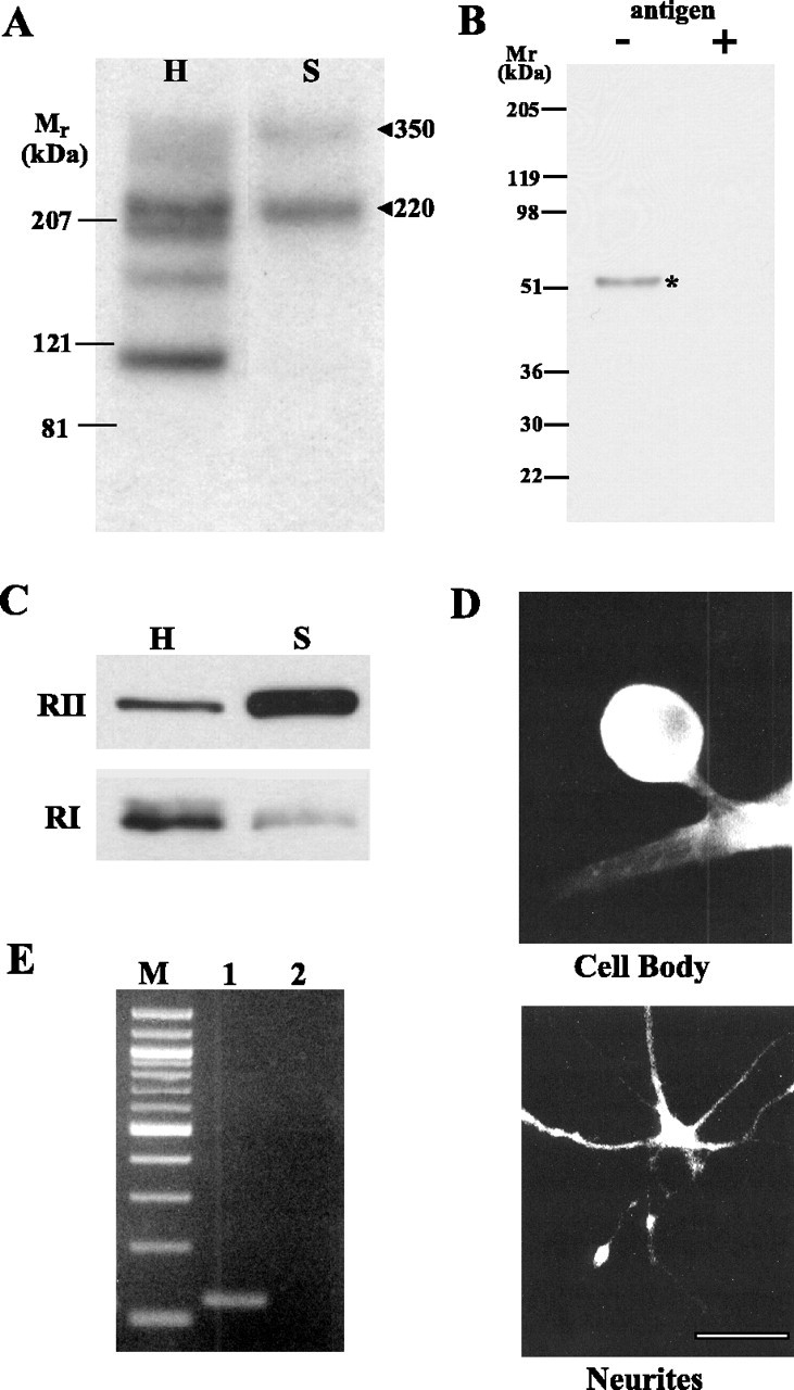
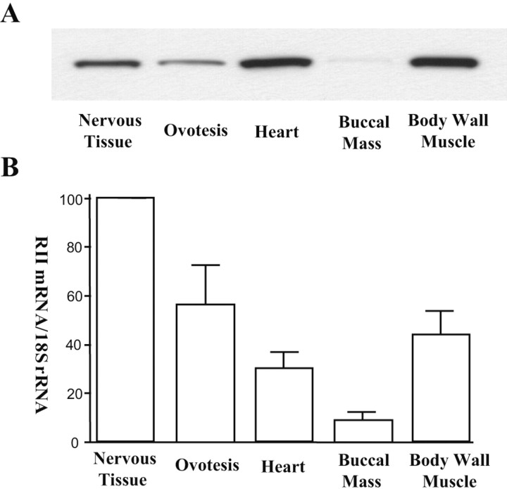
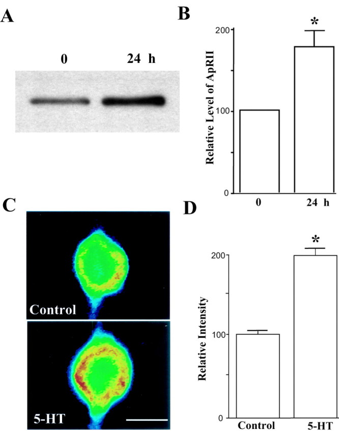
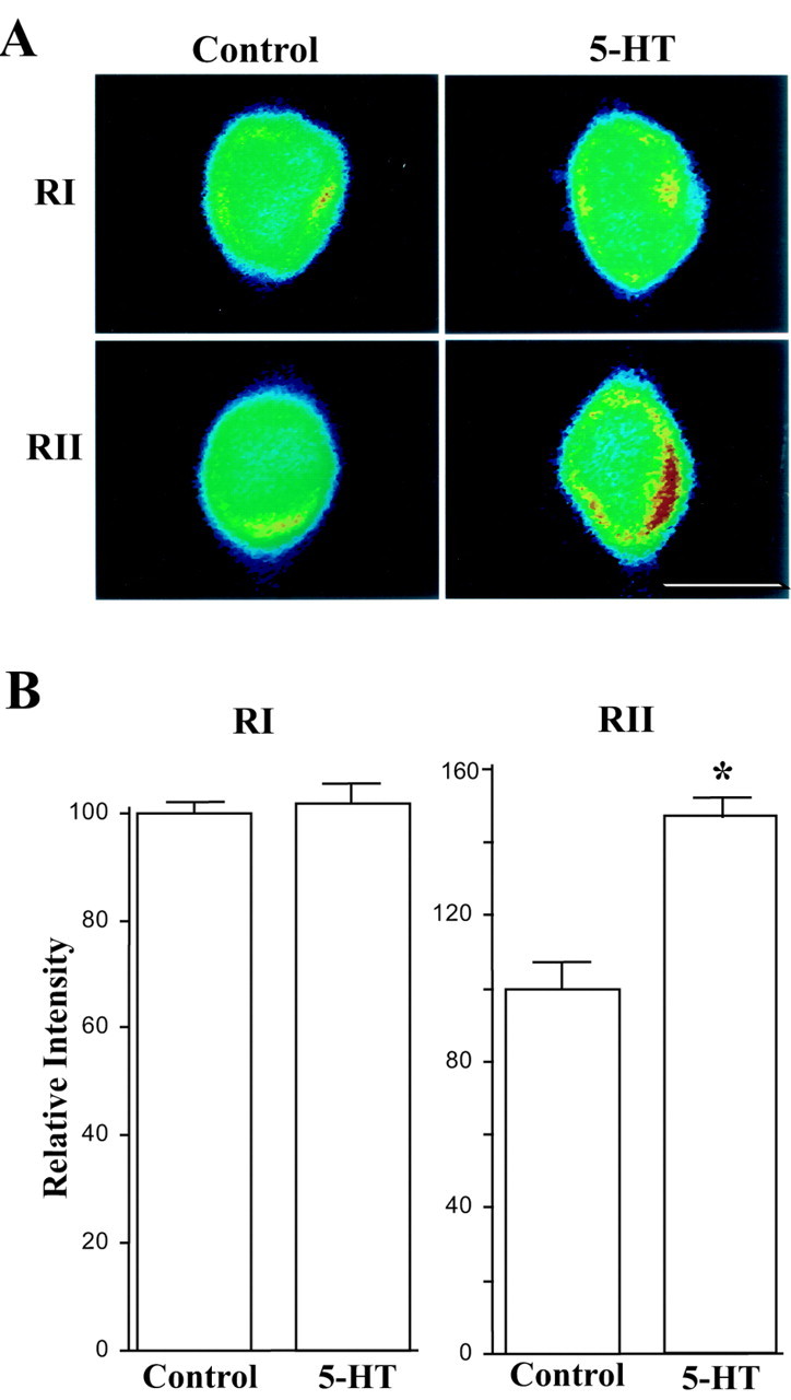
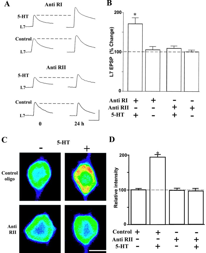
Activation of the cAMP-dependent protein kinase (PKA) is critical for both short- and long-term facilitation in Aplysia sensory neurons. There are two types of the kinase, I and II, differing in their regulatory (R) subunits. We cloned Aplysia RII; RI was cloned previously. Type I PKA is mostly soluble in the cell body whereas type II is enriched at nerve endings where it is bound to two prominent A kinase-anchoring-proteins (AKAPs). Disruption of the binding of RII to AKAPs by Ht31, an inhibitory peptide derived from a human thyroid AKAP, prevents both the short- and the long-term facilitation produced by serotonin (5-HT). During long-term facilitation, RII is transcriptionally upregulated; in contrast, the amount of RI subunits decreases, and previous studies have indicated that the decrease is through ubiquitin-proteosome-mediated proteolysis. Experiments with antisense oligonucleotides injected into the sensory neuron cell body show that the increase in RII protein is essential for the production of long-term facilitation. Using synaptosomes, we found that 5-HT treatment causes RII protein to increase at nerve endings. In addition, using reverse transcription-PCR, we found that RII mRNA is transported from the cell body to nerve terminals. Our results suggest that type I operates in the nucleus to maintain cAMP response element-binding protein-dependent gene expression, and type II PKA acts at sensory neuron synapses phosphorylating proteins to enhance release of neurotransmitter. Thus, the two types of the kinase have distinct but complementary functions in the production of facilitation at synapses of an identified neuron.


Figures

Figure 1.
Figure 1.
Disrupting ApRII-AKAP binding interferes with both short- and long-term facilitation. A, Incubation with S-Ht31 interferes with short-term facilitation. Representative EPSP traces were recorded in motor neuron L7 immediately before and 30 sec after the 5-HT treatment in the presence of the inactive (S-Ht31P) or inhibitory (S-Ht31) peptide. ANOVA indicates a significant effect of the treatment (df = 1, 18; F = 12.412; *p < 0.003). Individual comparison (Scheffé F test) indicates that S-Ht31 significantly reduced the change in EPSP amplitude (F = 15.597; p < 0.01). B, Incubation with S-Ht31 interferes with long-term facilitation. EPSP traces were recorded in L7 before and 24 hr after the 5-HT treatment in the presence of the control peptide (S-Ht31P) or the inhibitory peptide (S-Ht31). ANOVA indicates a significant effect of treatment (df = 1, 16; F = 32.02; p < 0.001), and individual comparison was significantly different (F = 17.063; *p < 0.01). C, Intracellular injection of Ht31 into sensory neurons blocks long-term facilitation produced by 5-HT. EPSP traces were recorded in L7 before and 24 hr after the treatment with 5-HT and sensory neurons injected with either buffer or Ht31. ANOVA indicates a significant effect of the treatment (df = 3, 24; F = 11.325; *p < 0.001). Individual comparisons (Scheffé F test) indicate that long-term facilitation was produced when 5-HT was applied to cultures whose sensory neurons were injected with buffer only (F = 3.230; p < 0.05 vs no treatment; F = 3.702; p < 0.05 vs injection of Ht31 only; and F = 4.152; p < 0.05 vs 5-HT and injection of Ht31). Calibration: 20 mV, 25 msec. The average amplitudes of the EPSP before each treatment were set as 100%.
Figure 2.
Figure 2.
The Aplysia RII gene. A, Nucleotide and predicted amino acid sequence of Aplysia RII. Autophosphorylation sites are highlighted. B, Phylogenetic analysis of R subunits. The predicated ApRII amino acid sequence was aligned using the MegAlign program from DNAStar with those of Drosophila RII (GenBank accession number AAF58862), human RIIα (P13861) and RIIβ (P31323), yeast R (CAA28726), Aplysia RI (CAA44246), human RIα (CAA01027), Caenorhabditis elegans RI (NP_508999), and Escherichia coli CAP (P03020) using the ClustralW method.
Figure 3.
Figure 3.
ApRII is enriched in neurites and synaptosomes along with two AKAPs. A, Autoradiographs showing the distribution of AKAPs in the nervous tissue homogenate (H) and synaptosomes (S). Synaptosomes were prepared as described by Chin et al. (1989). Proteins (80 μg) were resolved by SDS-PAGE on a 4-15% gradient gel and transferred to nitrocellulose membranes for the overlay assay. B, An affinity-purified antibody raised against KLSGALRFQENDTVNI, a peptide from ApRII, recognizes a single Mr 52,000 component in homogenates of Aplysia nervous tissue. Preincubation of the antibody with the antigen peptide (10 μm final concentration, ∼200 times excess of the ApRII antibody) abolishes its interaction with the ApRII protein. C, Immunoblots show that ApRII is enriched in synaptosomes (S), and type I R is not. The amount of ApRII in synaptosomes is ∼2.5 times (267 ± 38%; n = 3; p < 0.05, paired Student's t test) that in the homogenate, whereas the amount of RI in synaptosomes is about one-fourth (23 ± 2.7%; n = 3; p < 0.001, paired t test) that in homogenate. H, Total homogenate from which the synaptosomes were prepared. Equal amounts of protein (20 μg) from each fraction were separated by SDS-PAGE and transferred to nitrocellulose membranes for immunoblotting with specific antibodies against ApRII. The same sample was also probed using Aplysia RI antibodies. These blots are representative of three similar experiments. D, Immunocytochemistry showed that ApRII protein immunoreactivity is present in both the cell body and neurites. Sensory neurons were cultured with either L7 (cell body) or alone (neurites). Detection of ApRII in neurites of sensory neurons in the presence of L7 is difficult because neurites of L7 also stain for ApRII. Scale bar, 50 μm. E, ApRII mRNA is present in neurites. Lane 1, ApRII; lane 2, ApCAM (Aplysia cell adhesion protein); M, 100 bp ladder. Total RNA was isolated from dissected neurites of cultured sensory neurons. After the synthesis of cDNA, PCR was performed with gene-specific primers. The ApCAM message was used as a negative control because it does not enter the neurites (Schacher et al., 1999; Liu and Schwartz, 2003). Its absence indicates that the RNA in a sample is not contaminated with components from the cell body.
Figure 4.
Figure 4.
Tissue distribution of ApRII mRNA and protein. A, ApRII protein. Equal amounts of total protein (20 μg) from extracts of each tissue were separated by SDS-PAGE and transferred to nitrocellulose membranes for immunoblotting using the antibody against ApRII. The gel shown is representative of four similar experiments. Compared with its level in nervous tissue (set as 100), ApRII in protein is as follows: ovotestis, 61.5 ± 14.4; heart, 157.7 ± 17.0; buccal mass, 3.6 ± 1.1; and body wall muscle, 166.0 ± 19.9. B, ApRII mRNA. ApRII mRNA was assayed by quantitative RT-PCR. The amounts of RII mRNA in each tissue were normalized with an internal standard (18S ribosomal RNA). Error bars indicate SD (n = 3).
Figure 5.
Figure 5.
ApRII protein increases after 5-HT treatment and long-term facilitation. A, ApRII increases in synaptosomes after the treatment of 5-HT. An immunoblot of ApRII in synaptosomes of pleural-pedal ganglia is shown. Samples prepared from untreated animals served as controls. The gels shown are representative of three similar experiments. B, Quantitation of relative changes in ApRII (mean ± SEM) in synaptosomes. The amounts of ApRII in controls were set as 100 (*p < 0.02, paired t test). Immunoblots were quantified using Eastman Kodak Co. (Rochester, NY) 1D gel image analysis software. C, ApRII immunoreactivity increases in sensory neurons 24 hr after treatment with 5-HT. Cocultures were fixed 24 hr after the start of the treatment and immunostained with the anti-ApRII antibody and Cy3-conjugated anti-rabbit secondary antibody. Each micrograph shows the intensity of the fluorescence signals (false colors: red, high intensity; blue, low intensity). Scale bar, 50 μm. D, Quantitation of changes in ApRII (mean ± SEM) in cell bodies during long-term facilitation. The average intensity in controls was set as 100. Staining intensity after the 5-HT treatment was greater than that for the control (Scheffé F test, F = 81.094; *p < 0.01).
Figure 6.
Figure 6.
ApRII mRNA increases with long-term facilitation. A, In situ hybridization of Aplysia RII and RI mRNAs in sensory neurons cocultured with motor neurons. Cells were fixed 6 hr after the start of the 5-HT treatment and probed using biotin-labeled Aplysia RII or RI antisense oligonucleotides. The pseudocolor micrograph shows the intensity of fluorescence (false colors: red, high; blue, low). B, Quantitation of changes in the in situ hybridization signal for ApRII and RI mRNAs during long-term facilitation. Average intensity for control cultures was set as 100%. ANOVA indicates a significant effect of treatment (df = 3, 38; F = 36.119; *p < 0.001). Individual comparisons indicate that no change in RI mRNA resulted from the treatment with 5-HT. The amounts of ApRII mRNA increased significantly after treatment with 5-HT (F = 20.775; p < 0.01).
Figure 7.
Figure 7.
Inhibition of ApRII synthesis by antisense oligonucleotides blocks long-term Facilitation. A, Injection of antisense oligonucleotides of ApRII blocked long-term facilitation produced by 5-HT. Shown are representative EPSP traces before and 24 hr after the start of the 5-HT treatment. Treatment with 5-HT began 4-5 hr after injections of antisense oligonucleotides (anti-RI, corresponding to a gene-specific region of the cAMP-binding domain-A, or anti RII, corresponding to a gene-specific region between the PEST region and cAMP-binding domain-A) into sensory neurons. Calibration: 20 mV, 25 msec. B, Summary of changes in EPSP amplitude (mean ± SEM) in L7 at 24 hr produced by the indicated treatments. ANOVA indicates significant effects of antisense oligonucleotide injection (df = 3, 24; F = 11.724; p < 0.001). Long-term facilitation was produced when 5-HT was applied to cultures whose sensory neurons were injected with anti RI oligonucleotides (F = 5.976; p < 0.01 vs injection of anti RI alone). No long-term facilitation occurred by 5-HT when anti RII oligonucleotides were injected. C, Injection of antisense oligonucleotides of ApRII blocked the increase in ApRII protein immunoreactivity produced by 5-HT. Injection of the control oligonucleotide failed to block the increase. Cultures were treated with or without 5-HT 4-5 hr after injection of antisense or control oligonucleotide. Cultures were fixed 24 hr after the start of the treatment and immunostained with the anti-ApRII antibody and Cy3-conjugated anti-rabbit secondary antibody. Each micrograph represents the intensity of the fluorescence signals (false colors: red, high; blue, low). Long-term facilitation with 5-HT was detected only in cultures injected with the control oligonucleotide (data not shown). Scale bar, 50 μm. D, Quantitation of changes in ApRII immunoreactivity (mean ± SEM) in sensory neuron cell bodies during long-term facilitation. The average intensity in cultures injected with the control oligonucleotide was set as 100%. ANOVA indicated a significant effect of treatment (df = 3, 23; F = 88.194; p < 0.001). Cultures treated with 5-HT after injection with control oligonucleotides showed significantly greater signals than nontreated cultures (F = 62.615; p < 0.01) or cultures injected with antisense oligonucletides followed by 5-HT (F = 59.171; p < 0.01).

Similar articles

Cited by

References

    1. Alberini CM, Ghirardi M, Metz R, Kandel ER (1994) C/EBP is an immediate-early gene required for the consolidation of long-term facilitation in Aplysia. Cell 76: 1099-1114. - PubMed
    1. Amieux PS, McKnight GS (2002) The essential role of RI alpha in the maintenance of regulated PKA activity. Ann NY Acad Sci 968: 75-95. - PubMed
    1. Angers A, Fioravante D, Chin J, Cleary LJ, Bean AJ, Byrne JH (2002) Serotonin stimulates phosphorylation of Aplysia synapsin and alters its subcellular distribution in sensory neurons. J Neurosci 22: 5412-5422. - PMC - PubMed
    1. Bartsch D, Ghirardi M, Skehel PA, Karl KA, Herder SP, Chen M, Bailey CH, Kandel ER (1995) Aplysia CREB2 represses long-term facilitation: relief of repression converts transient facilitation into long-term functional and structural change. Cell 83: 979-992. - PubMed
    1. Bartsch D, Casadio A, Karl KA, Serodio P, Kandel ER (1998) CREB1 encodes a nuclear activator, a repressor, and a cytoplasmic modulator that form a regulatory unit critical for long-term facilitation. Cell 95: 211-223. - PubMed

Publication types

MeSH terms

LinkOut - more resources